 Explore
 Validate
 Learn
Learn Western blot
 Immunocytochemistry
Antibody data
- Antibody Data
- Antigen structure
- References [1]
- Comments [0]
- Validations
- Immunocytochemistry [1]
Submit
Validation data
Reference
Comment
Report error
- Product number
- BBA15 - Provider product page

- Provider
- R&D Systems
- Product name
- Human ICAM-3/CD50 Antibody
- Antibody type
- Monoclonal
- Description
- Protein A or G purified from hybridoma culture supernatant. Detects human ICAM-3/CD50 in ELISAs and Western blots. ICAM-3/CD50 was found not to recognize COS cells expressing the human ICAM-1 protein. The epitope for ICAM-3/CD50 has been found to be located on domain 1, or the interface between domains 1 and 2 of the human ICAM-3/CD50 molecule.
- Reactivity
- Human
- Host
- Mouse
- Conjugate
- Unconjugated
- Isotype
- IgG
- Antibody clone number
- ICAM-3.3
- Vial size
- 200 ug
- Concentration
- LYOPH
- Storage
- Use a manual defrost freezer and avoid repeated freeze-thaw cycles. 12 months from date of receipt, -20 to -70 °C as supplied. 1 month, 2 to 8 °C under sterile conditions after reconstitution. 6 months, -20 to -70 °C under sterile conditions after reconstitution.
Submitted references Macrophage recognition of ICAM-3 on apoptotic leukocytes.
Moffatt OD, Devitt A, Bell ED, Simmons DL, Gregory CD
Journal of immunology (Baltimore, Md. : 1950) 1999 Jun 1;162(11):6800-10
Journal of immunology (Baltimore, Md. : 1950) 1999 Jun 1;162(11):6800-10
No comments: Submit comment
Supportive validation
- Submitted by
- R&D Systems (provider)
- Main image
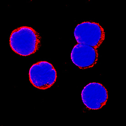
- Experimental details
- ICAM-3/CD50 in Human PBMCs. ICAM-3/CD50 was detected in immersion fixed human peripheral blood mononuclear cells (PBMCs) using Mouse Anti-Human ICAM-3/CD50 Monoclonal Antibody (Catalog # BBA15) at 15 µg/mL for 3 hours at room temperature. Cells were stained using the NorthernLights™ 557-conjugated Anti-Mouse IgG Secondary Antibody (red; Catalog # NL007) and counterstained with DAPI (blue). Specific staining was localized to cell surfaces. View our protocol for Fluorescent ICC Staining of Non-adherent Cells.
