Explore
 Validate
 Learn
Learn Immunocytochemistry
Antibody data
- Antibody Data
- Antigen structure
- References [0]
- Comments [0]
- Validations
- Immunocytochemistry [1]
Submit
Validation data
Reference
Comment
Report error
- Product number
- MAB6698 - Provider product page

- Provider
- R&D Systems
- Product name
- Rat beta-1,3-Glucuronyltransferase 1/B3GAT1 Antibody
- Antibody type
- Monoclonal
- Description
- Protein A or G purified from hybridoma culture supernatant. Detects rat beta-1,3-Glucuronyltransferase 1/B3GAT1 in ELISA.
- Reactivity
- Rat
- Host
- Mouse
- Conjugate
- Unconjugated
- Antigen sequence
NP_445455- Isotype
- IgG
- Antibody clone number
- 882302
- Vial size
- 100 ug
- Concentration
- LYOPH
- Storage
- Use a manual defrost freezer and avoid repeated freeze-thaw cycles. 12 months from date of receipt, -20 to -70 °C as supplied. 1 month, 2 to 8 °C under sterile conditions after reconstitution. 6 months, -20 to -70 °C under sterile conditions after reconstitution.
No comments: Submit comment
Supportive validation
- Submitted by
- R&D Systems (provider)
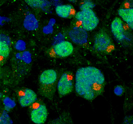
- Main image

- Experimental details
- beta-1,3-Glucuronyltransferase 1/B3GAT1 in Rat Cortical Stem Cells. beta-1,3-Glucuronyltransferase 1/B3GAT1 was detected in immersion fixed differentiated rat cortical stem cells using Mouse Anti-Rat beta-1,3-Glucuronyltransferase 1/B3GAT1 Monoclonal Antibody (Catalog # MAB6698) at 10 µg/mL for 3 hours at room temperature. Cells were stained using the NorthernLights™ 557-conjugated Anti-Mouse IgG Secondary Antibody (red; Catalog # NL007). Oligo2 was also detected in the cells using Human Olig2 Antigen Affinity-purified Polyclonal Antibody (Catalog # AF2418) and NorthernLights™ 637-conjugated Anti-Goat IgG Secondary Antibody (green; Catalog # NL003). Cells were counterstained with DAPI (blue). Specific staining of B3GAT1 was localized to transmembrane Golgi. View our protocol for Fluorescent ICC Staining of Stem Cells on Coverslips.