 Explore
 Validate
 Learn
Learn Western blot
Antibody data
- Antibody Data
- Antigen structure
- References [14]
- Comments [0]
- Validations
- Western blot [1]
- Immunohistochemistry [1]
- Flow cytometry [2]
Submit
Validation data
Reference
Comment
Report error
- Product number
- MA5-11605 - Provider product page

- Provider
- Invitrogen Antibodies
- Product name
- CD57 Monoclonal Antibody (HNK-1 (Leu-7))
- Antibody type
- Monoclonal
- Antigen
- Other
- Description
- MA5-11605 targets CD57 in FACS, WB and IHC (P) applications and shows reactivity with Human samples.
- Antibody clone number
- HNK-1 (Leu-7)
- Concentration
- 0.2 mg/mL
Submitted references Neural Crest Stem Cells in Juvenile Angiofibromas.
Senescent T Cells Predict the Development of Hyperglycemia in Humans.
Histologic and Immunohistochemical Analyses of Soft Tissue Sarcomas From brca2-Mutant/ tp53-Mutant Zebrafish Are Consistent With Neural Crest (Schwann Cell) Origin.
Enteric neural crest-derived cells promote their migration by modifying their microenvironment through tenascin-C production.
Expression pattern of the homeotic gene Bapx1 during early chick gastrointestinal tract development.
Immunophenotypic characterization of enteric neural crest cells in the developing avian colorectum.
Spatial differences in the presence of FOXP3+ and GranzymeB+ T cells between the intra- and extravascular compartments in renal allograft vasculopathy.
Innate and adaptive immunity during epileptogenesis and spontaneous seizures: evidence from experimental models and human temporal lobe epilepsy.
Hyalinizing spindle cell tumor with giant rosettes arising in the lung: report of a case with FUS-CREB3L2 fusion transcripts.
Isolation of neural crest-derived stem cells from rat embryonic mandibular processes.
Enteric nervous system patterning in the avian hindgut.
Primary acinic cell carcinoma of the lung with lymph node metastasis.
Epstein-Barr virus-associated lymphoproliferative disease occurring in a patient with sarcoidosis treated by methotrexate and methylprednisolone.
Epstein-Barr virus-associated lymphoproliferative disease occurring in a patient with sarcoidosis treated by methotrexate and methylprednisolone.
Schick B, Pillong L, Wenzel G, Wemmert S
International journal of molecular sciences 2022 Feb 9;23(4)
International journal of molecular sciences 2022 Feb 9;23(4)
Senescent T Cells Predict the Development of Hyperglycemia in Humans.
Lee YH, Kim SR, Han DH, Yu HT, Han YD, Kim JH, Kim SH, Lee CJ, Min BH, Kim DH, Kim KH, Cho JW, Lee WW, Shin EC, Park S
Diabetes 2019 Jan;68(1):156-162
Diabetes 2019 Jan;68(1):156-162
Histologic and Immunohistochemical Analyses of Soft Tissue Sarcomas From brca2-Mutant/ tp53-Mutant Zebrafish Are Consistent With Neural Crest (Schwann Cell) Origin.
White LA, Sexton JM, Shive HR
Veterinary pathology 2017 Mar;54(2):320-327
Veterinary pathology 2017 Mar;54(2):320-327
Enteric neural crest-derived cells promote their migration by modifying their microenvironment through tenascin-C production.
Akbareian SE, Nagy N, Steiger CE, Mably JD, Miller SA, Hotta R, Molnar D, Goldstein AM
Developmental biology 2013 Oct 15;382(2):446-56
Developmental biology 2013 Oct 15;382(2):446-56
Expression pattern of the homeotic gene Bapx1 during early chick gastrointestinal tract development.
Faure S, Georges M, McKey J, Sagnol S, de Santa Barbara P
Gene expression patterns : GEP 2013 Dec;13(8):287-92
Gene expression patterns : GEP 2013 Dec;13(8):287-92
Immunophenotypic characterization of enteric neural crest cells in the developing avian colorectum.
Nagy N, Burns AJ, Goldstein AM
Developmental dynamics : an official publication of the American Association of Anatomists 2012 May;241(5):842-51
Developmental dynamics : an official publication of the American Association of Anatomists 2012 May;241(5):842-51
Spatial differences in the presence of FOXP3+ and GranzymeB+ T cells between the intra- and extravascular compartments in renal allograft vasculopathy.
de Boer OJ, Teeling P, Jansen M, Ploegmakers H, van der Loos CM, Kummer JA, Florquin S, van der Wal AC
PloS one 2011 Apr 6;6(4):e18656
PloS one 2011 Apr 6;6(4):e18656
Innate and adaptive immunity during epileptogenesis and spontaneous seizures: evidence from experimental models and human temporal lobe epilepsy.
Ravizza T, Gagliardi B, Noé F, Boer K, Aronica E, Vezzani A
Neurobiology of disease 2008 Jan;29(1):142-60
Neurobiology of disease 2008 Jan;29(1):142-60
Hyalinizing spindle cell tumor with giant rosettes arising in the lung: report of a case with FUS-CREB3L2 fusion transcripts.
Kim L, Yoon YH, Choi SJ, Han JY, Park IS, Kim JM, Chu YC, Kim YJ
Pathology international 2007 Mar;57(3):153-7
Pathology international 2007 Mar;57(3):153-7
Isolation of neural crest-derived stem cells from rat embryonic mandibular processes.
Zhang J, Duan X, Zhang H, Deng Z, Zhou Z, Wen N, Smith AJ, Zhao W, Jin Y
Biology of the cell 2006 Oct;98(10):567-75
Biology of the cell 2006 Oct;98(10):567-75
Enteric nervous system patterning in the avian hindgut.
Doyle AM, Roberts DJ, Goldstein AM
Developmental dynamics : an official publication of the American Association of Anatomists 2004 Mar;229(3):708-12
Developmental dynamics : an official publication of the American Association of Anatomists 2004 Mar;229(3):708-12
Primary acinic cell carcinoma of the lung with lymph node metastasis.
Lee HY, Mancer K, Koong HN
Archives of pathology & laboratory medicine 2003 Apr;127(4):e216-9
Archives of pathology & laboratory medicine 2003 Apr;127(4):e216-9
Epstein-Barr virus-associated lymphoproliferative disease occurring in a patient with sarcoidosis treated by methotrexate and methylprednisolone.
Theate I, Michaux L, Dardenne S, Guiot Y, Brière J, Emile FJ, Fabiani B, Detry R, Gaulard P, Groupe d'Etude des Lymphomes de l'Adulte (GELA).
European journal of haematology 2002 Oct;69(4):248-53
European journal of haematology 2002 Oct;69(4):248-53
Epstein-Barr virus-associated lymphoproliferative disease occurring in a patient with sarcoidosis treated by methotrexate and methylprednisolone.
Theate I, Michaux L, Dardenne S, Guiot Y, Brière J, Emile FJ, Fabiani B, Detry R, Gaulard P, Groupe d'Etude des Lymphomes de l'Adulte (GELA)
European journal of haematology 2002 Oct;69(4):248-53
European journal of haematology 2002 Oct;69(4):248-53
No comments: Submit comment
Supportive validation
- Submitted by
- Invitrogen Antibodies (provider)
- Main image
- Experimental details
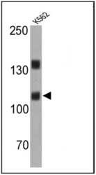
- Western blot analysis of CD57 was performed by loading 25 µg of K562 cell lysates onto an SDS polyacrylamide gel. Proteins were transferred to a PVDF membrane and blocked at 4ºC overnight. The membrane was probed with a CD57 monoclonal antibody (Product # MA5-11605) at a dilution of 1:200 overnight at 4°C, washed in TBST, and probed with an HRP-conjugated secondary antibody for 1 hr at room temperature in the dark. Chemiluminescent detection was performed using Pierce ECL Plus Western Blotting Substrate (Product # 32132). Results show a band at ~110 kDa.

Supportive validation
- Submitted by
- Invitrogen Antibodies (provider)
- Main image
- Experimental details
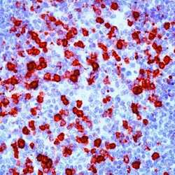
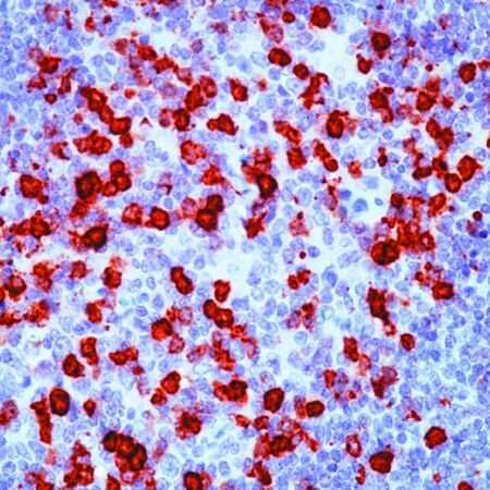
- Formalin-fixed, paraffin-embedded human tonsil stained with CD57 antibody using peroxidase-conjugate and AEC chromogen. Note staining of natural killer cells.
Supportive validation
- Submitted by
- Invitrogen Antibodies (provider)
- Main image

- Experimental details
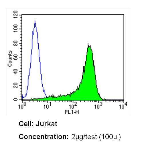
- Flow cytometry analysis of CD57 in Jurkat cells (green) compared to an isotype control (blue). Cells were harvested, adjusted to a concentration of 1-5x10^6 cells/mL, fixed with 2% paraformaldehyde and washed with PBS. Cells were blocked with a 2% solution of BSA-PBS for 30 min at room temperature and incubated with a CD57 monoclonal antibody (Product # MA5-11605) at a dilution of 2 µg/test for 60 min at room temperature. Cells were then incubated for 40 min at room temperature in the dark using a Dylight 488-conjugated secondary antibody and re-suspended in PBS for FACS analysis.
- Submitted by
- Invitrogen Antibodies (provider)
- Main image
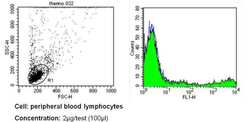
- Experimental details
- Flow cytometry analysis of CD57 in PBMC cells (green) compared to an isotype control (blue). Human blood was collected, combined with a hydrophilic polysaccharide, centrifuged, transferred to a conical tube and washed with PBS. 50 µL of cell solution was added to each tube at a dilution of 2x10^7 cells/mL, followed by the addition of 50 µL of isotype control and primary antibody (Product # MA5-11605) at a dilution of 2 µg/test. Cells were incubated for 30 min at 4ºC and washed with a cell buffer, followed by incubation with a DyLight 488-conjugated secondary antibody for 30 min at 4ºC in the dark. FACS analysis was performed using 400 µL of cell buffer.
