Explore
 Validate
 Learn
Learn Flow cytometry
 Other assay
Antibody data
- Antibody Data
- Antigen structure
- References [9]
- Comments [0]
- Validations
- Other assay [3]
Submit
Validation data
Reference
Comment
Report error
- Product number
- MHCD6904 - Provider product page

- Provider
- Invitrogen Antibodies
- Product name
- CD69 Monoclonal Antibody (CH/4), PE
- Antibody type
- Monoclonal
- Antigen
- Other
- Description
- R-phycoerythrin (PE) is a stable and highly soluble phycobiliprotein which provides maximal absorbance and fluorescence without susceptibility to internal or external fluorescence quenching, thus providing an exceptional quantum yields and molar extinction coefficients.
- Reactivity
- Human
- Host
- Mouse
- Conjugate
- Yellow dye
- Isotype
- IgG
- Antibody clone number
- CH/4
- Vial size
- 500 µL
- Storage
- 4° C, store in dark
Submitted references Synergy between B cell receptor/antigen uptake and MHCII peptide editing relies on HLA-DO tuning.
The impact of Nucleofection® on the activation state of primary human CD4 T cells.
Activation of human NK cells by Plasmodium-infected red blood cells.
Role of CD8(+) T cells in triggering reversal reaction in HIV/leprosy patients.
Distinct expression patterns of CD69 in mucosal and systemic lymphoid tissues in primary SIV infection of rhesus macaques.
NK cells as effectors of acquired immune responses: effector CD4+ T cell-dependent activation of NK cells following vaccination.
The chemokine interleukin-8 and the surface activation protein CD69 are markers for Bcr-Abl activity in chronic myeloid leukemia.
Influence of interleukin-15 on CD8+ natural killer cells in human immunodeficiency virus type 1-infected chimpanzees.
Patterns of A2A extracellular adenosine receptor expression in different functional subsets of human peripheral T cells. Flow cytometry studies with anti-A2A receptor monoclonal antibodies.
Jiang W, Adler LN, Macmillan H, Mellins ED
Scientific reports 2019 Sep 25;9(1):13877
Scientific reports 2019 Sep 25;9(1):13877
The impact of Nucleofection® on the activation state of primary human CD4 T cells.
Zhang M, Ma Z, Selliah N, Weiss G, Genin A, Finkel TH, Cron RQ
Journal of immunological methods 2014 Jun;408:123-31
Journal of immunological methods 2014 Jun;408:123-31
Activation of human NK cells by Plasmodium-infected red blood cells.
Horowitz A, Riley EM
Methods in molecular biology (Clifton, N.J.) 2013;923:447-64
Methods in molecular biology (Clifton, N.J.) 2013;923:447-64
Role of CD8(+) T cells in triggering reversal reaction in HIV/leprosy patients.
de Oliveira AL, Amadeu TP, de França Gomes AC, Menezes VM, da Costa Nery JA, Pinheiro RO, Sarno EN
Immunology 2013 Sep;140(1):47-60
Immunology 2013 Sep;140(1):47-60
Distinct expression patterns of CD69 in mucosal and systemic lymphoid tissues in primary SIV infection of rhesus macaques.
Wang X, Xu H, Alvarez X, Pahar B, Moroney-Rasmussen T, Lackner AA, Veazey RS
PloS one 2011;6(11):e27207
PloS one 2011;6(11):e27207
NK cells as effectors of acquired immune responses: effector CD4+ T cell-dependent activation of NK cells following vaccination.
Horowitz A, Behrens RH, Okell L, Fooks AR, Riley EM
Journal of immunology (Baltimore, Md. : 1950) 2010 Sep 1;185(5):2808-18
Journal of immunology (Baltimore, Md. : 1950) 2010 Sep 1;185(5):2808-18
The chemokine interleukin-8 and the surface activation protein CD69 are markers for Bcr-Abl activity in chronic myeloid leukemia.
Hantschel O, Gstoettenbauer A, Colinge J, Kaupe I, Bilban M, Burkard TR, Valent P, Superti-Furga G
Molecular oncology 2008 Oct;2(3):272-81
Molecular oncology 2008 Oct;2(3):272-81
Influence of interleukin-15 on CD8+ natural killer cells in human immunodeficiency virus type 1-infected chimpanzees.
Rodriguez AR, Arulanandam BP, Hodara VL, McClure HM, Cobb EK, Salas MT, White R, Murthy KK
The Journal of general virology 2007 Feb;88(Pt 2):641-651
The Journal of general virology 2007 Feb;88(Pt 2):641-651
Patterns of A2A extracellular adenosine receptor expression in different functional subsets of human peripheral T cells. Flow cytometry studies with anti-A2A receptor monoclonal antibodies.
Koshiba M, Rosin DL, Hayashi N, Linden J, Sitkovsky MV
Molecular pharmacology 1999 Mar;55(3):614-24
Molecular pharmacology 1999 Mar;55(3):614-24
No comments: Submit comment
Supportive validation
- Submitted by
- Invitrogen Antibodies (provider)
- Main image

- Experimental details
- NULL
- Conjugate
- Yellow dye
- Submitted by
- Invitrogen Antibodies (provider)
- Main image

- Experimental details
- NULL
- Conjugate
- Yellow dye
- Submitted by
- Invitrogen Antibodies (provider)
- Main image
- Experimental details
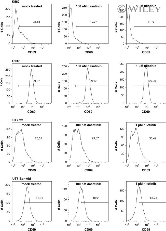
- FACS analysis of CD69 expression. FACS analysis of different cell lines showed Bcr-Abl dependent CD69 expression. Cells were either untreated or treated with 100 nm Dasatinib and 1muM Nilotinib for 16h. Percentage of CD69-positive cells is indicated in each histogram. Thirty-six percent of K562 cells showed CD69 expression, which was reduced upon treatment with Dasatinib and Nilotinib CD69 expression to 11% and 12%, respectively (top panel). U937, a Bcr-Abl negative cell line, did not alter their CD69 expression pattern after treatment with either of the drugs (second panel). 26% of untransduced UT-7 cells expressed CD69, whereas Bcr-Abl transduction in these cells increased CD69 expression to 82%. After treatment with either Dasatinib or Nilotinib CD69 expression in untransduced UT-7 remained unchanged, whereas it was decreased in UT7-Bcr-Abl cells (lower two panels).
- Conjugate
- Yellow dye