 Explore
 Validate
 Learn
Learn Western blot
 Immunocytochemistry
Antibody data
- Antibody Data
- Antigen structure
- References [7]
- Comments [0]
- Validations
- Immunocytochemistry [1]
- Flow cytometry [2]
Submit
Validation data
Reference
Comment
Report error
- Product number
- MA1-207 - Provider product page

- Provider
- Invitrogen Antibodies
- Product name
- CD69 Monoclonal Antibody (H1.2F3)
- Antibody type
- Monoclonal
- Antigen
- Other
- Description
- MA1-207 detects CD69 from human samples. MA1-207 has been successfully tested in western blot, immunofluorescence and flow cytometry applications. In western blot, MA1-207 detects CXCR1/IL8RA (human) at approximate molecular weight of 22.5 kDa.
- Reactivity
- Human, Mouse
- Isotype
- IgG
- Antibody clone number
- H1.2F3
- Vial size
- 100 μg
- Concentration
- 1 mg/mL
- Storage
- -20°C, Avoid Freeze/Thaw Cycles
Submitted references NK cells regulate CXCR2+ neutrophil recruitment during acute lung injury.
Expression of a Chimeric Antigen Receptor Specific for Donor HLA Class I Enhances the Potency of Human Regulatory T Cells in Preventing Human Skin Transplant Rejection.
C1q acts in the tumour microenvironment as a cancer-promoting factor independently of complement activation.
Aberrant germinal center formation, follicular T-helper cells, and germinal center B-cells were involved in chronic graft-versus-host disease.
Effector CD4 T-cell transition to memory requires late cognate interactions that induce autocrine IL-2.
Lysophosphatidic acid receptor 5 inhibits B cell antigen receptor signaling and antibody response.
Arhgef1 is required by T cells for the development of airway hyperreactivity and inflammation.
Hoegl S, Ehrentraut H, Brodsky KS, Victorino F, Golden-Mason L, Eltzschig HK, McNamee EN
Journal of leukocyte biology 2017 Feb;101(2):471-480
Journal of leukocyte biology 2017 Feb;101(2):471-480
Expression of a Chimeric Antigen Receptor Specific for Donor HLA Class I Enhances the Potency of Human Regulatory T Cells in Preventing Human Skin Transplant Rejection.
Boardman DA, Philippeos C, Fruhwirth GO, Ibrahim MA, Hannen RF, Cooper D, Marelli-Berg FM, Watt FM, Lechler RI, Maher J, Smyth LA, Lombardi G
American journal of transplantation : official journal of the American Society of Transplantation and the American Society of Transplant Surgeons 2017 Apr;17(4):931-943
American journal of transplantation : official journal of the American Society of Transplantation and the American Society of Transplant Surgeons 2017 Apr;17(4):931-943
C1q acts in the tumour microenvironment as a cancer-promoting factor independently of complement activation.
Bulla R, Tripodo C, Rami D, Ling GS, Agostinis C, Guarnotta C, Zorzet S, Durigutto P, Botto M, Tedesco F
Nature communications 2016 Feb 1;7:10346
Nature communications 2016 Feb 1;7:10346
Aberrant germinal center formation, follicular T-helper cells, and germinal center B-cells were involved in chronic graft-versus-host disease.
Shao L, Lie AK, Zhang Y, Wong CH, Kwong YL
Annals of hematology 2015 Sep;94(9):1493-504
Annals of hematology 2015 Sep;94(9):1493-504
Effector CD4 T-cell transition to memory requires late cognate interactions that induce autocrine IL-2.
McKinstry KK, Strutt TM, Bautista B, Zhang W, Kuang Y, Cooper AM, Swain SL
Nature communications 2014 Nov 5;5:5377
Nature communications 2014 Nov 5;5:5377
Lysophosphatidic acid receptor 5 inhibits B cell antigen receptor signaling and antibody response.
Hu J, Oda SK, Shotts K, Donovan EE, Strauch P, Pujanauski LM, Victorino F, Al-Shami A, Fujiwara Y, Tigyi G, Oravecz T, Pelanda R, Torres RM
Journal of immunology (Baltimore, Md. : 1950) 2014 Jul 1;193(1):85-95
Journal of immunology (Baltimore, Md. : 1950) 2014 Jul 1;193(1):85-95
Arhgef1 is required by T cells for the development of airway hyperreactivity and inflammation.
Brown JP, Taube C, Miyahara N, Koya T, Pelanda R, Gelfand EW, Torres RM
American journal of respiratory and critical care medicine 2007 Jul 1;176(1):10-9
American journal of respiratory and critical care medicine 2007 Jul 1;176(1):10-9
No comments: Submit comment
Supportive validation
- Submitted by
- Invitrogen Antibodies (provider)
- Main image
- Experimental details
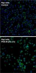
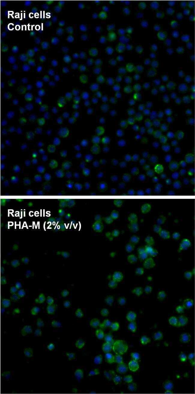
- Immunofluorescent analysis of CD69 (green) in PHA-M (2% v/v, Product # 10576-015) treated Raji cells. Cells were blocked with blocking buffer (Product # 37525) for 30 min at room temparture and then stained with CD69 hamster monoclonal antibody (Product # MA1-207) at a dilution of 1:100 for 1 hour on ice. After washing with ice-cold PBS for 3 times, the cells were then stained with DyLight 488 Goat anti-hamster (Armenian) IgG antibody for 1 hour on ice. The cells were fixed with 4% paraformaldehyde for 30 minutes at room temperature. Nuclei (blue) were counterstained with Hoechst 33342 dye (Product # 62249). Images were taken on a Thermo Scientific ToxInsight Instrument at 20X magnification.
Supportive validation
- Submitted by
- Invitrogen Antibodies (provider)
- Main image
- Experimental details
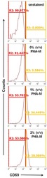
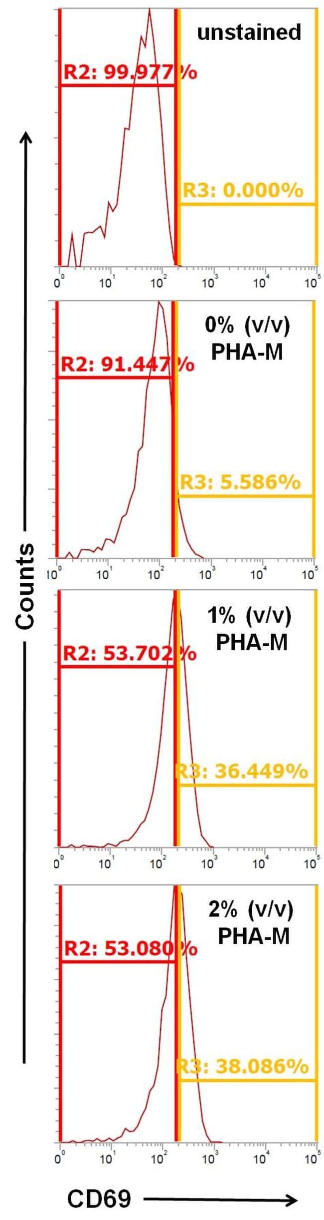
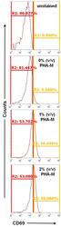
- Flow cytometry analysis of CD69 on Raji cells with/without the treatment of PHA-M (Product # 10576-015) as indicated for overnight. Cells were blocked with blocking buffer (Product # 37525) for 30 min at room temparture and then stained with CD69 hamster monoclonal antibody (Product # MA1-207) at a dilution of 1:100 for 1 hour on ice. After washing with ice-cold PBS for 3 times, the cells were stained with DyLight 488 Goat anti-hamster (Armenian) IgG antibody for 1 hour on ice. A representative 10,000 cells were acquired for each sample.
- Submitted by
- Invitrogen Antibodies (provider)
- Main image
- Experimental details
- Flow cytometry analysis of CD69 on Raji cells with/without the treatment of PHA-M (Product # 10576-015) as indicated for overnight. Cells were blocked with blocking buffer (Product # 37525) for 30 min at room temparture and then stained with CD69 hamster monoclonal antibody (Product # MA1-207) at a dilution of 1:100 for 1 hour on ice. After washing with ice-cold PBS for 3 times, the cells were stained with DyLight 488 Goat anti-hamster (Armenian) IgG antibody for 1 hour on ice. A representative 10,000 cells were acquired for each sample.
