Explore
 Validate
 Learn
Learn Western blot
 ELISA
ELISAAntibody data
- Antibody Data
- Antigen structure
- References [2]
- Comments [0]
- Validations
- Western blot [1]
Submit
Validation data
Reference
Comment
Report error
- Product number
- A00529-2 - Provider product page

- Provider
- Boster Biological Technology
- Product name
- Anti-CD69 Antibody Picoband™
- Antibody type
- Polyclonal
- Description
- Rabbit IgG polyclonal antibody for CD69 detection. Tested with WB, IHC-P, FCM, ICC/IF, Direct ELISA in Human;Mouse;Rat.
- Reactivity
- Human, Mouse, Rat
- Host
- Rabbit
- Vial size
- 100μg/vial
- Concentration
- Add 0.2ml of distilled water will yield a concentration of 500ug/ml.
- Storage
- At -20°C for one year. After reconstitution, at 4°C for one month. It can also be aliquoted and stored frozen at -20°C for a longer time. Avoid repeated freezing and thawing.
- Handling
- Add 0.2ml of distilled water will yield a concentration of 500ug/ml.
Submitted references The involvement of Th1 cell differentiation in the anti-tumor effect of purified polysaccharide from Sanghuangporus vaninii in colorectal cancer via multi-omics analysis.
MiRNA-340-5p mediates the functional and infiltrative promotion of tumor-infiltrating CD8(+) T lymphocytes in human diffuse large B cell lymphoma.
Qu Y, Yang H, Li S, Li L, Li Y, Wang D
International journal of biological macromolecules 2023 May 15;237:123927
International journal of biological macromolecules 2023 May 15;237:123927
MiRNA-340-5p mediates the functional and infiltrative promotion of tumor-infiltrating CD8(+) T lymphocytes in human diffuse large B cell lymphoma.
Xu Y, Liu Z, Lv L, Li P, Xiu B, Qian W, Liang A
Journal of experimental & clinical cancer research : CR 2020 Nov 10;39(1):238
Journal of experimental & clinical cancer research : CR 2020 Nov 10;39(1):238
No comments: Submit comment
Supportive validation
- Submitted by
- Boster Biological Technology (provider)
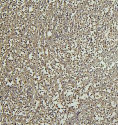
- Main image
- Experimental details
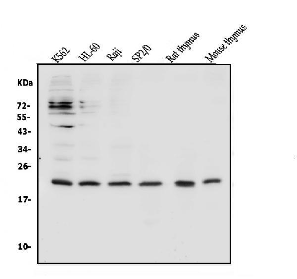
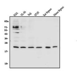
- Western blot analysis of CD69 using anti-CD69 antibody (A00529-2). Electrophoresis was performed on a 5-20% SDS-PAGE gel at 70V (Stacking gel) / 90V (Resolving gel) for 2-3 hours. The sample well of each lane was loaded with 50ug of sample under reducing conditions. Lane 1: human K562 whole cell lysates, Lane 2: human HL-60 whole cell lysates, Lane 3: human Raji whole cell lysates, Lane 4: mouse SP2/0 whole cell lysates, Lane 5: rat thymus tissue lysates, Lane 6: mouse thymus tissue lysates. After Electrophoresis, proteins were transferred to a Nitrocellulose membrane at 150mA for 50-90 minutes. Blocked the membrane with 5% Non-fat Milk/ TBS for 1.5 hour at RT. The membrane was incubated with rabbit anti-CD69 antigen affinity purified polyclonal antibody (Catalog # A00529-2) at 0.5 μg/mL overnight at 4°C, then washed with TBS-0.1%Tween 3 times with 5 minutes each and probed with a goat anti-rabbit IgG-HRP secondary antibody at a dilution of 1:10000 for 1.5 hour at RT. The signal is developed using an Enhanced Chemiluminescent detection (ECL) kit (Catalog # EK1002) with Tanon 5200 system. A specific band was detected for CD69 at approximately 22KD. The expected band size for CD69 is at 22KD.
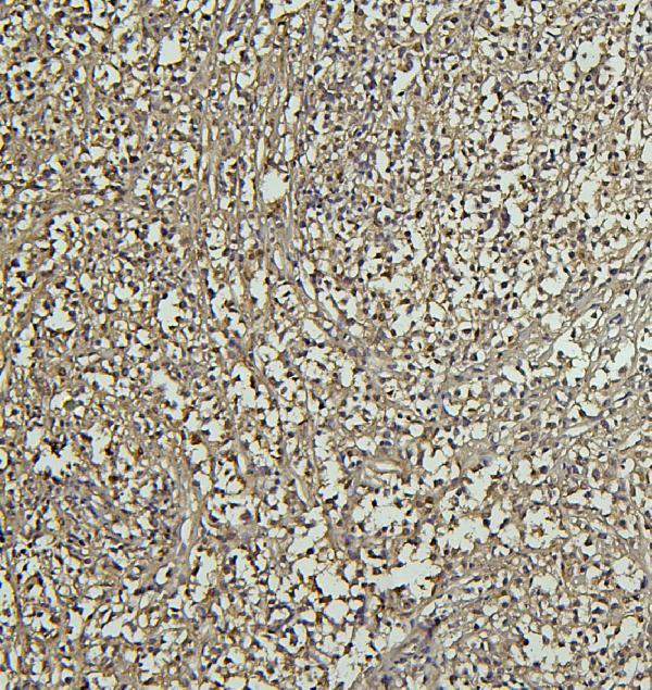
- Additional image