 Explore
 Validate
 Learn
Learn Western blot
Antibody data
- Antibody Data
- Antigen structure
- References [8]
- Comments [0]
- Validations
- Western blot [3]
- Immunohistochemistry [1]
Submit
Validation data
Reference
Comment
Report error
- Product number
- AF5058 - Provider product page

- Provider
- Novus Biologicals
- Product name
- Sheep Polyclonal Parvalbumin alpha Antibody
- Antibody type
- Polyclonal
- Description
- Antigen Affinity-purified. Detects human, mouse, and rat Parvalbumin alpha in direct ELISAs and Western blots.
- Reactivity
- Human, Mouse, Rat
- Host
- Sheep
- Conjugate
- Unconjugated
- Isotype
- IgG
- Vial size
- 100 ug
- Concentration
- LYOPH
- Storage
- Use a manual defrost freezer and avoid repeated freeze-thaw cycles. 12 months from date of receipt, -20 to -70 degreesC as supplied. 1 month, 2 to 8 degreesC under sterile conditions after reconstitution. 6 months, -20 to -70 degreesC under sterile conditions after reconstitution.
Submitted references Nogo receptor 1 is expressed by nearly all retinal ganglion cells.
Expression of the onconeural protein CDR1 in cerebellum and ovarian cancer.
Phenotypic and Functional Characterization of Peripheral Sensory Neurons derived from Human Embryonic Stem Cells.
Nogo Receptor 1 Confines a Disinhibitory Microcircuit to the Critical Period in Visual Cortex.
Unbiased classification of sensory neuron types by large-scale single-cell RNA sequencing.
Neuronal cell type-specific alternative splicing is regulated by the KH domain protein SLM1.
Plasticity of binocularity and visual acuity are differentially limited by nogo receptor.
Parvalbumin-containing chandelier and basket cell boutons have distinctive modes of maturation in monkey prefrontal cortex.
Solomon AM, Westbrook T, Field GD, McGee AW
PloS one 2018;13(5):e0196565
PloS one 2018;13(5):e0196565
Expression of the onconeural protein CDR1 in cerebellum and ovarian cancer.
Totland C, Kråkenes T, Mazengia K, Haugen M, Vedeler C
Oncotarget 2018 May 8;9(35):23975-23986
Oncotarget 2018 May 8;9(35):23975-23986
Phenotypic and Functional Characterization of Peripheral Sensory Neurons derived from Human Embryonic Stem Cells.
Alshawaf AJ, Viventi S, Qiu W, D'Abaco G, Nayagam B, Erlichster M, Chana G, Everall I, Ivanusic J, Skafidas E, Dottori M
Scientific reports 2018 Jan 12;8(1):603
Scientific reports 2018 Jan 12;8(1):603
Nogo Receptor 1 Confines a Disinhibitory Microcircuit to the Critical Period in Visual Cortex.
Stephany CÉ, Ikrar T, Nguyen C, Xu X, McGee AW
The Journal of neuroscience : the official journal of the Society for Neuroscience 2016 Oct 26;36(43):11006-11012
The Journal of neuroscience : the official journal of the Society for Neuroscience 2016 Oct 26;36(43):11006-11012
Unbiased classification of sensory neuron types by large-scale single-cell RNA sequencing.
Usoskin D, Furlan A, Islam S, Abdo H, Lönnerberg P, Lou D, Hjerling-Leffler J, Haeggström J, Kharchenko O, Kharchenko PV, Linnarsson S, Ernfors P
Nature neuroscience 2015 Jan;18(1):145-53
Nature neuroscience 2015 Jan;18(1):145-53
Neuronal cell type-specific alternative splicing is regulated by the KH domain protein SLM1.
Iijima T, Iijima Y, Witte H, Scheiffele P
The Journal of cell biology 2014 Feb 3;204(3):331-42
The Journal of cell biology 2014 Feb 3;204(3):331-42
Plasticity of binocularity and visual acuity are differentially limited by nogo receptor.
Stephany CÉ, Chan LL, Parivash SN, Dorton HM, Piechowicz M, Qiu S, McGee AW
The Journal of neuroscience : the official journal of the Society for Neuroscience 2014 Aug 27;34(35):11631-40
The Journal of neuroscience : the official journal of the Society for Neuroscience 2014 Aug 27;34(35):11631-40
Parvalbumin-containing chandelier and basket cell boutons have distinctive modes of maturation in monkey prefrontal cortex.
Fish KN, Hoftman GD, Sheikh W, Kitchens M, Lewis DA
The Journal of neuroscience : the official journal of the Society for Neuroscience 2013 May 8;33(19):8352-8
The Journal of neuroscience : the official journal of the Society for Neuroscience 2013 May 8;33(19):8352-8
No comments: Submit comment
Supportive validation
- Submitted by
- Novus Biologicals (provider)
- Main image
- Experimental details
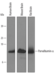
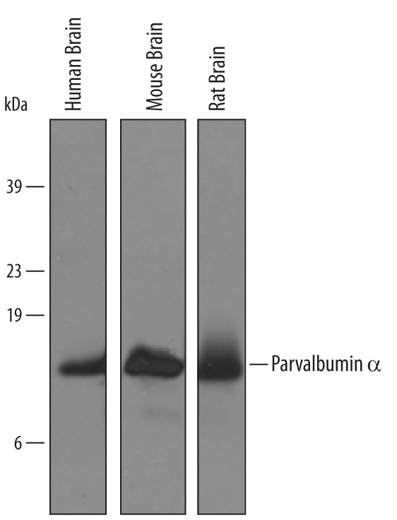
- Detection of Human/Mouse/Rat Parvalbumin alpha by Western Blot. Western blot shows lysates of human, mouse, and rat brain tissue. PVDF membrane was probed with 1 µg/mL of Sheep Anti-Human/Mouse/Rat Parvalbumin alpha Antigen Affinity-purified Polyclonal Antibody (Catalog # AF5058) followed by HRP-conjugated Anti-Sheep IgG Secondary Antibody (Catalog # HAF016). A specific band was detected for Parvalbumin alpha at approximately 12 kDa (as indicated). This experiment was conducted under reducing conditions and using Immunoblot Buffer Group 8.
- Submitted by
- Novus Biologicals (provider)
- Main image

- Experimental details
- Detection of Human and Mouse Parvalbumin alpha by Simple WesternTM. Simple Western lane view shows lysates of mouse brain (cortex) and human brain (cerebellum), loaded at 0.2 mg/mL. A specific band was detected for Parvalbumin alpha at approximately 14 and 17 kDa (as indicated) using 50 µg/mL of Sheep Anti-Human/Mouse/Rat Parvalbumin alpha Antigen Affinity-purified Polyclonal Antibody (Catalog # AF5058) followed by 1:50 dilution of HRP-conjugated Anti-Sheep IgG Secondary Antibody (Catalog # HAF016). This experiment was conducted under reducing conditions and using the 12-230 kDa separation system.

- Submitted by
- Novus Biologicals (provider)
- Main image

- Experimental details
- Detection of Rat Parvalbumin alpha by Simple WesternTM. Simple Western lane view shows lysates of rat brain tissue, loaded at 0.2 mg/mL. A specific band was detected for Parvalbumin alpha at approximately 15 kDa (as indicated) using 50 µg/mL of Sheep Anti-Human/Mouse/Rat Parvalbumin alpha Antigen Affinity-purified Polyclonal Antibody (Catalog # AF5058) followed by 1:50 dilution of HRP-conjugated Anti-Sheep IgG Secondary Antibody (Catalog # HAF016) . This experiment was conducted under reducing conditions and using the 12-230 kDa separation system.

Supportive validation
- Submitted by
- Novus Biologicals (provider)
- Main image

- Experimental details
- Parvalbumin alpha in Human Brain. Parvalbumin alpha was detected in immersion fixed paraffin-embedded sections of human brain (cortex) using 5 µg/mL Sheep Anti-Human/Mouse/Rat Parvalbumin alpha Antigen Affinity-purified Polyclonal Antibody (Catalog # AF5058) overnight at 4 °C. Tissue was stained with the Anti-Sheep HRP-DAB Cell & Tissue Staining Kit (brown; Catalog # CTS019) and counterstained with hematoxylin (blue). View our protocol for Chromogenic IHC Staining of Paraffin-embedded Tissue Sections.
