Explore
 Validate
 Learn
Learn Western blot
 Immunocytochemistry
Antibody data
- Antibody Data
- Antigen structure
- References [0]
- Comments [0]
- Validations
- Western blot [2]
- Other assay [1]
Submit
Validation data
Reference
Comment
Report error
- Product number
- NBP2-50036 - Provider product page

- Provider
- Novus Biologicals
- Product name
- Chicken Polyclonal Parvalbumin Antibody
- Antibody type
- Polyclonal
- Description
- Ammonium sulfate precipitation.
- Reactivity
- Human, Mouse, Rat
- Host
- Chicken/Avian
- Isotype
- IgY
- Vial size
- 0.1 ml
- Storage
- Store at 4C short term. Aliquot and store at -20C long term. Avoid freeze-thaw cycles.
No comments: Submit comment
Supportive validation
- Submitted by
- Novus Biologicals (provider)
- Main image
- Experimental details
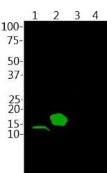
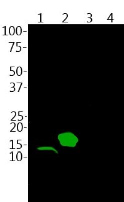
- Western Blot: Parvalbumin Antibody [NBP2-50036] - Analysis of mouse skeletal muscle lysate (lane 1) and His-tagged recombinant proteins: parvalbumin (lane 2), calretinin (lane 3) and calbindin (lane 4) were probed with NBP2-50036 at 1:2,500.
- Submitted by
- Novus Biologicals (provider)
- Main image

- Experimental details
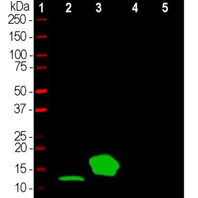
- Western Blot: Parvalbumin Antibody [NBP2-50036] - Analysis of skeletal muscle lysates and His-tagged recombinant human proteins using chicken parvalbumin pAb, dilution 1:2,000 (Green): [1] protein standard (Red), [2] mouse muscle, [3] recombinant full length human parvalbumin, [4] recombinant full length human calretinin and [5] recombinant full length human calbindin. Band at 12kDa in muscle lysate is native parvalbumin and 18kDa band in the next lane is His-tagged recombinant parvalbumin. The parvalbumin antibody binds parvalbumin strongly but is not cross-reactive with the related calcium binding proteins calretinin and calbindin.
Supportive validation
- Submitted by
- Novus Biologicals (provider)
- Main image

- Experimental details
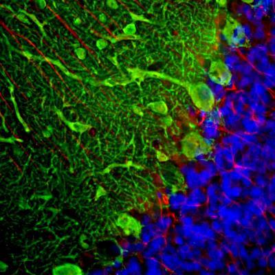
- Immunohistochemistry Free-Floating: Parvalbumin Antibody [NBP2-50036] - Analysis of a rat cerebellum section stained with chicken pAb to parvalbumin, NBP2-50036, dilution 1:2,000, in green, and costained with mouse mAb to GFAP, dilution 1:500, in red. The blue is DAPI staining of nuclear DNA. Following transcardial perfusion of rat with 4% paraformaldehyde, brain was post fixed for 24 hours, cut to 45uM, and free floating sections were stained with above antibodies. NBP2-50036 antibody labels the perikarya and dendrites of Purkinje cells and interneurons in the molecular layer of the cerebellum. The GFAP antibody stains the processes of Bergmann glia and astrocytes.