Explore
 Validate
 Learn
Learn Western blot
 ELISA
ELISAAntibody data
- Antibody Data
- Antigen structure
- References [2]
- Comments [0]
- Validations
- Western blot [2]
- Blocking/Neutralizing [1]
Submit
Validation data
Reference
Comment
Report error
- Product number
- MAB1786-100 - Provider product page

- Provider
- R&D Systems
- Product name
- Human Serpin E1/PAI-1 Antibody
- Antibody type
- Monoclonal
- Description
- Protein A or G purified from hybridoma culture supernatant. Detects human Serpin E1 in direct ELISAs and Western blots. In direct ELISAs, no cross-reactivity with recombinant human Serpin A1, A3, A4, A8, C1, F1, F2, I1, I2, recombinant mouse Serpin D1 or E2 is observed.
- Reactivity
- Human
- Host
- Mouse
- Conjugate
- Unconjugated
- Antigen sequence
P05121- Isotype
- IgG
- Antibody clone number
- 242816
- Vial size
- 100 ug
- Concentration
- LYOPH
- Storage
- Use a manual defrost freezer and avoid repeated freeze-thaw cycles. 12 months from date of receipt, -20 to -70 °C as supplied. 1 month, 2 to 8 °C under sterile conditions after reconstitution. 6 months, -20 to -70 °C under sterile conditions after reconstitution.
Submitted references Human CD4- 8- T cells are a distinctive immunoregulatory subset.
A multiplex immunoassay for human adipokine profiling.
Huang MC, Patel K, Taub DD, Longo DL, Goetzl EJ
FASEB journal : official publication of the Federation of American Societies for Experimental Biology 2010 Jul;24(7):2558-66
FASEB journal : official publication of the Federation of American Societies for Experimental Biology 2010 Jul;24(7):2558-66
A multiplex immunoassay for human adipokine profiling.
Schipper HS, de Jager W, van Dijk ME, Meerding J, Zelissen PM, Adan RA, Prakken BJ, Kalkhoven E
Clinical chemistry 2010 Aug;56(8):1320-8
Clinical chemistry 2010 Aug;56(8):1320-8
No comments: Submit comment
Supportive validation
- Submitted by
- R&D Systems (provider)
- Main image
- Experimental details
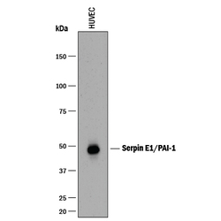
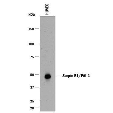
- Detection of Human Serpin E1/PAI-1 by Western Blot. Western blot shows lysates of HUVEC human umbilical vein endothelial cells. PVDF membrane was probed with 1 µg/mL of Mouse Anti-Human Serpin E1/PAI-1 Monoclonal Antibody (Catalog # MAB1786) followed by HRP-conjugated Anti-Mouse IgG Secondary Antibody (Catalog # HAF018). A specific band was detected for Serpin E1/PAI-1 at approximately 48 kDa (as indicated). This experiment was conducted under reducing conditions and using Immunoblot Buffer Group 1.
- Submitted by
- R&D Systems (provider)
- Main image

- Experimental details
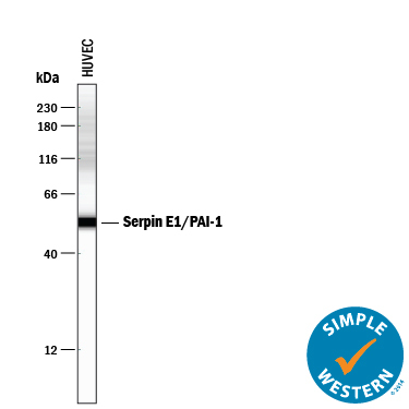
- Detection of Human Serpin E1/PAI-1 by Simple WesternTM. Simple Western lane view shows lysates of HUVEC human umbilical vein endothelial cells, loaded at 0.2 mg/mL. A specific band was detected for Serpin E1/PAI-1 at approximately 54 kDa (as indicated) using 2.5 µg/mL of Mouse Anti-Human Serpin E1/PAI-1 Monoclonal Antibody (Catalog # MAB1786) . This experiment was conducted under reducing conditions and using the 12-230 kDa separation system.
Supportive validation
- Submitted by
- R&D Systems (provider)
- Main image
- Experimental details
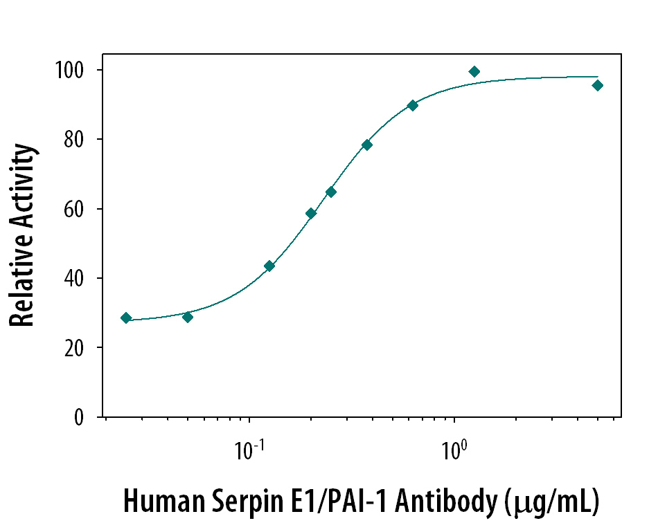
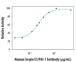
- Neutralization of Serpin E1/ PAI-1 Activity by Human Serpin E1/PAI-1 Antibody. Recombinant Human u-Plasminogen Activator (uPA)/Urokinase (0.1 µg/mL, Catalog # 1310-SE) activity is measured in the presence of Recombinant Human Serpin E1/PAI-1 (0.25 µg/mL, Catalog # 1786-PI) that has been preincubated with increasing concentrations of Mouse Anti-Human Serpin E1/PAI-1 Monoclonal Antibody (Catalog # MAB1786). The ND50 is typically 0.3 µg/mL.