 Explore
 Validate
 Learn
Learn Western blot
Antibody data
- Antibody Data
- Antigen structure
- References [7]
- Comments [0]
- Validations
- Western blot [4]
- Immunocytochemistry [1]
- Immunohistochemistry [5]
Submit
Validation data
Reference
Comment
Report error
- Product number
- GTX113016 - Provider product page

- Provider
- GeneTex
- Proper citation
- GeneTex Cat#GTX113016, RRID:AB_1952230
- Product name
- Tyrosine Hydroxylase antibody
- Antibody type
- Polyclonal
- Reactivity
- Human, Mouse, Rat
- Host
- Rabbit
Submitted references Understanding the Mechanism of Antidepressant-Related Sexual Dysfunction: Inhibition of Tyrosine Hydroxylase in Dopaminergic Neurons after Treatment with Paroxetine but Not with Agomelatine in Male Rats.
Cervical ganglioneuroblastoma in a new born Japanese Black calf.
Src-dependent phosphorylation of μ-opioid receptor at Tyr(336) modulates opiate withdrawal.
Pre-administration of BAX-inhibiting peptides decrease the loss of the nigral dopaminergic neurons in rats.
A single prolonged stress paradigm produces enduring impairments in social bonding in monogamous prairie voles.
Transplanted Neural Stem Cells: Playing a Neuroprotective Role by Ceruloplasmin in the Substantia Nigra of PD Model Rats?
Nicotinamide N-methyltransferase expression in SH-SY5Y neuroblastoma and N27 mesencephalic neurones induces changes in cell morphology via ephrin-B2 and Akt signalling.
Santana Y, Montejo AL, Martín J, LLorca G, Bueno G, Blázquez JL
Journal of clinical medicine 2019 Jan 23;8(2)
Journal of clinical medicine 2019 Jan 23;8(2)
Cervical ganglioneuroblastoma in a new born Japanese Black calf.
Park CH, Shiwa N, Kimitsuki K, Kakizaki T, Watanabe D
The Journal of veterinary medical science 2018 May 18;80(5):755-759
The Journal of veterinary medical science 2018 May 18;80(5):755-759
Src-dependent phosphorylation of μ-opioid receptor at Tyr(336) modulates opiate withdrawal.
Zhang L, Kibaly C, Wang YJ, Xu C, Song KY, McGarrah PW, Loh HH, Liu JG, Law PY
EMBO molecular medicine 2017 Nov;9(11):1521-1536
EMBO molecular medicine 2017 Nov;9(11):1521-1536
Pre-administration of BAX-inhibiting peptides decrease the loss of the nigral dopaminergic neurons in rats.
Ma C, Pan Y, Yang Z, Meng Z, Sun R, Wang T, Fei Y, Fan W
Life sciences 2016 Jan 1;144:113-20
Life sciences 2016 Jan 1;144:113-20
A single prolonged stress paradigm produces enduring impairments in social bonding in monogamous prairie voles.
Arai A, Hirota Y, Miyase N, Miyata S, Young LJ, Osako Y, Yuri K, Mitsui S
Behavioural brain research 2016 Dec 15;315:83-93
Behavioural brain research 2016 Dec 15;315:83-93
Transplanted Neural Stem Cells: Playing a Neuroprotective Role by Ceruloplasmin in the Substantia Nigra of PD Model Rats?
Xiao JJ, Yin M, Wang ZJ, Wang XP
Oxidative medicine and cellular longevity 2015;2015:618631
Oxidative medicine and cellular longevity 2015;2015:618631
Nicotinamide N-methyltransferase expression in SH-SY5Y neuroblastoma and N27 mesencephalic neurones induces changes in cell morphology via ephrin-B2 and Akt signalling.
Thomas MG, Saldanha M, Mistry RJ, Dexter DT, Ramsden DB, Parsons RB
Cell death & disease 2013 Jun 13;4(6):e669
Cell death & disease 2013 Jun 13;4(6):e669
No comments: Submit comment
Supportive validation
- Submitted by
- GeneTex (provider)
- Main image
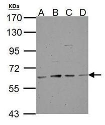
- Experimental details
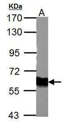
- Tyrosine Hydroxylase antibody detects TH protein by western blot analysis.A. 30 ?g NT2D1 whole cell lysate/extractB. 30 ?g PC-3 whole cell lysate/extractC. 30 ?g U87-MG whole cell lysate/extractD. 30 ?g SK-N-SH whole cell lysate/extract7.5% SDS-PAGETyrosine Hydroxylase antibody (GTX113016) dilution: 1:500 The HRP-conjugated anti-rabbit IgG antibody (GTX213110-01) was used to detect the primary antibody.
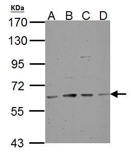
- Submitted by
- GeneTex (provider)
- Main image
- Experimental details
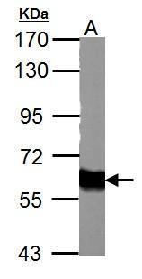
- Tyrosine Hydroxylase antibody detects TH protein by western blot analysis.A. 50 ?g mouse brain lysate/extract7.5% SDS-PAGETyrosine Hydroxylase antibody (GTX113016) dilution: 1:500 The HRP-conjugated anti-rabbit IgG antibody (GTX213110-01) was used to detect the primary antibody.
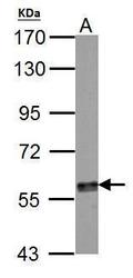
- Submitted by
- GeneTex (provider)
- Main image
- Experimental details
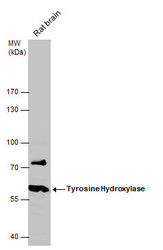
- Tyrosine Hydroxylase antibody detects TH protein by western blot analysis. A. 30 ?g PC-12 whole cell lysate/extract 7.5% SDS-PAGE Tyrosine Hydroxylase antibody (GTX113016) dilution: 1:1000 The HRP-conjugated anti-rabbit IgG antibody (GTX213110-01) was used to detect the primary antibody.
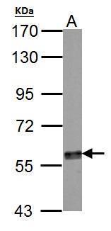
- Submitted by
- GeneTex (provider)
- Main image
- Experimental details
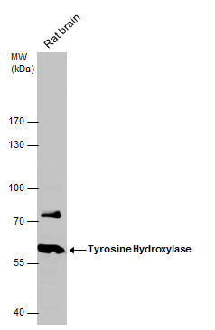
- Tyrosine Hydroxylase antibody detects Tyrosine Hydroxylase protein by western blot analysis. Rat tissue extracts (50 ?g) was separated by 10% SDS-PAGE, and the membrane was blotted with Tyrosine Hydroxylase antibody (GTX113016) diluted at 1:2000. The HRP-conjugated anti-rabbit IgG antibody (GTX213110-01) was used to detect the primary antibody.
Supportive validation
- Submitted by
- GeneTex (provider)
- Main image
- Experimental details
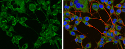
- Tyrosine Hydroxylase antibody detects Tyrosine Hydroxylase protein at cytoplasm by immunofluorescent analysis.Sample: U-87 MG cells were fixed in 4% paraformaldehyde at RT for 15 min.Green: Tyrosine Hydroxylase protein stained by Tyrosine Hydroxylase antibody (GTX113016) diluted at 1:400.Red: beta Tubulin 3/ TUJ1 protein stained by beta Tubulin 3/ TUJ1 antibody (GTX631836) diluted at 1:200.Blue: Hoechst 33342 staining.
Supportive validation
- Submitted by
- GeneTex (provider)
- Main image
- Experimental details
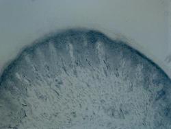
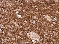
- Immunohistochemical analysis of Rat hindlimb pad skin tissue (paraformaldehyde-fixed frozen sections), using Tyrosine Hydroxylase(GTX113016) antibody at 1:100 dilution.

- Submitted by
- GeneTex (provider)
- Main image

- Experimental details
- Immunohistochemical analysis of Rat hindlimb pad skin tissue (paraformaldehyde-fixed frozen sections), using Tyrosine Hydroxylase(GTX113016) antibody at 1:100 dilution.

- Submitted by
- GeneTex (provider)
- Main image
- Experimental details
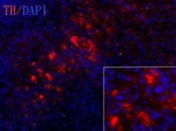
- Tyrosine hydroxylase antibody detects tyrosine hydroxylase protein on embryonic mouse brain by immunohistochemical analysis. Sample: Frozen section of embryonic mouse brain (mE18.5). Red: Tyrosine hydroxylase antibody [GTX113016] diluted at 1:250. Blue: DAPI

- Submitted by
- GeneTex (provider)
- Main image
- Experimental details
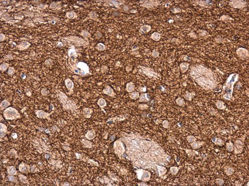
- Tyrosine Hydroxylase antibody detects Tyrosine Hydroxylase protein at cytoplasm in rat brain by immunohistochemical analysis. Sample: Paraffin-embedded rat brain. Tyrosine Hydroxylase antibody (GTX113016) diluted at 1:2500.
- Submitted by
- GeneTex (provider)
- Main image
- Experimental details
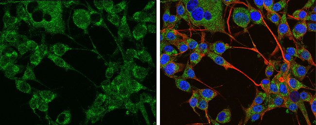
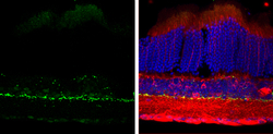
- Tyrosine Hydroxylase antibody detects Tyrosine Hydroxylase protein by immunohistochemical analysis.Sample: Frozen sectioned adult mouse retina. Green: Tyrosine Hydroxylase protein stained by Tyrosine Hydroxylase antibody (GTX113016) diluted at 1:250.Red: beta Tubulin 3/ TUJ1, stained by beta Tubulin 3/ TUJ1 antibody [GT11710] (GTX631836) diluted at 1:250.Blue: Fluoroshield with DAPI (GTX30920).
