 Explore
 Validate
 Learn
Learn Immunohistochemistry
Antibody data
- Antibody Data
- Antigen structure
- References [2]
- Comments [0]
- Validations
- Immunohistochemistry [1]
Submit
Validation data
Reference
Comment
Report error
- Product number
- HPA061003 - Provider product page

- Provider
- Atlas Antibodies
- Proper citation
- Atlas Antibodies Cat#HPA061003, RRID:AB_2630374
- Product name
- Anti-TH
- Antibody type
- Polyclonal
- Description
- Polyclonal Antibody against Human TH, Gene description: tyrosine hydroxylase, Alternative Gene Names: DYT5b, Validated applications: IHC, Uniprot ID: P07101, Storage: Store at +4°C for short term storage. Long time storage is recommended at -20°C.
- Reactivity
- Human, Mouse
- Host
- Rabbit
- Conjugate
- Unconjugated
- Isotype
- IgG
- Vial size
- 100 µl
- Concentration
- 0.2 mg/ml
- Storage
- Store at +4°C for short term storage. Long time storage is recommended at -20°C.
- Handling
- The antibody solution should be gently mixed before use.
Submitted references Peak density of immature nerve cells occurs with high‐grade dysplasia in intraductal papillary mucinous neoplasms of the pancreas
Astrocytes Modulate Baroreflex Sensitivity at the Level of the Nucleus of the Solitary Tract
Trinh V, Roland J, Wong J, Revetta F, Patel K, Shi C, DelGiorno K, Carter B, Tan M
The Journal of Pathology 2022;258(1):69-82
The Journal of Pathology 2022;258(1):69-82
Astrocytes Modulate Baroreflex Sensitivity at the Level of the Nucleus of the Solitary Tract
Mastitskaya S, Turovsky E, Marina N, Theparambil S, Hadjihambi A, Kasparov S, Teschemacher A, Ramage A, Gourine A, Hosford P
The Journal of Neuroscience 2020;40(15):3052-3062
The Journal of Neuroscience 2020;40(15):3052-3062
No comments: Submit comment
Supportive validation
- Submitted by
- Atlas Antibodies (provider)
- Enhanced method
- Orthogonal validation
- Main image
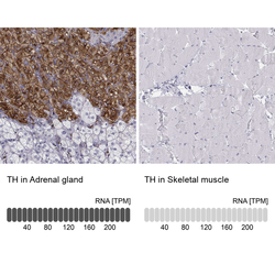
- Experimental details
- Immunohistochemistry analysis in human adrenal gland and skeletal muscle tissues using HPA061003 antibody. Corresponding TH RNA-seq data are presented for the same tissues.
- Sample type
- Human
- Protocol
- Protocol
