 Explore
 Validate
 Learn
Learn Western blot
Antibody data
- Antibody Data
- Antigen structure
- References [0]
- Comments [0]
- Validations
- Western blot [1]
- Immunohistochemistry [19]
Submit
Validation data
Reference
Comment
Report error
- Product number
- NBP2-38903 - Provider product page

- Provider
- Novus Biologicals
- Product name
- Rabbit Polyclonal Tyrosine Hydroxylase Antibody
- Antibody type
- Polyclonal
- Description
- Immunogen affinity purified. Specificity of human Tyrosine Hydroxylase antibody verified on a Protein Array containing target protein plus 383 other non-specific proteins.
- Reactivity
- Human, Mouse
- Host
- Rabbit
- Isotype
- IgG
- Vial size
- 0.1 ml
- Storage
- Store at 4C short term. Aliquot and store at -20C long term. Avoid freeze-thaw cycles.
No comments: Submit comment
Supportive validation
- Submitted by
- Novus Biologicals (provider)
- Main image

- Experimental details
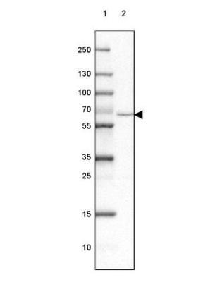
- Western Blot: Tyrosine Hydroxylase Antibody [NBP2-38903] - Lane 1: Marker [kDa] 250, 130, 100, 70, 55, 35, 25, 15, 10. Lane 2 : Human Adrenal Gland tissue.
Supportive validation
- Submitted by
- Novus Biologicals (provider)
- Main image

- Experimental details
- Immunohistochemistry: Tyrosine Hydroxylase Antibody [NBP2-38903] - Staining of mouse hypothalamus shows immunoreactivity in a subset of neurons in the paraventricular nucleaus.

- Submitted by
- Novus Biologicals (provider)
- Main image

- Experimental details
- Immunohistochemistry: Tyrosine Hydroxylase Antibody [NBP2-38903] - Staining of mouse basal forebrain shows positivity in the core of nucleus accumbens.

- Submitted by
- Novus Biologicals (provider)
- Main image
- Experimental details
- Immunohistochemistry: Tyrosine Hydroxylase Antibody [NBP2-38903] - Staining of mouse olfactory bulb shows immunoreactivity in the glomerular layer.

- Submitted by
- Novus Biologicals (provider)
- Main image

- Experimental details
- Immunohistochemistry: Tyrosine Hydroxylase Antibody [NBP2-38903] - Staining of mouse hindbrain shows positivity in the locus coeruleus.

- Submitted by
- Novus Biologicals (provider)
- Main image

- Experimental details
- Immunohistochemistry: Tyrosine Hydroxylase Antibody [NBP2-38903] - Staining of mouse Caudate putamen shows synaptic immunoreactivity.

- Submitted by
- Novus Biologicals (provider)
- Main image
- Experimental details
- Immunohistochemistry-Paraffin: Tyrosine Hydroxylase Antibody [NBP2-38903] - Staining of human lateral ventricle walls shows strong immunoreactivity in neural fibers.

- Submitted by
- Novus Biologicals (provider)
- Main image

- Experimental details
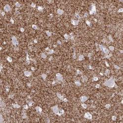
- Immunohistochemistry-Paraffin: Tyrosine Hydroxylase Antibody [NBP2-38903] - Staining of adrenal gland shows strong cytoplasmic positivity in medullary cells.

- Submitted by
- Novus Biologicals (provider)
- Main image

- Experimental details
- Immunohistochemistry-Paraffin: Tyrosine Hydroxylase Antibody [NBP2-38903] - Staining of human cerebral cortex shows positivity in scattered neural fibers.

- Submitted by
- Novus Biologicals (provider)
- Main image

- Experimental details
- Immunohistochemistry-Paraffin: Tyrosine Hydroxylase Antibody [NBP2-38903] - Staining of human adrenal gland shows strong cytoplasmic positivity in glandular cells.

- Submitted by
- Novus Biologicals (provider)
- Main image

- Experimental details
- Immunohistochemistry-Paraffin: Tyrosine Hydroxylase Antibody [NBP2-38903] - Staining of human pancreas shows no positivity in exocrine glandular cells as expected.

- Submitted by
- Novus Biologicals (provider)
- Main image

- Experimental details
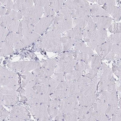
- Immunohistochemistry-Paraffin: Tyrosine Hydroxylase Antibody [NBP2-38903] - Staining of human skeletal muscle shows no positivity in myocytes as expected.
- Submitted by
- Novus Biologicals (provider)
- Main image

- Experimental details
- Immunohistochemistry-Paraffin: Tyrosine Hydroxylase Antibody [NBP2-38903] - Staining of mouse hypothalamus shows immunoreactivity in a subset of neurons in the paraventricular nucleus.

- Submitted by
- Novus Biologicals (provider)
- Main image

- Experimental details
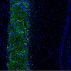
- Immunohistochemistry-Paraffin: Tyrosine Hydroxylase Antibody [NBP2-38903] - Analysis in human adrenal gland and stomach tissues. Corresponding TH RNA-seq data are presented for the same tissues.

- Submitted by
- Novus Biologicals (provider)
- Main image

- Experimental details
- Immunohistochemistry-Paraffin: Tyrosine Hydroxylase Antibody [NBP2-38903] - Staining of mouse amygdala shows positive processes.

- Submitted by
- Novus Biologicals (provider)
- Main image

- Experimental details
- Immunohistochemistry-Paraffin: Tyrosine Hydroxylase Antibody [NBP2-38903] - Staining of mouse hindbrain shows positivity in the locus coeruleus.

- Submitted by
- Novus Biologicals (provider)
- Main image

- Experimental details
- Immunohistochemistry-Paraffin: Tyrosine Hydroxylase Antibody [NBP2-38903] - Staining of mouse caudate putamen shows synaptic immunoreactivity.

- Submitted by
- Novus Biologicals (provider)
- Main image

- Experimental details
- Immunohistochemistry-Paraffin: Tyrosine Hydroxylase Antibody [NBP2-38903] - Staining of human caudate shows moderate positivity in neuropil.

- Submitted by
- Novus Biologicals (provider)
- Main image

- Experimental details
- Immunohistochemistry-Paraffin: Tyrosine Hydroxylase Antibody [NBP2-38903] - Staining of mouse midbrain shows positive processes and cell bodies.

- Submitted by
- Novus Biologicals (provider)
- Main image

- Experimental details
- Immunohistochemistry-Paraffin: Tyrosine Hydroxylase Antibody [NBP2-38903] - Staining of human stomach shows no positivity in glandular cells as expected.
