 Explore
 Validate
 Learn
Learn Western blot
Antibody data
- Antibody Data
- Antigen structure
- References [3]
- Comments [0]
- Validations
- Western blot [2]
- Immunocytochemistry [2]
Submit
Validation data
Reference
Comment
Report error
- Product number
- PA5-17800 - Provider product page

- Provider
- Invitrogen Antibodies
- Product name
- Tyrosine Hydroxylase Polyclonal Antibody
- Antibody type
- Polyclonal
- Antigen
- Synthetic peptide
- Description
- It is not recommended to aliquot this antibody.
- Reactivity
- Human, Mouse, Rat
- Host
- Rabbit
- Isotype
- IgG
- Vial size
- 100 µL
- Concentration
- 581 µg/mL
- Storage
- -20°C
Submitted references LRRK2 kinase activity regulates GCase level and enzymatic activity differently depending on cell type in Parkinson's disease.
Glycoproteomic analysis of the changes in protein N-glycosylation during neuronal differentiation in human-induced pluripotent stem cells and derived neuronal cells.
Neuroprotective effects of pomegranate (Punica granatum L.) juice and seed extract in paraquat-induced mouse model of Parkinson's disease.
Kedariti M, Frattini E, Baden P, Cogo S, Civiero L, Ziviani E, Zilio G, Bertoli F, Aureli M, Kaganovich A, Cookson MR, Stefanis L, Surface M, Deleidi M, Di Fonzo A, Alcalay RN, Rideout H, Greggio E, Plotegher N
NPJ Parkinson's disease 2022 Jul 19;8(1):92
NPJ Parkinson's disease 2022 Jul 19;8(1):92
Glycoproteomic analysis of the changes in protein N-glycosylation during neuronal differentiation in human-induced pluripotent stem cells and derived neuronal cells.
Kimura K, Koizumi T, Urasawa T, Ohta Y, Takakura D, Kawasaki N
Scientific reports 2021 May 27;11(1):11169
Scientific reports 2021 May 27;11(1):11169
Neuroprotective effects of pomegranate (Punica granatum L.) juice and seed extract in paraquat-induced mouse model of Parkinson's disease.
Fathy SM, El-Dash HA, Said NI
BMC complementary medicine and therapies 2021 Apr 26;21(1):130
BMC complementary medicine and therapies 2021 Apr 26;21(1):130
No comments: Submit comment
Supportive validation
- Submitted by
- Invitrogen Antibodies (provider)
- Main image
- Experimental details
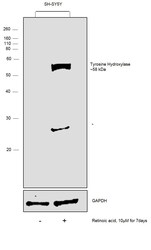
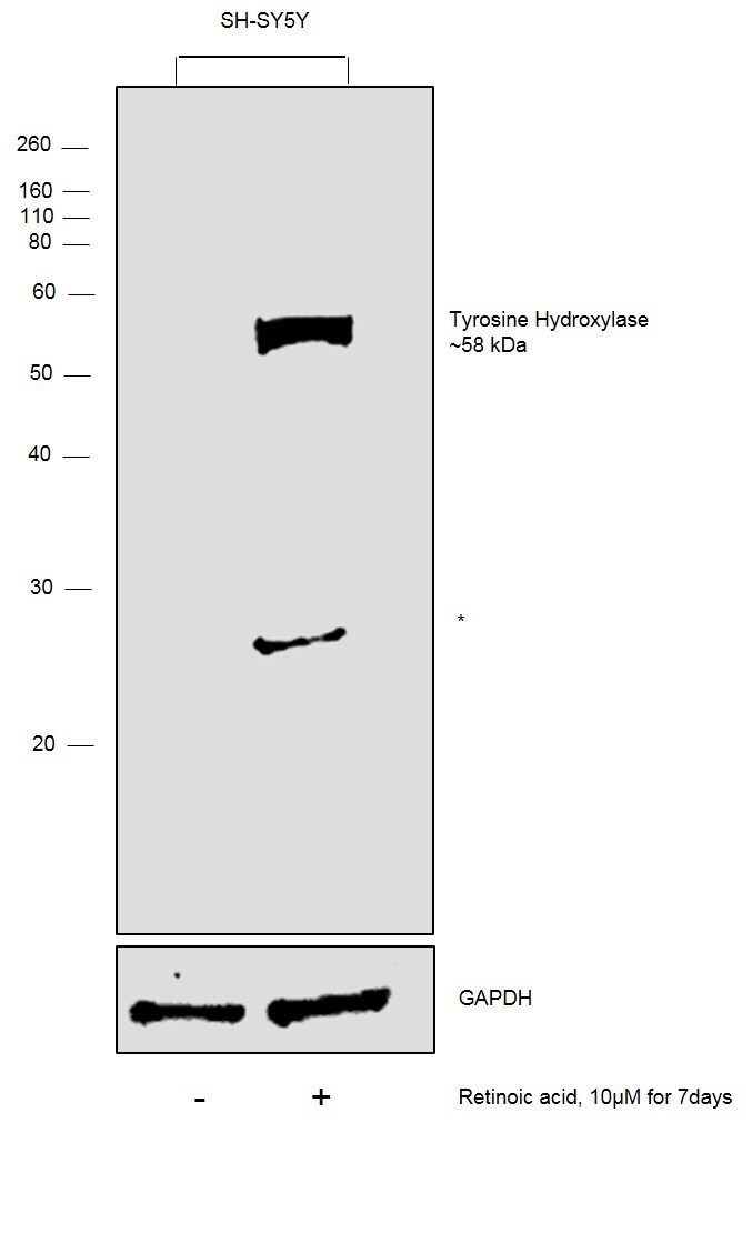
- Western blot was performed using anti-Tyrosine Hydroxylase Polyclonal Antibody, (Product # PA5-17800) and band at ~58 kDa corresponding to Tyrosine Hydroxylase was observed specifically in SH-SY5Y cells treated with Retinoic acid. An uncharacterized band at ~28 kDa (*) was also observed in treated SH-SY5Y cells. Whole cell extracts (30 µg lysate) of SH-SY5Y (Lane 1) and SH-SY5Y treated with Retinoic Acid (10uM for 7days) (Lane 2) were electrophoresed using Novex® NuPAGE® 4-12% % Bis-Tris gel (Product # NP0322BOX). Resolved proteins were then transferred onto a nitrocellulose membrane (Product # IB23001) by iBlot® 2 Dry Blotting System (Product # IB21001). The blot was probed with the primary antibody (1:1000 dilution) and detected by chemiluminescence with Goat anti-Rabbit IgG (H+L) Superclonal™ Recombinant Secondary Antibody, HRP (Product # A27036, 1:4000 dilution) using the iBright FL 1000 (Product # A32752). Chemiluminescent detection was performed using Novex® ECL Chemiluminescent Substrate Reagent Kit (Product # WP20005).
- Submitted by
- Invitrogen Antibodies (provider)
- Main image

- Experimental details
- Western blot analysis was performed on Whole cell extracts (30 µg lysate) of PC12 (Lane 1) and PC12 treated with Dexamethasone (6 µg/mL for 6 days) (Lane 2). The blot was probed with Anti-Tyrosine Hydroxylase Polyclonal Antibody (Product # PA5-17800, 1:1000 dilution) and detected by chemiluminescence using Goat anti-Rabbit IgG (H+L) Superclonal™ Secondary Antibody, HRP conjugate (Product # A27036, 0.25 µg/mL, 1:4000 dilution). A 58.6 kDa band corresponding to Tyrosine Hydroxylase was observed to be overexpressed upon Dexamethasone treatment of PC12 cells.

Supportive validation
- Submitted by
- Invitrogen Antibodies (provider)
- Main image
- Experimental details
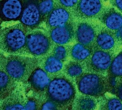
- Immunofluorescent analysis of Tyrosine Hydroxylase using a polyclonal antibody (Product # PA5-17800).

- Submitted by
- Invitrogen Antibodies (provider)
- Main image

- Experimental details
- Immunofluorescence analysis of Tyrosine Hydroxylase was performed using 70% confluent log phase PC-12 cells untreated or treated with 5 µg/mL Dexamethasone for 6 Days. The cells were fixed with 4% paraformaldehyde for 10 minutes, permeabilized with 0.1% Triton™ X-100 for 15 minutes, and blocked with 1% BSA for 1 hour at room temperature. The cells were labeled with anti-Tyrosine Hydroxylase Rabbit Monoclonal Antibody (Product # PA5-17800) at 1:200 dilution in 0.1% BSA, incubated at 4 degree Celsius overnight and then labeled with Goat anti-Rabbit IgG (H+L) Superclonal™ Secondary Antibody, Alexa Fluor® 488 conjugate (Product # A27034) at a dilution of 1:2000 for 45 minutes at room temperature (Panel a: green). Nuclei (Panel b: blue) were stained with ProLong™ Diamond Antifade Mountant with DAPI (Product # P36962). F-actin (Panel c: red) was stained with Rhodamine Phalloidin (Product # R415, 1:300). Panel d represents the merged image showing predominantly cytosolic localization. Panel e represents control cells with no treatment while panel f represents control cells with no primary antibody to assess background. The images were captured at 60X magnification.
