Explore
 Validate
 Learn
Learn Western blot
 Immunocytochemistry
Antibody data
- Antibody Data
- Antigen structure
- References [8]
- Comments [0]
- Validations
- Immunocytochemistry [2]
- Immunohistochemistry [1]
- Other assay [11]
Submit
Validation data
Reference
Comment
Report error
- Product number
- PA1-4679 - Provider product page

- Provider
- Invitrogen Antibodies
- Product name
- Tyrosine Hydroxylase Polyclonal Antibody
- Antibody type
- Polyclonal
- Antigen
- Purifed from natural sources
- Description
- This antibody is specific for the ~60 kDa Tyrosine Hydroxylase in Western blots of rat brain extracts.
- Reactivity
- Human, Mouse, Rat
- Host
- Sheep
- Isotype
- IgG
- Vial size
- 100 μL
- Concentration
- 0.15 mg/mL
- Storage
- -20°C, Avoid Freeze/Thaw Cycles
Submitted references Post-Ganglionic Sympathetic Neurons can Directly Sense Raised Extracellular Na(+) via SCN7a/Na(x).
Methanolic Extract of Boswellia serrata Gum Protects the Nigral Dopaminergic Neurons from Rotenone-Induced Neurotoxicity.
The Mesolimbic Dopamine Activity Signatures of Relapse to Alcohol-Seeking.
Parkin deficiency accelerates consequences of mitochondrial DNA deletions and Parkinsonism.
Loss of the transcription factor Meis1 prevents sympathetic neurons target-field innervation and increases susceptibility to sudden cardiac death.
Abnormal sinoatrial node development resulting from disturbed vascular endothelial growth factor signaling.
Astroglial heme oxygenase-1 and the origin of corpora amylacea in aging and degenerating neural tissues.
Catheter-based renal sympathetic denervation significantly inhibits atrial fibrillation induced by electrical stimulation of the left stellate ganglion and rapid atrial pacing.
Davis H, Paterson DJ, Herring N
Frontiers in physiology 2022;13:931094
Frontiers in physiology 2022;13:931094
Methanolic Extract of Boswellia serrata Gum Protects the Nigral Dopaminergic Neurons from Rotenone-Induced Neurotoxicity.
Shadfar S, Khanal S, Bohara G, Kim G, Sadigh-Eteghad S, Ghavami S, Choi H, Choi DY
Molecular neurobiology 2022 Sep;59(9):5874-5890
Molecular neurobiology 2022 Sep;59(9):5874-5890
The Mesolimbic Dopamine Activity Signatures of Relapse to Alcohol-Seeking.
Liu Y, Jean-Richard-Dit-Bressel P, Yau JO, Willing A, Prasad AA, Power JM, Killcross S, Clifford CWG, McNally GP
The Journal of neuroscience : the official journal of the Society for Neuroscience 2020 Aug 12;40(33):6409-6427
The Journal of neuroscience : the official journal of the Society for Neuroscience 2020 Aug 12;40(33):6409-6427
Parkin deficiency accelerates consequences of mitochondrial DNA deletions and Parkinsonism.
Song L, McMackin M, Nguyen A, Cortopassi G
Neurobiology of disease 2017 Apr;100:30-38
Neurobiology of disease 2017 Apr;100:30-38
Loss of the transcription factor Meis1 prevents sympathetic neurons target-field innervation and increases susceptibility to sudden cardiac death.
Bouilloux F, Thireau J, Ventéo S, Farah C, Karam S, Dauvilliers Y, Valmier J, Copeland NG, Jenkins NA, Richard S, Marmigère F
eLife 2016 Feb 8;5
eLife 2016 Feb 8;5
Abnormal sinoatrial node development resulting from disturbed vascular endothelial growth factor signaling.
Calkoen EE, Vicente-Steijn R, Hahurij ND, van Munsteren CJ, Roest AA, DeRuiter MC, Steendijk P, Schalij MJ, Gittenberger-de Groot AC, Blom NA, Jongbloed MR
International journal of cardiology 2015 Mar 15;183:249-57
International journal of cardiology 2015 Mar 15;183:249-57
Astroglial heme oxygenase-1 and the origin of corpora amylacea in aging and degenerating neural tissues.
Song W, Zukor H, Liberman A, Kaduri S, Arvanitakis Z, Bennett DA, Schipper HM
Experimental neurology 2014 Apr;254:78-89
Experimental neurology 2014 Apr;254:78-89
Catheter-based renal sympathetic denervation significantly inhibits atrial fibrillation induced by electrical stimulation of the left stellate ganglion and rapid atrial pacing.
Hou Y, Hu J, Po SS, Wang H, Zhang L, Zhang F, Wang K, Zhou Q
PloS one 2013;8(11):e78218
PloS one 2013;8(11):e78218
No comments: Submit comment
Supportive validation
- Submitted by
- Invitrogen Antibodies (provider)
- Main image
- Experimental details
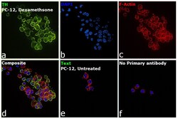
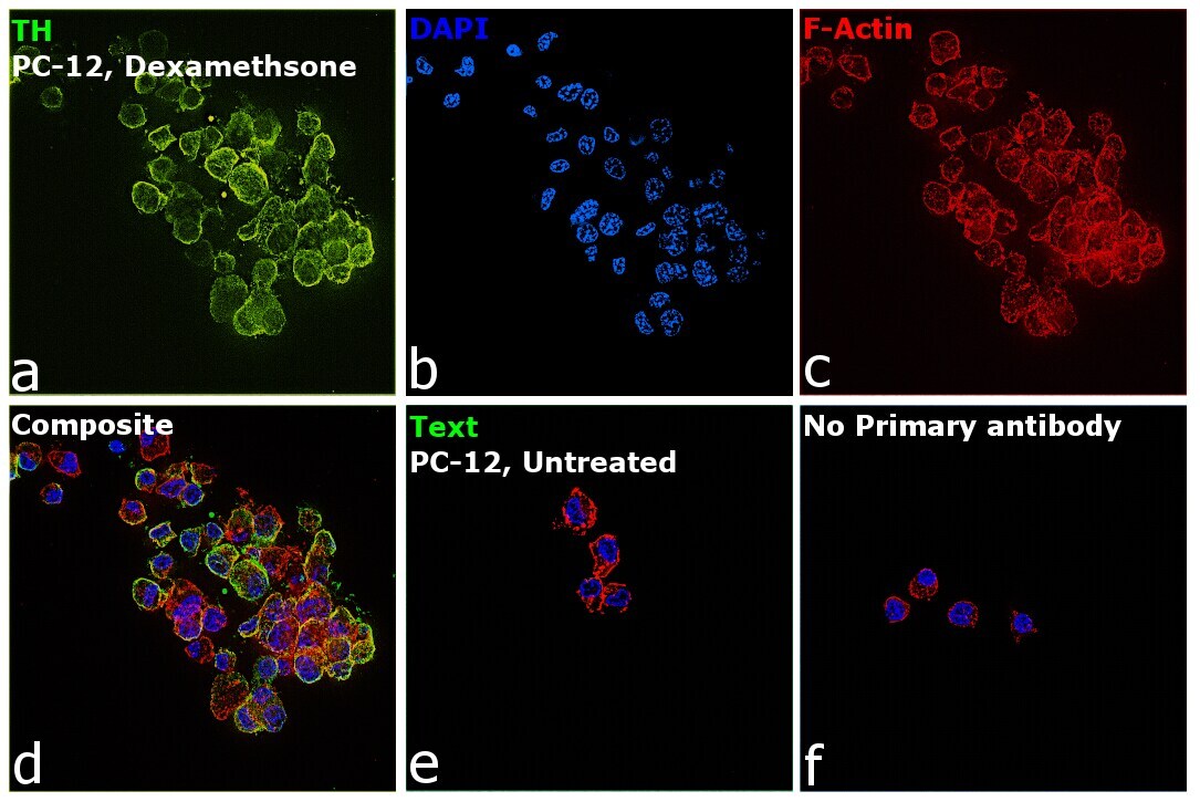
- Immunofluorescence analysis of Anti-Tyrosine Hydroxylase Polyclonal Antibody (Product # PA1-4679) was performed using 70% confluent log phase PC-12 cells treated with 5 µg/mL dexamethasone for 6 days. The cells were fixed with 4% Paraformaldehyde for 10 minutes, permeabilized with 0.1% Triton™ X-100 for 10 minutes, and blocked with 2% BSA for 10 minutes at room temperature. The cells were labeled with Anti-Tyrosine Hydroxylase Polyclonal Antibody (Product # PA1-4679) at 1:1000 dilution in 0.1% BSA, incubated at 4 degree Celsius overnight and then labeled with Donkey anti-Sheep IgG (H+L) Cross-Adsorbed Secondary Antibody, Alexa Fluor 488 (Product # A-11015, 1:2000 dilution) for 45 minutes at room temperature (Panel a: Green). Nuclei (Panel b: Blue) were stained with SlowFade® Gold Antifade Mountant with DAPI (Product # S36938). F-actin (Panel c: Red) was stained with Rhodamine Phalloidin (Product # R415, 1:300). Panel d represents the merged image showing Cytoplasmic localization. Panel e represents untreated PC-12 cells having low expression of TH. Panel f represents control cells with no primary antibody to assess background. The images were captured at 60X magnification.

- Submitted by
- Invitrogen Antibodies (provider)
- Main image

- Experimental details
- Immunofluorescence analysis of Anti-Tyrosine Hydroxylase Polyclonal Antibody (Product # PA1-4679) was performed using 70% confluent log phase PC-12 cells treated with 5 µg/mL dexamethasone for 6 days. The cells were fixed with 4% Paraformaldehyde for 10 minutes, permeabilized with 0.1% Triton™ X-100 for 10 minutes, and blocked with 2% BSA for 10 minutes at room temperature. The cells were labeled with Anti-Tyrosine Hydroxylase Polyclonal Antibody (Product # PA1-4679) at 1:1000 dilution in 0.1% BSA, incubated at 4 degree Celsius overnight and then labeled with Donkey anti-Sheep IgG (H+L) Cross-Adsorbed Secondary Antibody, Alexa Fluor 488 (Product # A-11015, 1:2000 dilution) for 45 minutes at room temperature (Panel a: Green). Nuclei (Panel b: Blue) were stained with SlowFade® Gold Antifade Mountant with DAPI (Product # S36938). F-actin (Panel c: Red) was stained with Rhodamine Phalloidin (Product # R415, 1:300). Panel d represents the merged image showing Cytoplasmic localization. Panel e represents untreated PC-12 cells having low expression of TH. Panel f represents control cells with no primary antibody to assess background. The images were captured at 60X magnification.
Supportive validation
- Submitted by
- Invitrogen Antibodies (provider)
- Main image
- Experimental details
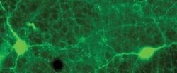
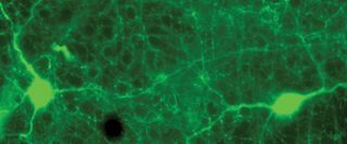
- Immunohistochemistry analysis of Tyrosine Hydroxylase in rabbit retina showing specific labeling of Phospho-NMDAR2B, (Tyr1336) polyclonal antibody (Product # PA1-4679) in green.
Supportive validation
- Submitted by
- Invitrogen Antibodies (provider)
- Main image
- Experimental details
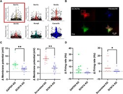
- FIGURE 5 Na x is responsible for sodium sensitivity in stellate ganglia neurons. (A) Single-cell RNA-sequencing highlighted expression of the Na x encoding gene SCN7A (Yellow, with red box) at a transcript level in the stellate ganglia sympathetic neurons. These data are shown against expression of other ion channel encoding genes, SCN1A - SCN3A (Na v ) (Red), KCNQ5 (K v ) (Green) and CACNA1B (Ca v ) (Blue). Here expression level indicates normalized count data. Here the count data per cell is divided by the total number of counts within that cell, multiplied by a scale factor and then natural-log transformed. (B) Immunohistochemistry highlights protein expression of Na x in tyrosine hydroxylase positive stellate ganglia neurons. Cultured stellate ganglia cells were co-labelled for Na x (Red), the sympathetic neuron marker, tyrosine hydroxylase (Green), and the nuclear stain Hoescht 33342 (Blue). (C) Knockdown of SCN7A significantly reduced the depolarization induced by an additional 30 mM NaCl, compared to GAPDH RNA knockdown (-2.87 +- 1.04 mV; n = 11-12; One-tailed unpaired t -test; p = 0.006) knockdown of a scrambled RNA control (-3.091 +- 0.8674 mV; n = 7-12; One-tailed unpaired t -test; p = 0.0012). (D) Knockdown of SCN7A caused a non-significant decrease in the increase in firing rate following 170 mM [NaCl] o (4.5-0 Hz; n = 8-10; One-tailed Mann Whitney test; p = 0.06). Similarly, SCN7A knockdown reduced the firing rate induced by 170 mM [NaCl] o when compared to a scr

- Submitted by
- Invitrogen Antibodies (provider)
- Main image
- Experimental details
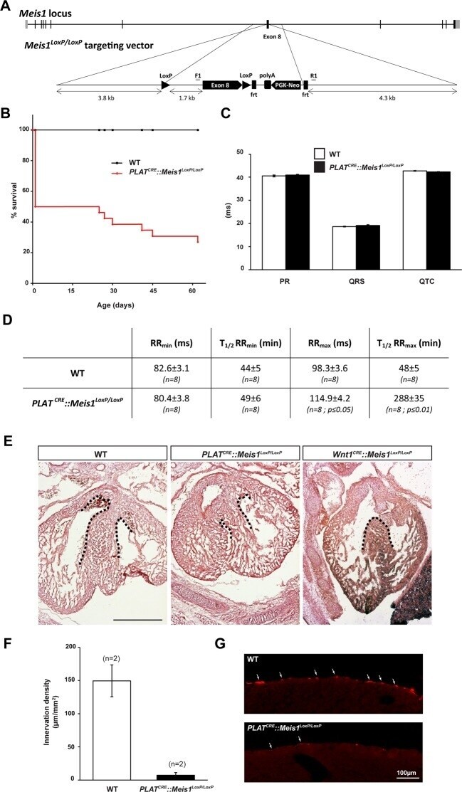
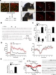
- Figure 1--figure supplement 1. Sympatho-vagal deregulation of cardiac functions following Meis1 inactivation in the PNS. ( A ) Targeting vector used for the generation of the Meis1 conditional knockout mouse strain ( Meis1 LoxP/LoxP ). The exon 8 of the gene encoding the beginning of the homeodomain has been flanked with LoxP sites. ( B ) Kaplan-Meier survival curve showing the premature death of PLAT CRE ::Meis1 LoxP/LoxP mice (in red) compared to WT mice (in black). ( C ) Measure of the PR, QRS and QTc intervals on a 24 hr ECG telemetric recording showed no statistically difference between groups, indicating that Meis1 invalidation in the PNS does not affect the intrinsic cardiac conduction properties. ( D ) Table recapitulating the minimum RR (RR min ), corresponding to the maximal effect of isoproterenol on heart rate and the time to lose half of the effect (T 1/2 RRmin), the maximum RR interval (RR max ), corresponding to the maximal effect of carbamoylcholine chloride and the duration to lose half of the effect (T 1/2 RR max ) in WT and PLAT CRE ::Meis1 LoxP/LoxP mice. Data are represented as mean +/- s.e.m., n = 8 in all experiments. ( E ) Histologic section of E14.5 WT, PLAT CRE ::Meis1 LoxP/LoxP and Wnt1 CRE ::Meis1 LoxP/LoxP hearts embryos. Whereas the cardiac septum was identical to that of WT in 3/3 analyzed PLAT CRE ::Meis1 LoxP/LoxP embryos, we observed a defect in the cardiac septum of 1/1 Wnt1 CRE ::Meis1 LoxP/LoxP embryo. Scale bar = 500 um. ( F ) Quantificat
- Submitted by
- Invitrogen Antibodies (provider)
- Main image
- Experimental details
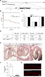
- Figure 1. Specific Meis1 inactivation in the PNS results in sympatho-vagal deregulation of cardiac functions. ( A ) Immunochemistry for Meis1 and the transcription factor Phox2b on sections of SCGs from E16.5 WT and PLAT CRE ::Meis1 LoxP/LoxP mice, and western blot analysis for Meis1 from adult stellate ganglia. Surviving adult mutants exhibited blepharoptosis (white dotted square). Immunostaining for TH + sympathetic fibers in the heart of newborn WT and PLAT CRE ::Meis1 LoxP/LoxP mice. VE = Ventricle, AR = Atrium. White arrows point at TH + sympathetic fibers. Scale bar = 20 um. ( B ) Example of an ECG trace recorded in an adult male PLAT CRE ::Meis1 LoxP/LoxP mouse. The black arrow indicates the start of a junctional bradycardia and the red asterisks mark abnormal P waves locations. Dotted black square delineates the power magnification of the ECG trace showed. ( C ) HRV analysis of the ECGs recordings of PLAT CRE ::Meis1 LoxP/LoxP and WT mice. Graphical representations of the mean RR interval, the standard deviation of all normal to normal beats (SDNN), the power of low frequency band (LF), the power of the high frequency band (HF) and the LF/HF ratio. ( D ) Time course of the mean RR interval during isoproterenol or carbamoylcholine chloride challenges in WT and PLAT CRE ::Meis1 LoxP/LoxP mice. ( E ) Typical sinus arrest (SA) trace obtained in a PLAT CRE ::Meis1 LoxP/LoxP mouse and quantification of the mean number of sinus arrest per hour over a 12 hr ECG recording in P

- Submitted by
- Invitrogen Antibodies (provider)
- Main image

- Experimental details
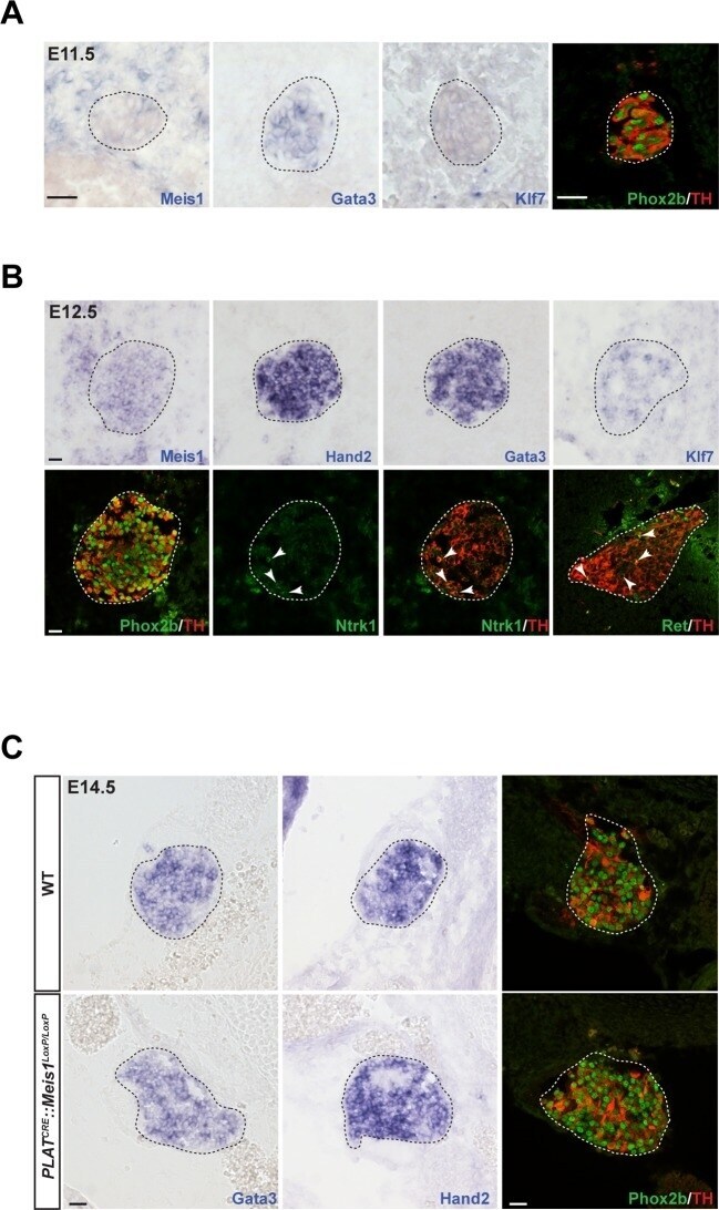
- Figure 2. Meis1 expression in sympathetic neurons is incompatible with early sympathetic specification but coincides with target-field innervation onset. ( A ) ISH for Meis1, Gata3 and Klf7, and immunochemistry for Phox2b and TH in WT E11.5 embryonic SCGs. ( B ) ISH for Meis1, Hand2, Gata3 and Klf7 and immunochemistry for Phox2b, TH, Ntrk1 and Ret in WT E12.5 embryonic SCGs. White arrows point at Ntrk1 or Ret neurons. ( C ) ISH for Gata3 and Hand2 and immunochemistry for Phox2b and TH on WT and PLAT CRE ::Meis1 LoxP/LoxP E14.5 embryonic SCGs. White and black dotted lines delineate the SCGs. Scale bar = 20 um. See also Figure 2--figure supplement 1 . DOI: http://dx.doi.org/ Figure 2--figure supplement 1. Meis1 is the only member of the Meis family to be expressed by embryonic sympathetic neurons and is indifferently expressed by noradrenergic and cholinergic sympathetic neurons. ( A ) In situ hybridization (ISH) for Meis1, Meis2 and Meis3 on E11.5, E13.5 and E14.5 WT embryos showed that Meis1 is the only member of the family to be expressed in sympathetic ganglia. Black dashed lines encircles the SCGs. ( B ) Double ISH for Meis1 (pseudo color in red) with Dopamine beta-Hydroxylase (DBH, pseudo color in green) or with Ret (pseudo color in red) indicated that both major subpopulations (noradrenergic and presumptive cholinergic) of sympathetic neurons expressed Meis1 at E16.5. White arrowheads point at double positive neurons. White dotted lines encircle the SCGs. ( C ) Immunoche
- Submitted by
- Invitrogen Antibodies (provider)
- Main image
- Experimental details
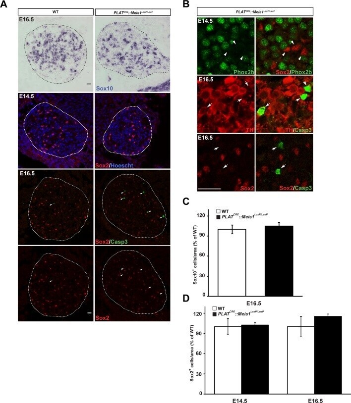
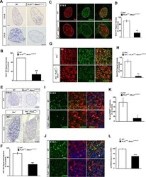
- Figure 3--figure supplement 1. Survival of glial progenitor cells is not affected following Meis1 inactivation. ( A ) ISH for Sox10 and immunochemistry for Sox2 and Casp3 on E14.5 and E16.5 SCGs from WT and PLAT CRE ::Meis1 LoxP/LoxP embryos. White arrows point at Casp3 + cells. ( B ) Immunochemistry for Sox2 and Phox2b on E14.5 SCGs showed that their expressions are mutually exclusive. Immunochemistry for Sox2 or TH with Casp3 showed that Casp3 immunoreactivity was present in TH + neurons but never in Sox2 + glial progenitor cells. ( C ) Quantification of the number of Sox10 + cells in E16.5 WT and PLAT CRE ::Meis1 LoxP/LoxP SCGs. ( D ) Quantification of the number of Sox2 + cells in E14.5 and E16.5 WT and PLAT CRE ::Meis1 LoxP/LoxP SCGs. Data are represented as mean +/- s.e.m. n = 3; Scale bars = 20 um in A and B. DOI: http://dx.doi.org/
- Submitted by
- Invitrogen Antibodies (provider)
- Main image
- Experimental details
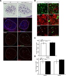
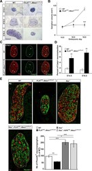
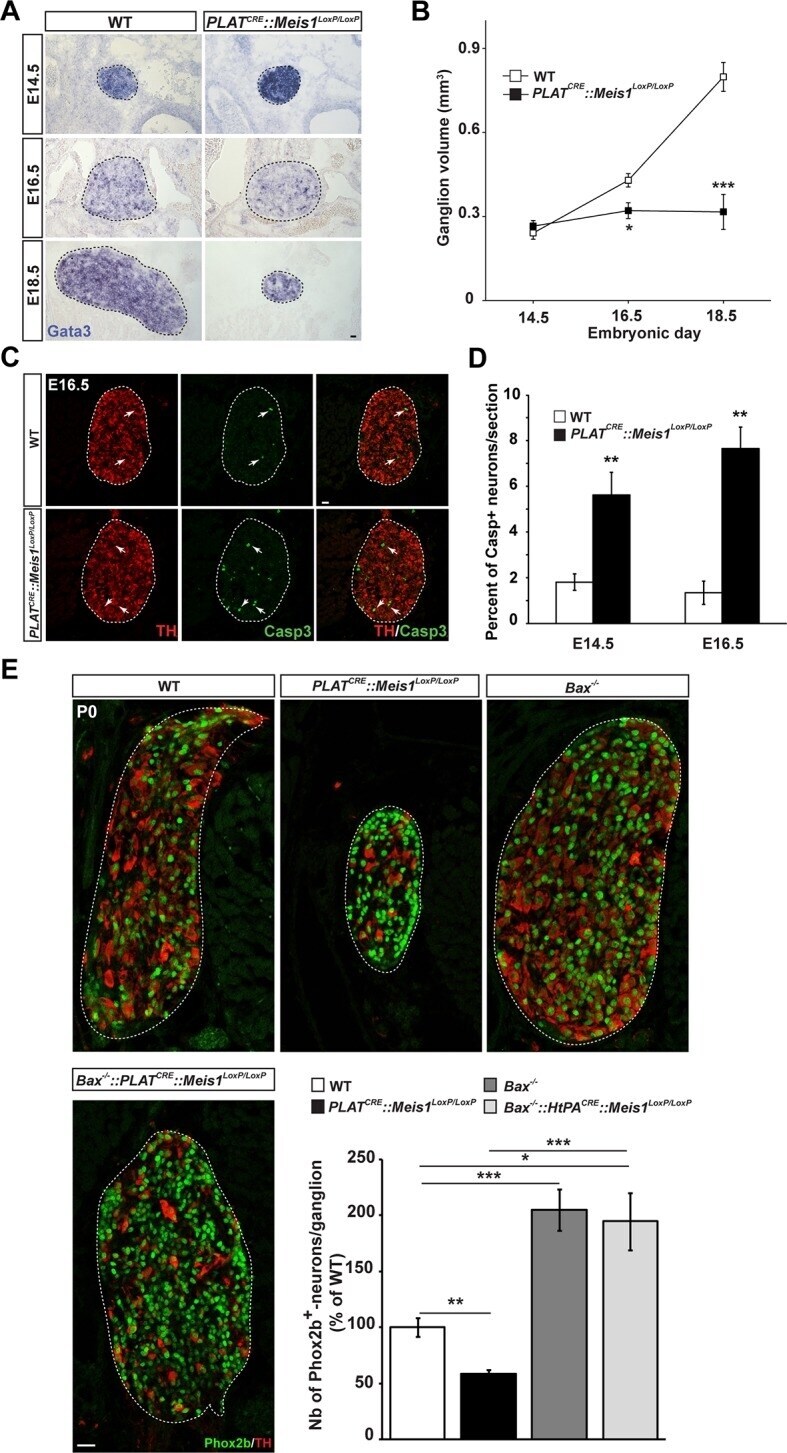
- Figure 3. Exacerbated neuronal apoptosis in Meis1 mutant sympathetic neurons. ( A ) ISH for Gata3 on SCGs of PLAT CRE ::Meis1 LoxP/LoxP and WT embryos at E14.5, 16.5 and E18.5. ( B ) Quantification of the volume of the SCGs in WT vs PLAT CRE ::Meis1 LoxP/LoxP mice at E14.5, E16.5 and E18.5. ( C ) Representative images of the SCGs visualized by immunochemistry for activated Caspase-3 (Casp3) and TH at E16.5 in WT and PLAT CRE ::Meis1 LoxP/LoxP mice. Arrows point at Casp3 + /TH + neurons. ( D ) Quantification of the average number of Casp3 + /TH + neurons per section in the SCGs of PLAT CRE ::Meis1 LoxP/LoxP and WT embryos at E14.5 and E16.5. ( E ) Immunochemistry for Phox2b and TH on SCGs of P0 WT, Bax -/- , PLAT CRE ::Meis1 LoxP/LoxP and PLAT CRE ::Meis1 LoxP/LoxP ::Bax -/- newborn pups, and quantification of the number of Phox2b + neurons per SCG in P0 WT, Bax -/- , PLAT CRE ::Meis1 LoxP/LoxP and Bax -/- ::PLAT CRE ::Meis1 LoxP/LoxP mice. Data are represented as mean +/- s.e.m. n = 3; *p
- Submitted by
- Invitrogen Antibodies (provider)
- Main image
- Experimental details
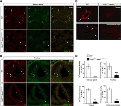
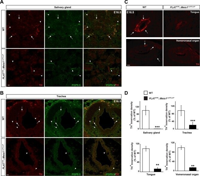
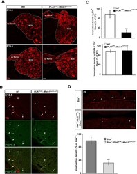
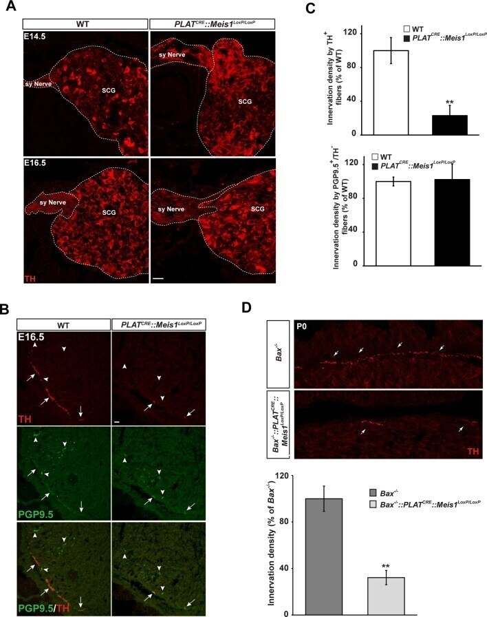
- Figure 4--figure supplement 1. Sympathetic target-field innervation of peripheral organs is compromised following Meis1 loss of function. ( A and B ) Visualization of TH + sympathetic fibers (in red) and others PGP9.5 + /TH - peripheral nervous projections (in green) by immunochemistry in the salivary glands ( A ) and the trachea ( B ) of PLAT CRE ::Meis1 LoxP/LoxP and WT embryos at E16.5. Only the TH + sympathetic innervation is affected in the mutant. Arrows point at TH + sympathetic fibers and arrowheads point at PGP9.5 + /TH - nerves. ( C ) Visualization by immunochemistry of TH + sympathetic fibers (in red) in the tongue and the vomeronasal organ of PLAT CRE ::Meis1 LoxP/LoxP and WT embryos at E16.5. Arrows point at TH + sympathetic fibers. ( D ) Quantification of the density of TH + sympathetic fibers on sections of the salivary glands, the vomeronasal organ, the tongue and the trachea in PLAT CRE ::Meis1 LoxP/LoxP and WT embryos at E16.5 revealed that SCG neurons failed to establish contact with their peripheral targets during embryogenesis. Data are represented as mean +/- s.e.m. n = 3; ** p
- Submitted by
- Invitrogen Antibodies (provider)
- Main image
- Experimental details
- Figure 4. Lack of terminal sympathetic target-field innervation in Meis1 mutant embryos. ( A ) Immunochemistry for TH on SCGs from E14.5 and E16.5 WT and PLAT CRE ::Meis1 LoxP/LoxP embryos showing that proximal axonal growth of sympathetic neurons is not affected. Sy Nerve = sympathetic nerve. ( B ) Visualization of TH + sympathetic fibers and others PGP9.5 + /TH - peripheral neurons projections in the heart of PLAT CRE ::Meis1 LoxP/LoxP and WT embryos at E16.5. Arrows point at TH + sympathetic fibers and arrowheads point at PGP9.5 + /TH - nerves. ( C ) Quantification of the density of TH + /PGP9.5 + sympathetic and TH - /PGP9.5 + sensory fibers on heart sections of PLAT CRE ::Meis1 LoxP/LoxP and WT embryos at E16.5. ( D ) Representative images and quantification of TH + sympathetic fibers in the heart of Bax -/- and PLAT CRE ::Meis1 LoxP/LoxP ::Bax -/- newborn pups. Data are represented as mean +/- s.e.m. n = 3; **p
- Submitted by
- Invitrogen Antibodies (provider)
- Main image
- Experimental details
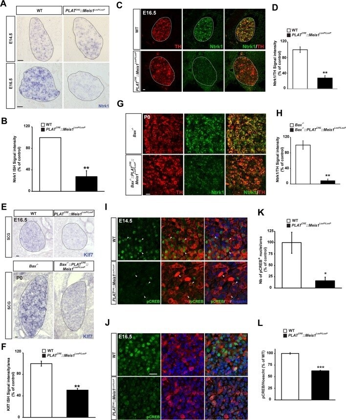
- Figure 5. Target-field innervation signaling pathways in Meis1 -inactivated sympathetic neurons. ( A ) ISH for the neurotrophins high affinity receptor Ntrk1 in E14.5 and E16.5 WT and PLAT CRE ::Meis1 LoxP/LoxP SCGs. ( B ) Quantification of Ntrk1 ISH signal intensity in E16.5 WT and PLAT CRE ::Meis1 LoxP/LoxP SCGs. ( C ) Immunochemistry for TH and Ntrk1 on E16.5 WT and PLAT CRE ::Meis1 LoxP/LoxP embryos. White dotted lines encircle the SCGs. ( D ) Quantification of the intensity of immuno-fluorescence for Ntrk1 in the SCGs of E16.5 WT and PLAT CRE ::Meis1 LoxP/LoxP embryos. ( E ) Representative images of ISH for Klf7 on SCGs of E16.5 WT and PLAT CRE ::Meis1 LoxP/LoxP embryos and on SCGs of P0 Bax -/- and PLAT CRE ::Bax -/- ::Meis1 LoxP/LoxP mice. ( F ) Quantification of Klf7 ISH signal intensity in SCGs of E16.5 WT and PLAT CRE ::Meis1 LoxP/LoxP embryos. ( G ) Immunochemistry for TH and Ntrk1 on P0 Bax -/- and PLAT CRE ::Meis1 LoxP/LoxP ::Bax -/- mice. ( H ) Quantification of the intensity of Ntrk1 immuno-fluorescence in the SCGs of P0 Bax -/- and PLAT CRE ::Meis1 LoxP/LoxP ::Bax -/- mice. ( I ) Immunochemistry for pCREB and TH on E14.5 SCGs from WT and PLAT CRE ::Meis1 LoxP/LoxP embryos and quantification of the number of pCREB + nuclei. ( J ) Immunochemistry for pCREB and TH on E16.5 SCGs from WT and PLAT CRE ::Meis1 LoxP/LoxP embryos and quantification of pCREB signal intensity in neuronal nuclei. Data are represented as mean +/- s.e.m. n = 3; *p
- Submitted by
- Invitrogen Antibodies (provider)
- Main image
- Experimental details
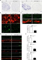
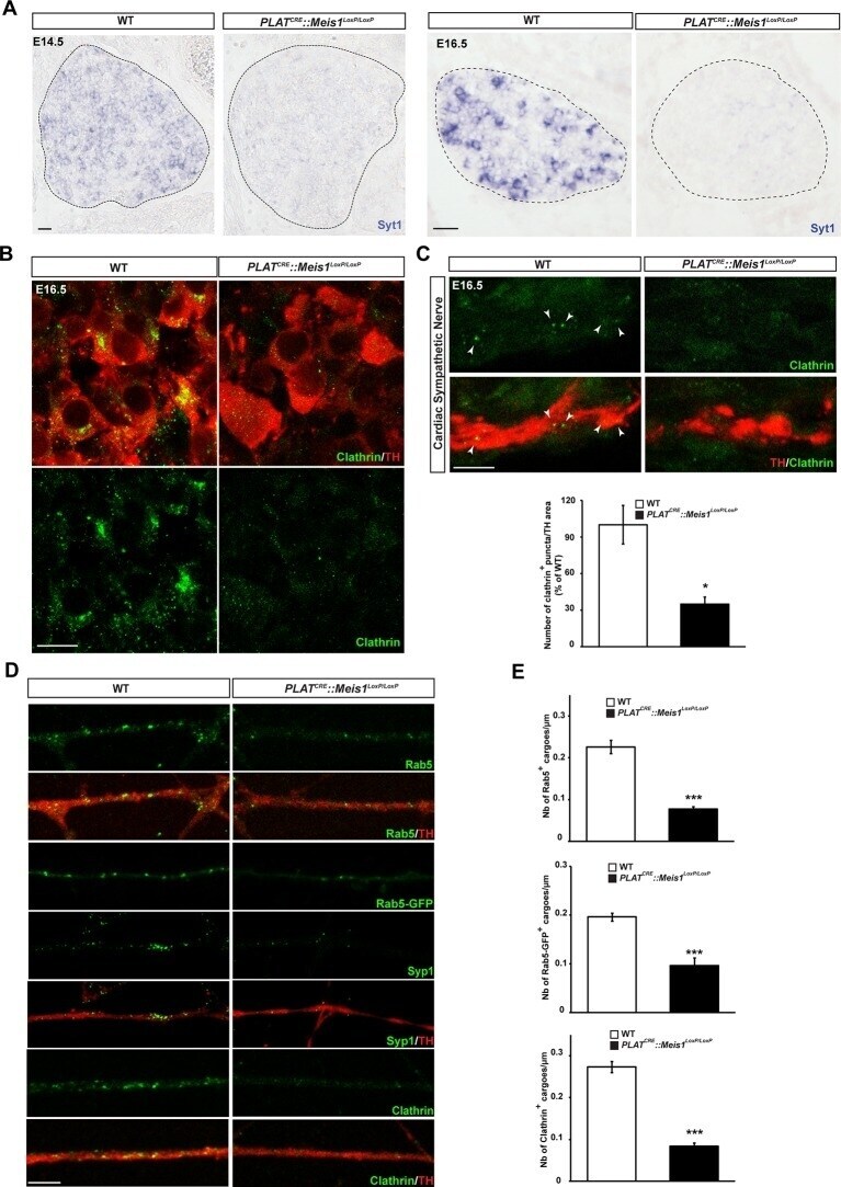
- Figure 6. Meis1 target genes encode proteins necessary for early endosomes formation. ( A ) ISH for Syt1 on SCGs of E14.5 and E16.5 WT and PLAT CRE ::Meis1 LoxP/LoxP embryos. ( B ) Immunochemistry for clathrin and TH on SCGs of E16.5 WT and PLAT CRE ::Meis1 LoxP/LoxP embryos. ( C ) Representative images and quantification of immunochemistry for clathrin and TH on the sympathetic nerves within the heart of E16.5 WT and PLAT CRE ::Meis1 LoxP/LoxP embryos. White arrowheads point at clathrin-coated pits and cargoes. ( D ) Immunochemistry for clathrin, TH, Syp1 and Rab5 and overexpression of a Rab5-GFP construct in cultured explants of SCGs from WT and PLAT CRE ::Meis1 LoxP/LoxP embryos. ( E ) Quantification of the numbers of Rab5, Rab5-GFP and clathrin + cargoes in cultured explants of SCGs. Dotted lines encircle the SCGs. Scale bar = 20 um in A, 10 um in B and 5 um in C and D . Data are represented as mean +/- s.e.m; n = 3; *p
- Submitted by
- Invitrogen Antibodies (provider)
- Main image
- Experimental details
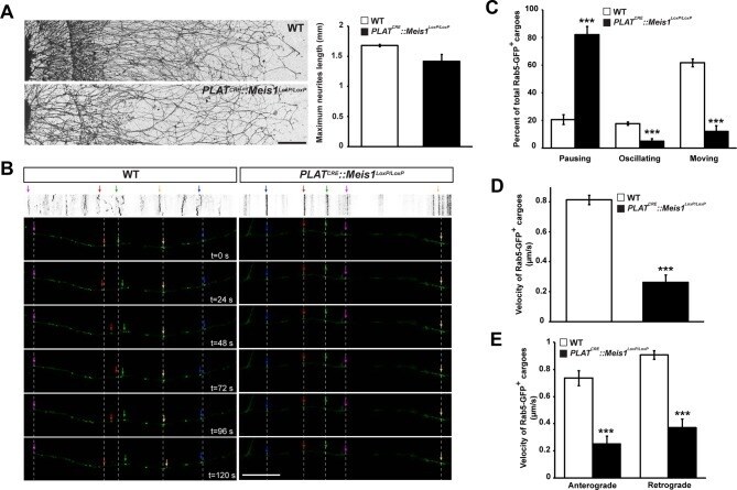
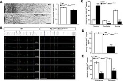
- Figure 7. Meis1 is necessary for early endosomes trafficking. ( A ) Camera lucida of TH-immunochemistry and quantification of neurites length on SCGs explants form WT and PLAT CRE ::Meis1 LoxP/LoxP embryos. ( B ) Individual frames and corresponding kymograph of a 2 min time lapse video showing the movement of Rab5-GFP + endosomes in WT ( Video 1 ) and PLAT CRE ::Meis1 LoxP/LoxP ( Video 3 ) cultured SCGs explants. ( C ) Analysis of the percentage of the number of Rab5-GFP + endosomes that are pausing, moving or oscillating in WT and PLAT CRE ::Meis1 LoxP/LoxP cultured SCGs explants. ( D ) Measure of the velocity of Rab5-GFP + endosomes that are moving in WT and mutant conditions. ( E ) Measure of the velocity of Rab5-GFP + endosomes that are moving retrogradely and anterogradely in WT and PLAT CRE ::Meis1 LoxP/LoxP cultured SCGs explants. Data are represented as mean +/- s.e.m; n = 3; ***p