Explore
 Validate
 Learn
LearnPA1-30399
antibody from Invitrogen Antibodies
Targeting: CDKN1A
CAP20, CDKN1, CIP1, P21, p21CIP1, p21Cip1/Waf1, SDI1, WAF1
 Western blot
 Immunoprecipitation
Antibody data
- Antibody Data
- Antigen structure
- References [3]
- Comments [0]
- Validations
- Western blot [1]
- Other assay [3]
Submit
Validation data
Reference
Comment
Report error
- Product number
- PA1-30399 - Provider product page

- Provider
- Invitrogen Antibodies
- Product name
- p21 Polyclonal Antibody
- Antibody type
- Polyclonal
- Antigen
- Synthetic peptide
- Description
- Recommended positive controls: C32.
- Reactivity
- Human, Mouse, Rat
- Host
- Rabbit
- Isotype
- IgG
- Vial size
- 1 mL
- Concentration
- 0.2 mg/mL
- Storage
- 4° C, do not freeze
Submitted references HDAC inhibitors suppress the proliferation, migration and invasiveness of human head and neck squamous cell carcinoma cells via p63‑mediated tight junction molecules and p21‑mediated growth arrest.
Senescent cells promote tissue NAD(+) decline during ageing via the activation of CD38(+) macrophages.
IL-6 deficiency attenuates p53 protein accumulation in aged male mouse hippocampus.
Kakiuchi A, Kakuki T, Ohwada K, Kurose M, Kondoh A, Obata K, Nomura K, Miyata R, Kaneko Y, Konno T, Kohno T, Himi T, Takano KI, Kojima T
Oncology reports 2021 Apr;45(4)
Oncology reports 2021 Apr;45(4)
Senescent cells promote tissue NAD(+) decline during ageing via the activation of CD38(+) macrophages.
Covarrubias AJ, Kale A, Perrone R, Lopez-Dominguez JA, Pisco AO, Kasler HG, Schmidt MS, Heckenbach I, Kwok R, Wiley CD, Wong HS, Gibbs E, Iyer SS, Basisty N, Wu Q, Kim IJ, Silva E, Vitangcol K, Shin KO, Lee YM, Riley R, Ben-Sahra I, Ott M, Schilling B, Scheibye-Knudsen M, Ishihara K, Quake SR, Newman J, Brenner C, Campisi J, Verdin E
Nature metabolism 2020 Nov;2(11):1265-1283
Nature metabolism 2020 Nov;2(11):1265-1283
IL-6 deficiency attenuates p53 protein accumulation in aged male mouse hippocampus.
Bialuk I, Cieślińska M, Kowalczuk O, Bonda TA, Nikliński J, Winnicka MM
Biogerontology 2020 Feb;21(1):29-43
Biogerontology 2020 Feb;21(1):29-43
No comments: Submit comment
Supportive validation
- Submitted by
- Invitrogen Antibodies (provider)
- Main image
- Experimental details
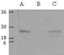
- Western blot of p21/Cip1 in 15 µg of (A) A431 lysate (B) HL-60 lysate and (C) Hela lysate using a p21/Cip1 polyclonal antibody (Product # PA1-30399) at a dilution of '1:200.

Supportive validation
- Submitted by
- Invitrogen Antibodies (provider)
- Main image
- Experimental details
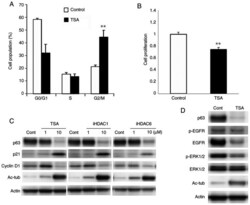
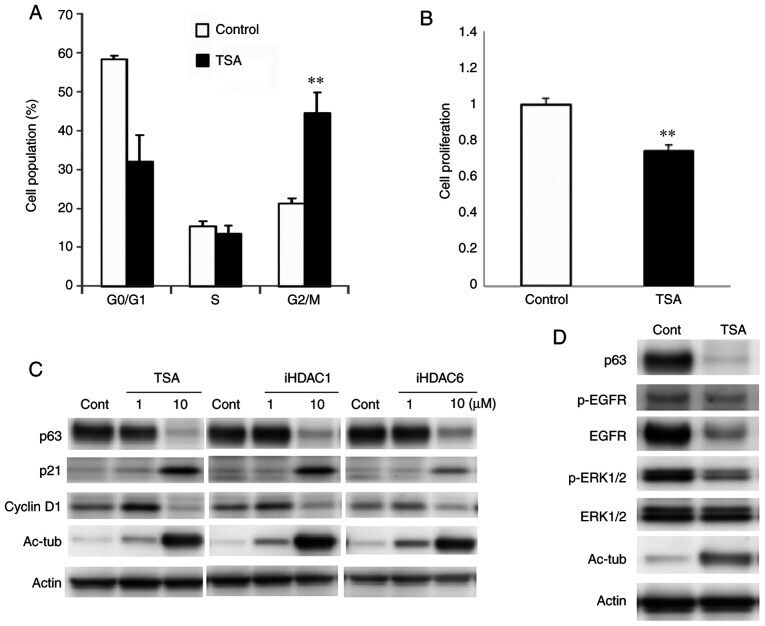
- Figure 4. (A) Cell cycle and (B) cell counting assays using Detroit 562 cells treated with TSA at 10 uM. (C) Western blot analysis for p63, p21 and cyclin D1 in Detroit 562 cells treated with 1 or 10 uM HDAC inhibitors. (D) Western blot analysis of p63, EGFR, phosphorylated-EGFR, ERK1/2 and phosphorylated-ERK1/2 in Detroit 562 cells treated with 10 uM TSA. **P
- Submitted by
- Invitrogen Antibodies (provider)
- Main image

- Experimental details
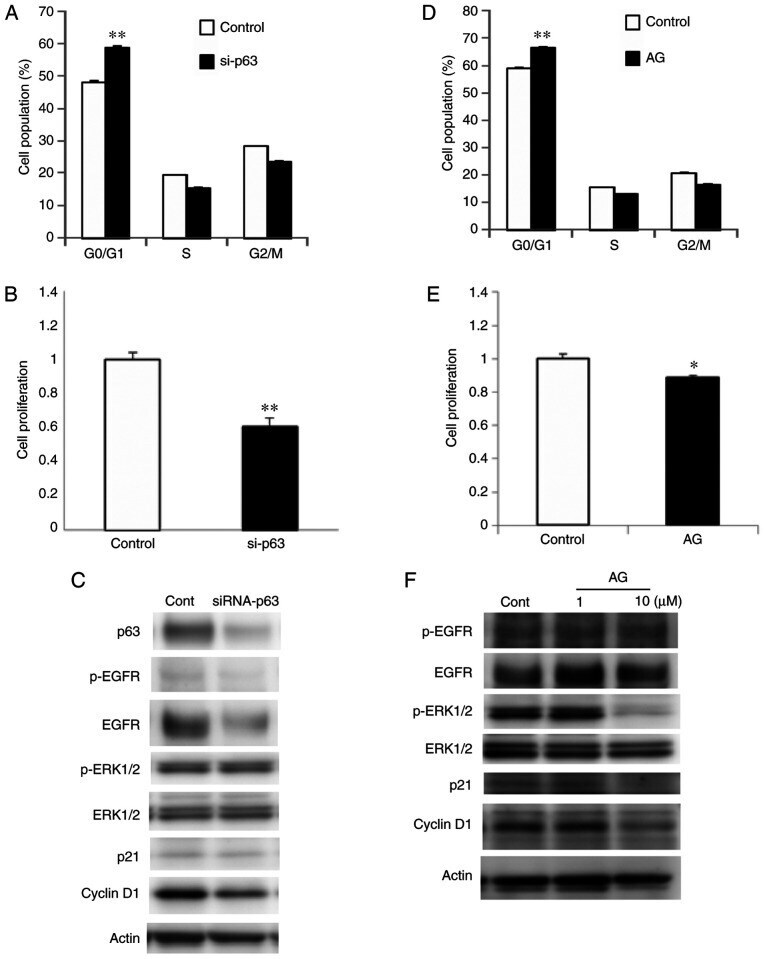
- Figure 5. (A) Cell cycle and (B) cell counting assays, (C) and western blot analysis for p63, EGFR, phosphorylated-EGFR, ERK1/2, phosphorylated-ERK1/2, p21 and cyclin D1 in Detroit 562 cells transfected with p63 siRNA. (D) Cell cycle assay, (E) cell counting assay and (F) western blotting for EGFR, phosphorylated-EGFR, ERK1/2, phosphorylated-ERK1/2, p21 and cyclin D1 in Detroit 562 cells treated with the EGFR inhibitor AG1478. *P
- Submitted by
- Invitrogen Antibodies (provider)
- Main image

- Experimental details
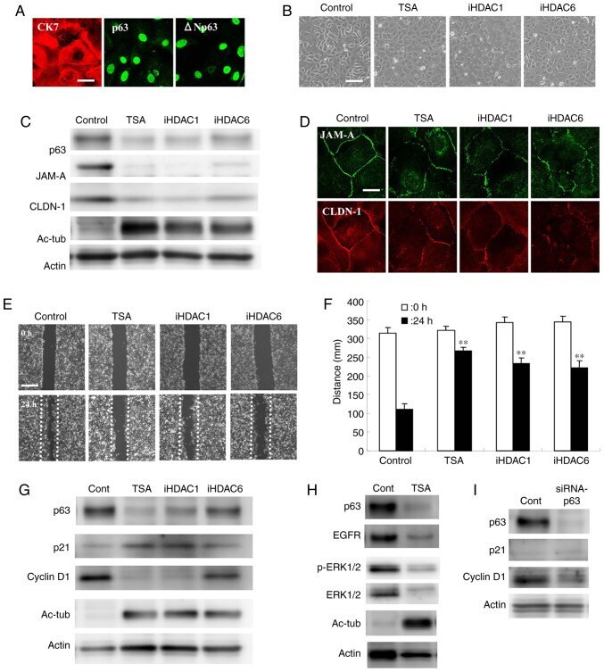
- Figure 6. (A) Immunocytochemical staining for CK7, p63 and DeltaNp63 in primary cultured cancer cells derived from human head and neck squamous cell carcinoma tissues. Scale bar, 10 um. (B) Phase-contrast images. Scale bar, 50 um. (C) Western blotting for p63, JAM-A and claudin-1, and (D) immunocytochemical staining for JAM-A and claudin-1 in primary cultured cancer cells treated with 10 uM HDAC inhibitors. Scale bar, 10 um. (E) Wound-healing assays and (G) western blotting for p63, p21 and cyclin D1 in primary cultured cancer cells treated with HDAC inhibitors at 10 uM. Scale bar, 200 um. (F) Quantification of the results in (E). (H) Western blotting for p63, EGFR, phosphorylated-EGFR, ERK1/2 and phosphorylated-ERK1/2 in primary cultured cancer cells treated with 10 uM TSA. (I) Western blotting for p63, p21 and cyclin D1 in primary cultured cancer cells transfected with p63siRNA of. **P