Explore
 Validate
 Learn
LearnAF1047
antibody from Novus Biologicals
Targeting: CDKN1A
CAP20, CDKN1, CIP1, P21, p21CIP1, p21Cip1/Waf1, SDI1, WAF1
 Western blot
 Immunocytochemistry
Antibody data
- Antibody Data
- Antigen structure
- References [1]
- Comments [0]
- Validations
- Western blot [2]
- Immunohistochemistry [1]
- Flow cytometry [1]
Submit
Validation data
Reference
Comment
Report error
- Product number
- AF1047 - Provider product page

- Provider
- Novus Biologicals
- Product name
- Goat Polyclonal p21/CIP1/CDKN1A Antibody
- Antibody type
- Polyclonal
- Description
- Antigen Affinity-purified. Detects human p21 in Western blots.
- Reactivity
- Human
- Host
- Goat
- Conjugate
- Unconjugated
- Isotype
- IgG
- Vial size
- 100 ug
- Concentration
- LYOPH
- Storage
- Use a manual defrost freezer and avoid repeated freeze-thaw cycles. 12 months from date of receipt, -20 to -70 degreesC as supplied. 1 month, 2 to 8 degreesC under sterile conditions after reconstitution. 6 months, -20 to -70 degreesC under sterile conditions after reconstitution.
Submitted references Induction of alternative lengthening of telomeres-associated PML bodies by p53/p21 requires HP1 proteins.
Jiang WQ, Zhong ZH, Nguyen A, Henson JD, Toouli CD, Braithwaite AW, Reddel RR
The Journal of cell biology 2009 Jun 1;185(5):797-810
The Journal of cell biology 2009 Jun 1;185(5):797-810
No comments: Submit comment
Supportive validation
- Submitted by
- Novus Biologicals (provider)
- Main image
- Experimental details
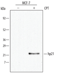
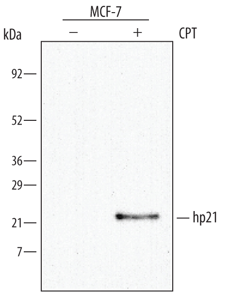
- Detection of Human p21/CIP1/CDKN1A by Western Blot. Western blot shows lysates of MCF-7 human breast cancer cell line untreated (-) or treated (+) with 1 μM camptothecin (CPT) for 16 hours. PVDF membrane was probed with 0.5 µg/mL of Goat Anti-Human p21/CIP1/CDKN1A Antigen Affinity-purified Polyclonal Antibody (Catalog # AF1047), followed by HRP-conjugated Anti-Goat IgG Secondary Antibody (Catalog # HAF017). A specific band was detected for p21/CIP1/CDKN1A at approximately 21 kDa (as indicated). This experiment was conducted under reducing conditions and using Immunoblot Buffer Group 1.
- Submitted by
- Novus Biologicals (provider)
- Main image
- Experimental details
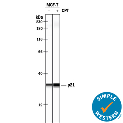
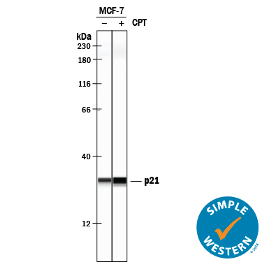
- Detection of Human p21/CIP1/CDKN1A by Simple WesternTM. Simple Western lane view shows lysates of MCF-7 human breast cancer cell line untreated (-) or treated (+) with 1 µM Camptothecin (CPT) for 16 hours, loaded at 0.2 mg/mL. A specific band was detected for p21/CIP1/CDKN1A at approximately 30 kDa (as indicated) using 5 µg/mL of Goat Anti-Human p21/CIP1/CDKN1A Antigen Affinity-purified Polyclonal Antibody (Catalog # AF1047) followed by 1:50 dilution of HRP-conjugated Anti-Goat IgG Secondary Antibody (Catalog # HAF109). This experiment was conducted under reducing conditions and using the 12-230 kDa separation system.
Supportive validation
- Submitted by
- Novus Biologicals (provider)
- Main image
- Experimental details
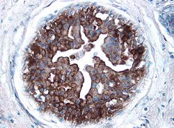
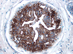
- p21/CIP1/CDKN1A in Human Breast Cancer Tissue. p21/CIP1/CDKN1A was detected in immersion fixed paraffin-embedded sections of human breast cancer tissue using 1.7 µg/mL Goat Anti-Human p21/CIP1/CDKN1A Antigen Affinity-purified Polyclonal Antibody (Catalog # AF1047) overnight at 4 °C. Tissue was stained with the Anti-Goat HRP-DAB Cell & Tissue Staining Kit (brown; Catalog # CTS008) and counterstained with hematoxylin (blue). View our protocol for Chromogenic IHC Staining of Paraffin-embedded Tissue Sections.
Supportive validation
- Submitted by
- Novus Biologicals (provider)
- Main image

- Experimental details
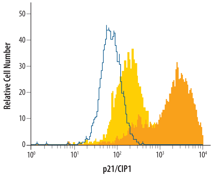
- Detection of p21/CIP1/CDKN1A in MCF-7 Human Cell Line by Flow Cytometry. MCF-7 human breast cancer cell line was unstimulated (light orange filled histogram) or treated with 1 μM camphtothecin for 16 hours, then stained with Goat Anti-Human p21/CIP1/CDKN1A Antigen Affinity-purified Polyclonal Antibody (Catalog # AF1047, dark orange filled histogram) or isotype control antibody (Catalog # AB-108-C, open histogram), followed by Allophycocyanin-conjugated Anti-Goat IgG Secondary Antibody (Catalog # F0108). To facilitate intracellular staining, cells were fixed with paraformaldehyde and permeabilized with methanol.