Explore
 Validate
 Learn
LearnMA5-13293
antibody from Invitrogen Antibodies
Targeting: CDKN1A
CAP20, CDKN1, CIP1, P21, p21CIP1, p21Cip1/Waf1, SDI1, WAF1
 Western blot
 Immunocytochemistry
 Immunoprecipitation Immunohistochemistry Flow cytometry Other assay
Antibody data
- Antibody Data
- Antigen structure
- References [13]
- Comments [0]
- Validations
- Western blot [1]
- Immunohistochemistry [1]
- Flow cytometry [3]
- Other assay [3]
Submit
Validation data
Reference
Comment
Report error
- Product number
- MA5-13293 - Provider product page

- Provider
- Invitrogen Antibodies
- Product name
- Anti-p21 Monoclonal Antibody (HZ52)
- Antibody type
- Monoclonal
- Antigen
- Recombinant full-length protein
- Description
- MA5-13293 targets p21WAF1 in WB, FACS, ICC/IF, IHC (P), and IP applications and shows reactivity with Bovine, Human, and Rat samples. This antibody detects a non-specific band at approx. 18 kDa in U87-MG and C6 cell lysates. Staining of formalin-fixed tissues requires boiling tissue sections in 10mM citrate buffer, pH 6.0 for 10-20 minutes. The MA5-13293 immunogen is full length human recombinant p21 protein.
- Reactivity
- Human, Rat, Bovine
- Host
- Mouse
- Isotype
- IgG
- Antibody clone number
- HZ52
- Vial size
- 500 µL
- Concentration
- 0.2 mg/mL
- Storage
- 4° C
Submitted references Phenotype profiling of primary testicular diffuse large B-cell lymphomas.
Bexarotene induces cellular senescence in MMTV-Neu mouse model of mammary carcinogenesis.
Immunohistochemistry with apoptotic-antiapoptotic proteins (p53, p21, bax, bcl-2), c-kit, telomerase, and metallothionein as a diagnostic aid in benign, borderline, and malignant serous and mucinous ovarian tumors.
Prevalence of human papillomavirus, Epstein-Barr virus, p21, and p53 expression in sinonasal inverted papilloma, nasal polyp, and hypertrophied turbinate in Hong Kong patients.
Efficacy and drawbacks of neoadjuvant chemoradiotherapy in squamous cell carcinoma of the thoracic esophagus.
Large-scale analysis of cell cycle regulators in urothelial bladder cancer identifies p16 and p27 as potentially useful prognostic markers.
Ribosomal protein S9 is a novel B23/NPM-binding protein required for normal cell proliferation.
The ubiquitin-proteasome system regulates p53-mediated transcription at p21waf1 promoter.
Cell death induced by taxanes in breast cancer cells: cytochrome C is released in resistant but not in sensitive cells.
Azoxymethane-induced pre-adipocyte factor 1 (Pref-1) functions as a differentiation inhibitor in colonic epithelial cells.
Essential role of ribosomal protein L11 in mediating growth inhibition-induced p53 activation.
Mammary epithelial cells of PR-A transgenic mice exhibit distinct alterations in gene expression and growth potential associated with transformation.
Iron deprivation induces apoptosis independently of p53 in human and murine tumour cells.
Menter T, Ernst M, Drachneris J, Dirnhofer S, Barghorn A, Went P, Tzankov A
Hematological oncology 2014 Jun;32(2):72-81
Hematological oncology 2014 Jun;32(2):72-81
Bexarotene induces cellular senescence in MMTV-Neu mouse model of mammary carcinogenesis.
Shilkaitis A, Bratescu L, Green A, Yamada T, Christov K
Cancer prevention research (Philadelphia, Pa.) 2013 Apr;6(4):299-308
Cancer prevention research (Philadelphia, Pa.) 2013 Apr;6(4):299-308
Immunohistochemistry with apoptotic-antiapoptotic proteins (p53, p21, bax, bcl-2), c-kit, telomerase, and metallothionein as a diagnostic aid in benign, borderline, and malignant serous and mucinous ovarian tumors.
Ozer H, Yenicesu G, Arici S, Cetin M, Tuncer E, Cetin A
Diagnostic pathology 2012 Sep 20;7:124
Diagnostic pathology 2012 Sep 20;7:124
Prevalence of human papillomavirus, Epstein-Barr virus, p21, and p53 expression in sinonasal inverted papilloma, nasal polyp, and hypertrophied turbinate in Hong Kong patients.
Sham CL, To KF, Chan PK, Lee DL, Tong MC, van Hasselt CA
Head & neck 2012 Apr;34(4):520-33
Head & neck 2012 Apr;34(4):520-33
Efficacy and drawbacks of neoadjuvant chemoradiotherapy in squamous cell carcinoma of the thoracic esophagus.
Wolfárd A, Paszt A, Szentpáli K, Hideghéthy K, Uhercsák G, Németh I, Tiszlavicz L, Lázár G
Hepato-gastroenterology 2011 Jul-Aug;58(109):1214-9
Hepato-gastroenterology 2011 Jul-Aug;58(109):1214-9
Large-scale analysis of cell cycle regulators in urothelial bladder cancer identifies p16 and p27 as potentially useful prognostic markers.
Brunner A, Verdorfer I, Prelog M, Mayerl C, Mikuz G, Tzankov A
Pathobiology : journal of immunopathology, molecular and cellular biology 2008;75(1):25-33
Pathobiology : journal of immunopathology, molecular and cellular biology 2008;75(1):25-33
Ribosomal protein S9 is a novel B23/NPM-binding protein required for normal cell proliferation.
Lindström MS, Zhang Y
The Journal of biological chemistry 2008 Jun 6;283(23):15568-76
The Journal of biological chemistry 2008 Jun 6;283(23):15568-76
The ubiquitin-proteasome system regulates p53-mediated transcription at p21waf1 promoter.
Zhu Q, Wani G, Yao J, Patnaik S, Wang QE, El-Mahdy MA, Praetorius-Ibba M, Wani AA
Oncogene 2007 Jun 21;26(29):4199-208
Oncogene 2007 Jun 21;26(29):4199-208
Cell death induced by taxanes in breast cancer cells: cytochrome C is released in resistant but not in sensitive cells.
Ehrlichová M, Koc M, Truksa J, Naldová Z, Václavíková R, Kovárr J
Anticancer research 2005 Nov-Dec;25(6B):4215-24
Anticancer research 2005 Nov-Dec;25(6B):4215-24
Azoxymethane-induced pre-adipocyte factor 1 (Pref-1) functions as a differentiation inhibitor in colonic epithelial cells.
Dong M, Guda K, Nambiar PR, Nakanishi M, Lichtler AC, Nishikawa M, Giardina C, Rosenberg DW
Carcinogenesis 2004 Nov;25(11):2239-46
Carcinogenesis 2004 Nov;25(11):2239-46
Essential role of ribosomal protein L11 in mediating growth inhibition-induced p53 activation.
Bhat KP, Itahana K, Jin A, Zhang Y
The EMBO journal 2004 Jun 16;23(12):2402-12
The EMBO journal 2004 Jun 16;23(12):2402-12
Mammary epithelial cells of PR-A transgenic mice exhibit distinct alterations in gene expression and growth potential associated with transformation.
Chou YC, Uehara N, Lowry JR, Shyamala G
Carcinogenesis 2003 Mar;24(3):403-9
Carcinogenesis 2003 Mar;24(3):403-9
Iron deprivation induces apoptosis independently of p53 in human and murine tumour cells.
Truksa J, Kovár J, Valenta T, Ehrlichová M, Polák J, Naumann PW
Cell proliferation 2003 Aug;36(4):199-213
Cell proliferation 2003 Aug;36(4):199-213
No comments: Submit comment
Supportive validation
- Submitted by
- Invitrogen Antibodies (provider)
- Main image

- Experimental details
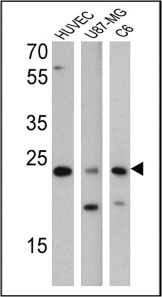
- Western blot analysis of p21WAF1 was performed by loading 25 ug of HUVEC (Lane 1), U87-MG (Lane 2), and C6 (Lane 3) cell lysates and a molecular weight protein ladder onto an SDS polyacrylamide gel. Proteins were transferred to a PVDF membrane and blocked with a blocking buffer at 4ºC overnight. The membrane was probed with a p21WAF1 monoclonal antibody (Product # MA5-13293) at a dilution of 1:200 overnight at 4°C, washed in TBST, and probed with an HRP-conjugated secondary antibody for 1 hr at room temperature in the dark. Results show a band at 21 kDa in all three cell lysates.
Supportive validation
- Submitted by
- Invitrogen Antibodies (provider)
- Main image
- Experimental details
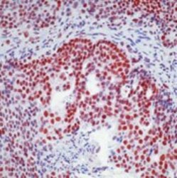
- Formalin-fixed, paraffin-embedded human colon cancer stained with p21 antibody using peroxidase-conjugate and AEC chromogen. Note nuclear staining of tumor cells.

Supportive validation
- Submitted by
- Invitrogen Antibodies (provider)
- Main image
- Experimental details
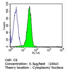
- Flow cytometry analysis of p21WAF1 in C6 cells compared to an isotype control (blue). Cells were harvested, adjusted to a concentration of 1-5x10^6 cells/ml, fixed with 2% paraformaldehyde, washed with PBS, and incubated with p21WAF1 monoclonal antibody (Product # MA5-13293) at a dilution of 0.5 ug/test for 60 min at room temperature. Cells were then blocked in a solution of 2% BSA-PBS for 30 min at room temperature, incubated for 40 min at room temperature in the dark using a Dylight 488-conjugated goat anti-mouse IgG (H+L) secondary antibody, and re-suspended in PBS for FACS analysis.

- Submitted by
- Invitrogen Antibodies (provider)
- Main image

- Experimental details
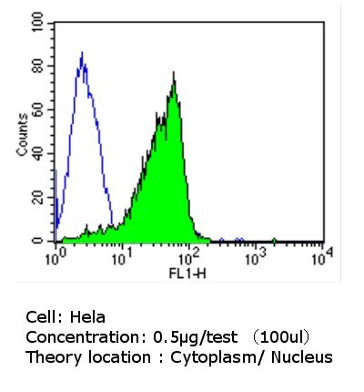
- Flow cytometry analysis of p21WAF1 in Hela cells compared to an isotype control (blue). Cells were harvested, adjusted to a concentration of 1-5x10^6 cells/ml, fixed with 2% paraformaldehyde, washed with PBS, and incubated with p21WAF1 monoclonal antibody (Product # MA5-13293) at a dilution of 0.5 ug/test for 60 min at room temperature. Cells were then blocked in a solution of 2% BSA-PBS for 30 min at room temperature, incubated for 40 min at room temperature in the dark using a Dylight 488-conjugated goat anti-mouse IgG (H+L) secondary antibody, and re-suspended in PBS for FACS analysis.
- Submitted by
- Invitrogen Antibodies (provider)
- Main image
- Experimental details
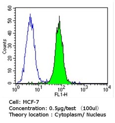
- Flow cytometry analysis of p21WAF1 in MCF-7 cells compared to an isotype control (blue). Cells were harvested, adjusted to a concentration of 1-5x10^6 cells/ml, fixed with 2% paraformaldehyde, washed with PBS, and incubated with p21WAF1 monoclonal antibody (Product # MA5-13293) at a dilution of 0.5 ug/test for 60 min at room temperature. Cells were then blocked in a solution of 2% BSA-PBS for 30 min at room temperature, incubated for 40 min at room temperature in the dark using a Dylight 488-conjugated goat anti-mouse IgG (H+L) secondary antibody, and re-suspended in PBS for FACS analysis.

Supportive validation
- Submitted by
- Invitrogen Antibodies (provider)
- Main image

- Experimental details
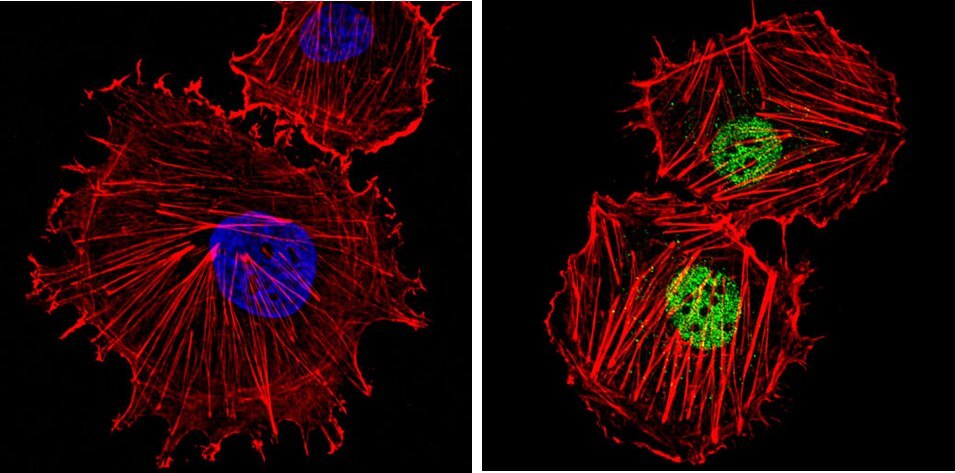
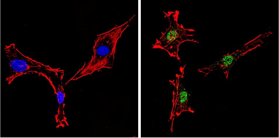
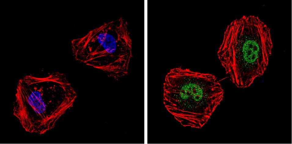
- Immunofluorescent analysis of p21WAF1 (green) showing staining in the cytoplasm and nucleus of C6 cells. Formalin-fixed cells were permeabilized with 0.1% Triton X-100 in TBS for 5-10 minutes and blocked with 3% BSA-PBS for 30 minutes at room temperature. Cells were probed with a p21WAF1 monoclonal antibody (Product # MA5-13293) in 3% BSA-PBS at a dilution of 1:50 and incubated overnight at 4 ºC in a humidified chamber. Cells were washed with PBST and incubated with a DyLight-conjugated secondary antibody in PBS at room temperature in the dark. F-actin (red) was stained with a fluorescent red phalloidin and nuclei (blue) were stained with Hoechst or DAPI. Images were taken at a magnification of 60x.
- Submitted by
- Invitrogen Antibodies (provider)
- Main image
- Experimental details
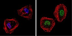
- Immunofluorescent analysis of p21WAF1 (green) showing staining in the cytoplasm and nucleus of HeLa cells. Formalin-fixed cells were permeabilized with 0.1% Triton X-100 in TBS for 5-10 minutes and blocked with 3% BSA-PBS for 30 minutes at room temperature. Cells were probed with a p21WAF1 monoclonal antibody (Product # MA5-13293) in 3% BSA-PBS at a dilution of 1:50 and incubated overnight at 4 ºC in a humidified chamber. Cells were washed with PBST and incubated with a DyLight-conjugated secondary antibody in PBS at room temperature in the dark. F-actin (red) was stained with a fluorescent red phalloidin and nuclei (blue) were stained with Hoechst or DAPI. Images were taken at a magnification of 60x.
- Submitted by
- Invitrogen Antibodies (provider)
- Main image
- Experimental details
- Immunofluorescent analysis of p21WAF1 (green) showing staining in the cytoplasm and nucleus of HUVEC cells. Formalin-fixed cells were permeabilized with 0.1% Triton X-100 in TBS for 5-10 minutes and blocked with 3% BSA-PBS for 30 minutes at room temperature. Cells were probed with a p21WAF1 monoclonal antibody (Product # MA5-13293) in 3% BSA-PBS at a dilution of 1:50 and incubated overnight at 4 ºC in a humidified chamber. Cells were washed with PBST and incubated with a DyLight-conjugated secondary antibody in PBS at room temperature in the dark. F-actin (red) was stained with a fluorescent red phalloidin and nuclei (blue) were stained with Hoechst or DAPI. Images were taken at a magnification of 60x.