 Explore
 Validate
 Learn
Learn Western blot
Antibody data
- Antibody Data
- Antigen structure
- References [4]
- Comments [0]
- Validations
- Western blot [2]
- Flow cytometry [1]
Submit
Validation data
Reference
Comment
Report error
- Product number
- 44-584G - Provider product page

- Provider
- Invitrogen Antibodies
- Product name
- Phospho-Rb (Ser249, Thr252) Polyclonal Antibody
- Antibody type
- Polyclonal
- Antigen
- Synthetic peptide
- Reactivity
- Human
- Host
- Rabbit
- Isotype
- IgG
- Vial size
- 100 µL
- Storage
- -20°C
Submitted references Hypophosphorylated pRb knock-in mice exhibit hallmarks of aging and vitamin C-preventable diabetes.
Dual targeting of CDK and tropomyosin receptor kinase families by the oral inhibitor PHA-848125, an agent with broad-spectrum antitumor efficacy.
Transient treatment with CDK inhibitors eliminates proliferative potential even when their abilities to evoke apoptosis and DNA damage are blocked.
Maid (GCIP) is involved in cell cycle control of hepatocytes.
Jiang Z, Li H, Schroer SA, Voisin V, Ju Y, Pacal M, Erdmann N, Shi W, Chung PED, Deng T, Chen NJ, Ciavarra G, Datti A, Mak TW, Harrington L, Dick FA, Bader GD, Bremner R, Woo M, Zacksenhaus E
The EMBO journal 2022 Feb 15;41(4):e106825
The EMBO journal 2022 Feb 15;41(4):e106825
Dual targeting of CDK and tropomyosin receptor kinase families by the oral inhibitor PHA-848125, an agent with broad-spectrum antitumor efficacy.
Albanese C, Alzani R, Amboldi N, Avanzi N, Ballinari D, Brasca MG, Festuccia C, Fiorentini F, Locatelli G, Pastori W, Patton V, Roletto F, Colotta F, Galvani A, Isacchi A, Moll J, Pesenti E, Mercurio C, Ciomei M
Molecular cancer therapeutics 2010 Aug;9(8):2243-54
Molecular cancer therapeutics 2010 Aug;9(8):2243-54
Transient treatment with CDK inhibitors eliminates proliferative potential even when their abilities to evoke apoptosis and DNA damage are blocked.
Scrace SF, Kierstan P, Borgognoni J, Wang LZ, Denny S, Wayne J, Bentley C, Cansfield AD, Jackson PS, Lockie AM, Curtin NJ, Newell DR, Williamson DS, Moore JD
Cell cycle (Georgetown, Tex.) 2008 Dec 15;7(24):3898-907
Cell cycle (Georgetown, Tex.) 2008 Dec 15;7(24):3898-907
Maid (GCIP) is involved in cell cycle control of hepatocytes.
Sonnenberg-Riethmacher E, Wüstefeld T, Miehe M, Trautwein C, Riethmacher D
Hepatology (Baltimore, Md.) 2007 Feb;45(2):404-11
Hepatology (Baltimore, Md.) 2007 Feb;45(2):404-11
No comments: Submit comment
Supportive validation
- Submitted by
- Invitrogen Antibodies (provider)
- Main image

- Experimental details
- Peptide Competition. Extracts of Jurkat cells in high growth phase were resolved by SDS-PAGE on a 10% Tris-glycine gel and transferred to PVDF. The membrane was blocked with a 5% milk-TBST buffer for one hour at room temperature, then incubated with the Rb (pSpT249/252) antibody for two hours at room temperature in a 1% milk-TBST buffer following its prior incubation with: no peptide (1), a generic phosphoserine-containing peptide (2), a generic phosphothreonine- containing peptide (3), the non-phosphopeptide corresponding to the phosphopeptide immunogen (4), or the phosphopeptide immunogen (5). After washing, the membrane was incubated with goat F (ab')2 anti-rabbit IgG alkaline phosphatase (Product # ALI4405) and signals were detected using the Tropix WesternStar™ detection method.The data show that only the phosphopeptide corresponding to Rb (pSpT249/252) blocks the antibody signal, demonstrating the specificity of the antibody.

- Submitted by
- Invitrogen Antibodies (provider)
- Main image
- Experimental details
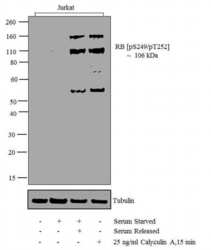
- Western blot analysis was performed on whole cell extracts (30 µg lysate) of Jurkat (Lane 1), Serum Starved Jurkat (Lane 2), Jurkat Serum Starved for overnight following by serum release (Lane 3) and Jurkat treated for 15 min with 25 ng/mL of Calyculin A (lane 4). The blots were probed with Anti-RB1 (pS249)/ (pT252) Rabbit Polyclonal Antibody (Product # 44-584G, 1:500-1:2000 µg/mL) and detected by chemiluminescence using Goat anti-Rabbit IgG (H+L) Secondary Antibody, HRP conjugate (Product # G-21234, 1:5000 dilution). A 106 kDa band corresponding to RB1 (pS249)/ (pT252) was observed across the treated cell lines tested. Two extra bands at 160 and 55 kDa were observed in treated cell lines. Known quantity of protein samples were electrophoresed using Novex® NuPAGE® 12 % Bis-Tris gel (Product # NP0342BOX), XCell SureLock™ Electrophoresis System (Product # EI0002) and Novex® Sharp Pre-Stained Protein Standard (Product # LC5800). Resolved proteins were then transferred onto a nitrocellulose membrane with iBlot® 2 Dry Blotting System (Product # IB21001). The membrane was probed with the relevant primary and secondary Antibody following blocking with 5 % skimmed milk. Chemiluminescent detection was performed using Pierce™ ECL Western Blotting Substrate (Product # 32106).

Supportive validation
- Submitted by
- Invitrogen Antibodies (provider)
- Main image

- Experimental details
- Flow cytometry analysis of RB1 [pSer249/pThr252] was done on COLO 205 cells (serum starved overnight followed by serum release for 4 hours). Cells were fixed with 70% ethanol for 10 minutes, permeabilized with 0.25% Triton™ X-100 for 20 minutes, and blocked with 5% BSA for 30 minutes at room temperature. Cells were labeled with RB1 [pSer249/pThr252] Rabbit Polyclonal Antibody (44584G, red histogram) or with rabbit isotype control (pink histogram) at 3-5 ug/million cells in 2.5% BSA. After incubation at room temperature for 2 hours, the cells were labeled with Alexa Fluor® 488 Goat Anti-Rabbit Secondary Antibody (A11008) at a dilution of 1:400 for 30 minutes at room temperature. The representative 10,000 cells were acquired and analyzed for each sample using an Attune® Acoustic Focusing Cytometer. The purple histogram represents unstained control cells and the green histogram represents no-primary-antibody control.
