 Explore
 Validate
 Learn
Learn Western blot
Antibody data
- Antibody Data
- Antigen structure
- References [1]
- Comments [0]
- Validations
- Western blot [1]
- Immunocytochemistry [1]
Submit
Validation data
Reference
Comment
Report error
- Product number
- 702097 - Provider product page

- Provider
- Invitrogen Antibodies
- Product name
- Phospho-Rb (Ser807, Ser811) Recombinant Rabbit Monoclonal Antibody (13H27L9)
- Antibody type
- Monoclonal
- Antigen
- Synthetic peptide
- Reactivity
- Human
- Host
- Rabbit
- Isotype
- IgG
- Antibody clone number
- 13H27L9
- Vial size
- 100 µg
- Concentration
- 0.5 mg/mL
- Storage
- Store at 4°C short term. For long term storage, store at -20°C, avoiding freeze/thaw cycles.
Submitted references Amplification of CDK4 and MDM2: a detailed study of a high-risk neuroblastoma subgroup.
Martinez-Monleon A, Kryh Öberg H, Gaarder J, Berbegall AP, Javanmardi N, Djos A, Ussowicz M, Taschner-Mandl S, Ambros IM, Øra I, Sandstedt B, Beiske K, Ladenstein R, Noguera R, Ambros PF, Gordon Murkes L, Ljungman G, Kogner P, Fransson S, Martinsson T
Scientific reports 2022 Jul 20;12(1):12420
Scientific reports 2022 Jul 20;12(1):12420
No comments: Submit comment
Supportive validation
- Submitted by
- Invitrogen Antibodies (provider)
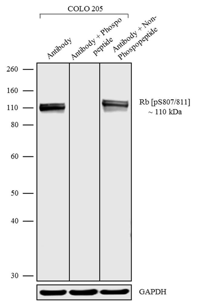
- Main image

- Experimental details
- Western blot analysis was performed on whole cell extracts (30 µg lysate) of COLO 205 (Lane 1, 2 & 3). The blots were probed with Anti-phospho-Rb (Ser807/Ser811) Recombinant Rabbit Monoclonal Antibody (Product # 702097, 1 µg/mL). To confirm the specificity of Phospho-Rb (Ser807/Ser811), competition was performed with the phosphopeptide (10 µg/mL) (Lane 2) and non phosphopeptide (10 µg/mL) (Lane 3). A 110 kDa band corresponding to Phospho-Rb (Ser807/Ser811) was observed as shown. The blots were detected by chemiluminescence using Goat anti-Rabbit IgG (H+L) Superclonal™ Secondary Antibody, HRP conjugate (Product # A27036, 0.4 µg/mL, 1:5000 dilution). Known quantity of protein samples were electrophoresed using Novex® NuPAGE® 4-12% Bis-Tris gel (Product # NP0321BOX), XCell SureLock™ Electrophoresis System (Product # EI0002) and Novex® Sharp Pre-Stained Protein Standard (Product # LC5800). Resolved proteins were then transferred onto a nitrocellulose membrane with wet transfer method. The membrane was probed with the relevant primary and secondary antibody following blocking with 5% skimmed milk. Chemiluminescent detection was performed using Pierce™ ECL Western blotting Substrate (Product # 32106).
Supportive validation
- Submitted by
- Invitrogen Antibodies (provider)
- Main image
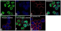
- Experimental details
- For immunofluorescence analysis, COLO 205 cells were fixed and permeabilized for detection of endogenous Phospho Rb (Ser807/Ser811) using Anti- Phospho Rb (Ser807/Ser811) Recombinant Rabbit Monoclonal Antibody (Product # 702097, 2 µg/mL) and labeled with Goat anti-Rabbit IgG (H+L) Superclonal™ Secondary Antibody, Alexa Fluor® 488 conjugate (Product # A27034, 1:2000). Panel a) shows representative cells that were stained for detection and localization of Phospho-Rb (Ser807/Ser811) protein (green), Panel b) is stained for nuclei (blue) using SlowFade® Gold Antifade Mountant with DAPI (Product # S36938). Panel c) represents cytoskeletal F-actin staining using Rhodamine Phalloidin (Product # R415, 1:300). Panel d) is a composite image of panels a, b and c clearly demonstrating nuclear localization of Phospho-Rb (Ser807/Ser811). Panel e) shows loss of signal by competition with the Phospho-Rb (Ser807/Ser811) phospho peptide, demonstrating antibody specificity, and panel f) demonstrates no competition with the non-phosphorylated peptide. Panel g) represents control cells with no primary antibody to assess background. The images were captured at 60X magnification.
