Explore
 Validate
 Learn
Learn Western blot
Antibody data
- Antibody Data
- Antigen structure
- References [0]
- Comments [0]
- Validations
- Western blot [1]
- Immunocytochemistry [1]
- Immunohistochemistry [1]
Submit
Validation data
Reference
Comment
Report error
- Product number
- PA5-12682 - Provider product page

- Provider
- Invitrogen Antibodies
- Product name
- Phospho-Rb (Ser788) Polyclonal Antibody
- Antibody type
- Polyclonal
- Antigen
- Synthetic peptide
- Reactivity
- Human
- Host
- Rabbit
- Isotype
- IgG
- Vial size
- 400 µL
- Concentration
- 0.5 mg/mL
- Storage
- Store at 4°C short term. For long term storage, store at -20°C, avoiding freeze/thaw cycles.
No comments: Submit comment
Supportive validation
- Submitted by
- Invitrogen Antibodies (provider)
- Main image

- Experimental details
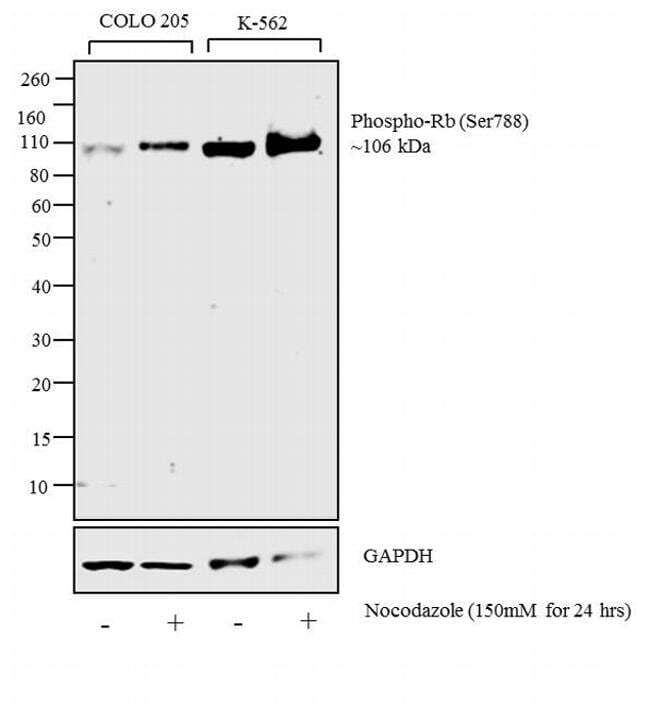
- Western blot analysis was performed on whole cell extracts (30 µg lysate) of COLO 205 (Lane 1), COLO 205 treated with Nocodazole (150 mM for 24hrs) (Lane 2), K-562 ( Lane 3) and K-562 treated with Nocodazole (150 mM for 24hrs) (Lane 4). The blot was probed with Anti-Phospho-Rb (Ser788) Rabbit Polyclonal Antibody (Product PA5-12682 #, 1:1000 dilution) and detected by chemiluminescence using Goat anti-Rabbit IgG (H+L) Superclonal™ Secondary Antibody, HRP conjugate (Product # A27036, 0.25 ug/ml, 1:4000 dilution). A 106 kDa band corresponding to Phospho-Rb (Ser788) was detected in the cell lines and was enhanced upon Nocodazole treatment.
Supportive validation
- Submitted by
- Invitrogen Antibodies (provider)
- Main image
- Experimental details
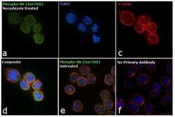
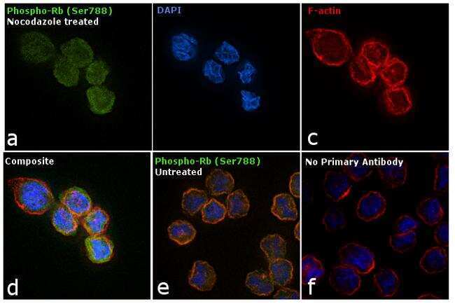
- Immunofluorescence analysis of Phospho Rb (Ser788) was performed using 70% confluent log phase COLO 205 cells treated with Nocodazole (150mM for 24hrs). The cells were fixed with 4% paraformaldehyde for 10 minutes, permeabilized with 0.1% Triton™ X-100 for 15 minutes, and blocked with 1% BSA for 1 hour at room temperature. The cells were labeled with Phospho Rb (Ser 788) Rabbit Polyclonal Antibody (Product # PA5-12682) at 1:250 dilution in 0.1% BSA, incubated at 4 degree Celsius overnight and then labeled with Goat anti-Rabbit IgG (H+L) Superclonal™ Secondary Antibody, Alexa Fluor® 488 conjugate (Product # A27034) at a dilution of 1:2000 for 45 minutes at room temperature (Panel a: green). Nuclei (Panel b: blue) were stained with ProLong™ Diamond Antifade Mountant with DAPI (Product # P36962). F-actin (Panel c: red) was stained with Rhodamine Phalloidin (Product # R415, 1:300). Panel d represents the merged image showing cytoplasmic localization. Panel e shows untreated cells with less signal as compared to the treated cells (Panel d). Panel f represents Isotype control cells to assess background. The images were captured at 60X magnification.
Supportive validation
- Submitted by
- Invitrogen Antibodies (provider)
- Main image

- Experimental details
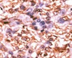
- Immunohistochemical analysis of formalin-fixed, paraffin-embedded human cancer tissue using a Phospho-Rb pSer788 polyclonal antibody (Product # PA5-12682), followed by HRP-conjugated secondary antibody and DAB staining.