Explore
 Validate
 Learn
Learn Western blot
Antibody data
- Antibody Data
- Antigen structure
- References [5]
- Comments [0]
- Validations
- Western blot [1]
- Immunocytochemistry [3]
- Other assay [1]
Submit
Validation data
Reference
Comment
Report error
- Product number
- MA5-12584 - Provider product page

- Provider
- Invitrogen Antibodies
- Product name
- Phospho-Rb (Ser608) Monoclonal Antibody (51B7)
- Antibody type
- Monoclonal
- Antigen
- Other
- Description
- This antibody was orginally validated as part of a Thermo Scientific Cellomics High Content Screening Kit. The antibody sold separately may have slightly different performance and may need to be further optimized for the best results.
- Reactivity
- Human, Mouse
- Host
- Mouse
- Isotype
- IgG
- Antibody clone number
- 51B7
- Vial size
- 500 µL
- Concentration
- 0.2 mg/mL
- Storage
- 4° C
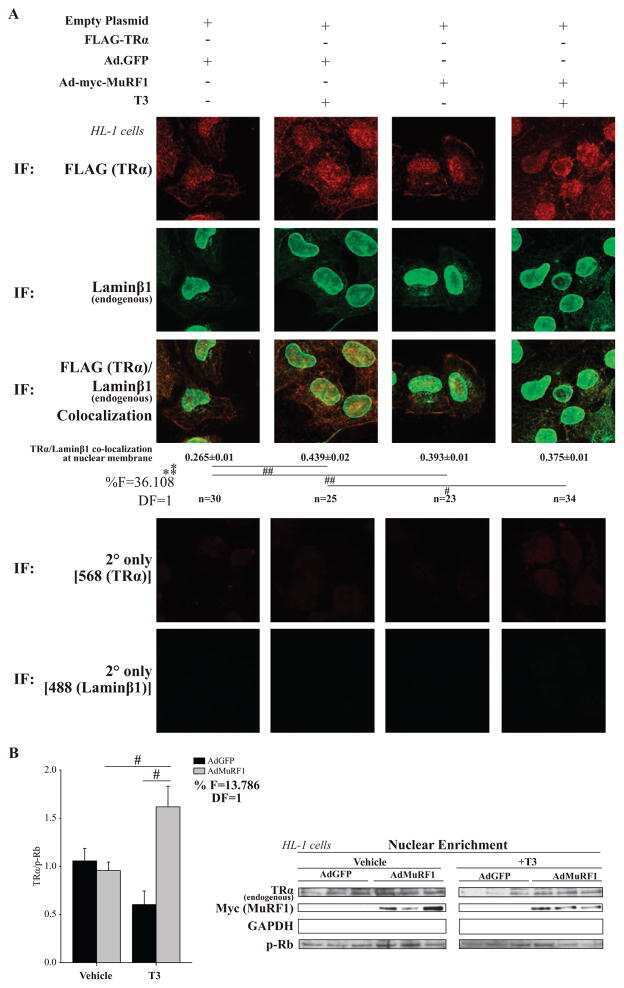
Submitted references MuRF1 mono-ubiquitinates TRα to inhibit T3-induced cardiac hypertrophy in vivo.
Bexarotene induces cellular senescence in MMTV-Neu mouse model of mammary carcinogenesis.
UM-SCC-104: a new human papillomavirus-16-positive cancer stem cell-containing head and neck squamous cell carcinoma cell line.
Selective inhibition of rRNA transcription downregulates E2F-1: a new p53-independent mechanism linking cell growth to cell proliferation.
Proliferation capacity of the renal proximal tubule involves the bulk of differentiated epithelial cells.
Wadosky KM, Berthiaume JM, Tang W, Zungu M, Portman MA, Gerdes AM, Willis MS
Journal of molecular endocrinology 2016 Apr;56(3):273-90
Journal of molecular endocrinology 2016 Apr;56(3):273-90
Bexarotene induces cellular senescence in MMTV-Neu mouse model of mammary carcinogenesis.
Shilkaitis A, Bratescu L, Green A, Yamada T, Christov K
Cancer prevention research (Philadelphia, Pa.) 2013 Apr;6(4):299-308
Cancer prevention research (Philadelphia, Pa.) 2013 Apr;6(4):299-308
UM-SCC-104: a new human papillomavirus-16-positive cancer stem cell-containing head and neck squamous cell carcinoma cell line.
Tang AL, Hauff SJ, Owen JH, Graham MP, Czerwinski MJ, Park JJ, Walline H, Papagerakis S, Stoerker J, McHugh JB, Chepeha DB, Bradford CR, Carey TE, Prince ME
Head & neck 2012 Oct;34(10):1480-91
Head & neck 2012 Oct;34(10):1480-91
Selective inhibition of rRNA transcription downregulates E2F-1: a new p53-independent mechanism linking cell growth to cell proliferation.
Donati G, Brighenti E, Vici M, Mazzini G, Treré D, Montanaro L, Derenzini M
Journal of cell science 2011 Sep 1;124(Pt 17):3017-28
Journal of cell science 2011 Sep 1;124(Pt 17):3017-28
Proliferation capacity of the renal proximal tubule involves the bulk of differentiated epithelial cells.
Vogetseder A, Picard N, Gaspert A, Walch M, Kaissling B, Le Hir M
American journal of physiology. Cell physiology 2008 Jan;294(1):C22-8
American journal of physiology. Cell physiology 2008 Jan;294(1):C22-8
No comments: Submit comment
Supportive validation
- Submitted by
- Invitrogen Antibodies (provider)
- Main image
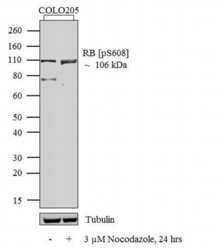
- Experimental details
- Western blot analysis was performed on whole cell extracts (30 µg lysate) of COLO205 (Lane 1) and COLO205 treated with 3 uM of Nocodazole for 24 hrs (Lane 2). The blots were probed with Anti-RB (pS608) Mouse Monoclonal Antibody (Product # MA5-12584, 1-2 µg/mL) and detected by chemiluminescence using Goat anti-Mouse IgG (H+L) Secondary Antibody, HRP conjugate (Product # 62-6520, 1:4000 dilution). A 106 kDa corresponding to RB (pS608) was observed across the cell lines tested and was enhanced upon treatment. Known quantity of protein samples were electrophoresed using Novex® NuPAGE® 12 % Bis-Tris gel (Product # NP0342BOX), XCell SureLock™ Electrophoresis System (Product # EI0002) and Novex® Sharp Pre-Stained Protein Standard (Product # LC5800). Resolved proteins were then transferred onto a nitrocellulose membrane with iBlot® 2 Dry Blotting System (Product # IB21001). The membrane was probed with the relevant primary and secondary Antibody following blocking with 5 % skimmed milk. Chemiluminescent detection was performed using Pierce™ ECL Western Blotting Substrate (Product # 32106).

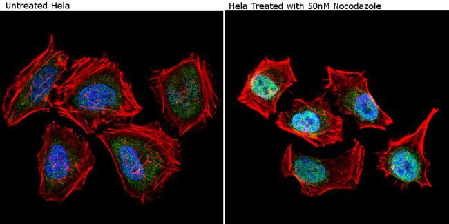
Supportive validation
- Submitted by
- Invitrogen Antibodies (provider)
- Main image

- Experimental details
- Immunofluorescent analysis of Phospho-Retinoblastoma pSer608 (green) showing staining in the nucleus of untreated Hela cells (left) and Hela cells treated with 50nM Nocodazole for 16 hrs (right). Formalin-fixed cells were permeabilized with 0.1% Triton X-100 in TBS for 5-10 minutes and blocked with 3% BSA-PBS for 30 minutes at room temperature. Cells were probed with a Phospho-Retinoblastoma pSer608 monoclonal antibody (Product # MA5-12584) in 3% BSA-PBS at a dilution of 1:20 and incubated overnight at 4ºC in a humidified chamber. Cells were washed with PBST and incubated with a DyLight-conjugated secondary antibody in PBS at room temperature in the dark. F-actin (red) was stained with a flourescent red phalloidin and nuclei (blue) were stained with Hoechst or DAPI. Images were taken at a magnification of 60x.
- Submitted by
- Invitrogen Antibodies (provider)
- Main image
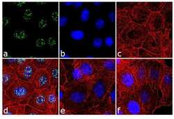
- Experimental details
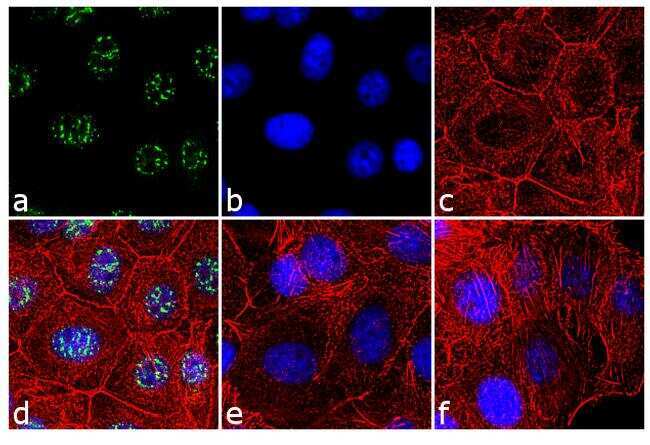
- Immunofluorescence analysis of Phospho-Retinoblastoma pSer608 was done on 70% confluent log phase Colo cells treated with serum starvation for 16 hours. The cells were fixed with 4% paraformaldehyde for 10 minutes, permeabilized with 0.1% Triton™ X-100 for 10 minutes, and blocked with 1% BSA for 1 hour at room temperature. The cells were labeled with Phospho-Retinoblastoma pSer608 Antibody (51B7) Mouse Monoclonal Antibody (Product # MA5-12584) at 2 µg/mL in 0.1% BSA and incubated for 3 hours at room temperature and then labeled with Goat anti-Mouse IgG (H+L) Superclonal™ Secondary Antibody, Alexa Fluor® 488 conjugate (Product # A28175) at a dilution of 1:2000 for 45 minutes at room temperature (Panel a: green). Nuclei (Panel b: blue) were stained with SlowFade® Gold Antifade Mountant with DAPI (Product # S36938). F-actin (Panel c: red) was stained with Alexa Fluor® 555 Rhodamine Phalloidin (Product # R415, 1:300).Panel d is a merged image showing punctate nuclear localization. Panel e is untreated cell with no signal. Panel f is a no primary antibody control. The images were captured at 60X magnification.
- Submitted by
- Invitrogen Antibodies (provider)
- Main image
- Experimental details
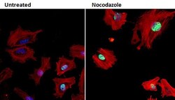
- Immunofluorescent analysis of Phospho-Retinoblastoma (Phospho-Rb) pSer608 (green) in HeLa cells either left untreated (left panel) or treated with 50nM Nocodazole (right panel) for 16 hours. Formalin fixed cells were permeabilized with 0.1% Triton X-100 in TBS for 10 minutes at room temperature and blocked with 1% Blocker BSA (Product # 37525) for 15 minutes at room temperature. Cells were probed with a Phospho-Rb pSer608 monoclonal antibody (Product # MA5-12584) at a dilution of 1:100 for at least 1 hour at room temperature, washed with PBS, and incubated with DyLight 488 goat anti-mouse IgG secondary antibody (Product # 35502) at a dilution of 1:400 for 30 minutes at room temperature. F-Actin (red) was stained with DyLight 554 Phalloidin (Product # 21834) and nuclei (blue) were stained with Hoechst 33342 dye (Product # 62249). Images were taken on a Thermo Scientific ArrayScan and ToxInsight Instrument at 20X magnification.

Supportive validation
- Submitted by
- Invitrogen Antibodies (provider)
- Main image

- Experimental details
- NULL