 Explore
 Validate
 Learn
Learn Western blot
 ELISA
ELISAAntibody data
- Antibody Data
- Antigen structure
- References [0]
- Comments [0]
- Validations
- Western blot [1]
- Immunohistochemistry [2]
Submit
Validation data
Reference
Comment
Report error
- Product number
- 600-401-889 - Provider product page

- Provider
- Invitrogen Antibodies
- Product name
- Cbl-c Polyclonal Antibody
- Antibody type
- Polyclonal
- Antigen
- Synthetic peptide
- Reactivity
- Human
- Host
- Rabbit
- Isotype
- IgG
- Vial size
- 100 µg
- Concentration
- 1.2 mg/mL
- Storage
- -20° C, Avoid Freeze/Thaw Cycles
No comments: Submit comment
Supportive validation
- Submitted by
- Invitrogen Antibodies (provider)
- Main image
- Experimental details
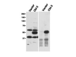
- Immunoprecipitation (right) and western blot (left) using Rocklands Affinity Purified anti-Cbl-c antibody shows detection of a predominant band at ~52 kDa corresponding to Cbl-c. Lysates used are from HEK293T cells transfected with empty vector or with Cbl-c and western blotting (left panel). The predicted size of Cbl-c is 52 kDa. Size markers in kDa are shown to the left of the panel. The (right panel) shows immunoprecipitation with Rabbit anti-Cbl-c followed by western blotting using a Goat anti-Cbl-c antibody. Personal Communication. Stan Lipkowitz, NCI, NIH, Bethesda, MD.

Supportive validation
- Submitted by
- Invitrogen Antibodies (provider)
- Main image
- Experimental details
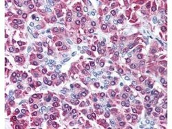
- Rocklands affinity purified anti-Cbl-c antibody was used at 5 µg/ml to detect signal in a variety of tissues including multi-human, multi-brain and multi-cancer slides. This image shows moderate intracellular positive staining of human pancreatic acinar epithelium at 40X. Tissue was formalin-fixed and paraffin embedded. The image shows localization of the antibody as the precipitated red signal, with a hematoxylin purple nuclear counterstain. Personal Communication, Tina Roush, LifeSpanBiosciences, Seattle, WA.

- Submitted by
- Invitrogen Antibodies (provider)
- Main image
- Experimental details
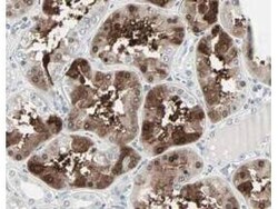
- Rocklands Affinity Purified anti-Cbl-c antibody shows strong nuclear and cytoplasmic staining of cells in tubuli in human kidney tissue. Tissue was formalin-fixed and paraffin embedded. Brown color indicates presence of protein, blue color shows cell nuclei. Personal Communication, Kenneth Wester, www.proteinatlas.org, Uppsala, Sweden.
