 Explore
 Validate
 Learn
Learn Western blot
 ELISA
ELISAAntibody data
- Antibody Data
- Antigen structure
- References [0]
- Comments [0]
- Validations
- Western blot [1]
- Immunohistochemistry [2]
Submit
Validation data
Reference
Comment
Report error
- Product number
- NBP1-78063 - Provider product page

- Provider
- Novus Biologicals
- Proper citation
- Novus Cat#NBP1-78063, RRID:AB_11017519
- Product name
- Rabbit Polyclonal Cbl-c Antibody
- Antibody type
- Polyclonal
- Description
- Immunogen affinity purified. A BLAST analysis was used to suggest that this antibody would react with Cbl-c from human and chimpanzee sources. Expect partial reactivity against mouse and rat sources of Cbl-c as approx 83% sequence homology is on record for the immunogen sequence. Reactivity with Cbl-c from other sources has not been determined. No reactivity is expected with Cbl-a or Cbl-b.
- Reactivity
- Human
- Host
- Rabbit
- Isotype
- IgG
- Vial size
- 0.1 mg
- Concentration
- 1.1 mg/ml
- Storage
- Store at -20C. Avoid freeze-thaw cycles.
No comments: Submit comment
Supportive validation
- Submitted by
- Novus Biologicals (provider)
- Main image
- Experimental details
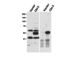
- Western Blot: Cbl-c Antibody [NBP1-78063] - dominant band at ~52 kDa corresponding to Cbl-c (arrowhead) in transfected cell lysates (left panel). Lysates are from Hek 293T cells transfected with empty vector or with Cbl-c. The predicted size of Cbl-c is 52 kDa. Size markers in kDa are shown to the left of the panel. The right panel shows western blotting after first immunoprecipitating with Rabbit anti-Cbl-c followed by western blotting using a Goat anti-Cbl-c antibody.

Supportive validation
- Submitted by
- Novus Biologicals (provider)
- Main image

- Experimental details
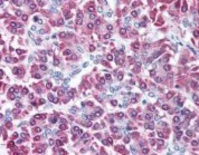
- Immunohistochemistry-Paraffin: Cbl-c Antibody [NBP1-78063] - 5 ug/ml to detect signal in a variety of tissues including multi-human, multi-brain and multi-cancer slides. This image shows moderate intracellular positive staining of human pancreatic acinar epithelium at 40X. The image shows localization of the antibody as the precipitated red signal, with a hematoxylin purple nuclear counterstain.
- Submitted by
- Novus Biologicals (provider)
- Main image
- Experimental details
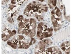
- Immunohistochemistry: Cbl-c Antibody [NBP1-78063] - Staining of cells in tubuli in human kidney tissue. Tissue was formalin-fixed and paraffin embedded. Brown color indicates presence of protein, blue color shows cell nuclei.
