Explore
 Validate
 Learn
Learn Western blot
 ELISA
ELISAAntibody data
- Antibody Data
- Antigen structure
- References [3]
- Comments [0]
- Validations
- Western blot [2]
- Immunohistochemistry [2]
Submit
Validation data
Reference
Comment
Report error
- Product number
- GTX27195 - Provider product page

- Provider
- GeneTex
- Proper citation
- GeneTex Cat#GTX27195, RRID:AB_386083
- Product name
- Gli2 antibody
- Antibody type
- Polyclonal
- Reactivity
- Human, Mouse
- Host
- Rabbit
Submitted references Identification of Human Cutaneous Basal Cell Carcinoma Cancer Stem Cells.
Crosstalk between Notch and Sonic hedgehog signaling in a mouse model of amyotrophic lateral sclerosis.
CD200-expressing human basal cell carcinoma cells initiate tumor growth.
Morgan H, Olivero C, Patel GK
Methods in molecular biology (Clifton, N.J.) 2019;1879:435-450
Methods in molecular biology (Clifton, N.J.) 2019;1879:435-450
Crosstalk between Notch and Sonic hedgehog signaling in a mouse model of amyotrophic lateral sclerosis.
Ma X, Drannik A, Jiang F, Peterson R, Turnbull J
Neuroreport 2017 Feb 8;28(3):141-148
Neuroreport 2017 Feb 8;28(3):141-148
CD200-expressing human basal cell carcinoma cells initiate tumor growth.
Colmont CS, Benketah A, Reed SH, Hawk NV, Telford WG, Ohyama M, Udey MC, Yee CL, Vogel JC, Patel GK
Proceedings of the National Academy of Sciences of the United States of America 2013 Jan 22;110(4):1434-9
Proceedings of the National Academy of Sciences of the United States of America 2013 Jan 22;110(4):1434-9
No comments: Submit comment
Supportive validation
- Submitted by
- GeneTex (provider)
- Main image
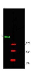
- Experimental details
- Western blot using anti-Gli-2 antibody (1:750 dilution) shows detection of a predominant band at ~190 kDa corresponding to Gli-2 (arrowhead) in mouse brain whole cell lysate (lane 1). Molecular weight markers are shown (M) using the 700 nm channel (red).
- Validation comment
- WB

- Submitted by
- GeneTex (provider)
- Main image
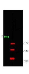
- Experimental details
- Western blot using GeneTex's Affinity Purified anti-Gli-2 antibody shows detection of a predominant band at ~190 kDa corresponding to Gli-2 (arrowhead) in mouse brain whole cell lysate (lane 1). Pre-incubation of antibody with immunizing peptide completely blocks staining of this band (lane 2). ~ 25 ?g of lysate was resolved on a 4-8% Tris-glycine gel by SDS-PAGE and transferred onto nitrocellulose. After blocking with 5% goat serum and 0.5% BLOTTO in PBS, the membrane was probed with the primary antibody diluted to 1:750. Incubation was at room temperature for 2 h followed by washes and reaction with a 1:10,000 dilution of IRDye? 800 conjugated Gt-a-Rabbit IgG (H&L) MX10 for 45 min at room temperature. Molecular weight markers are shown (M) using the 700 nm channel (red). IRDye? 800 fluorescence image was captured using the Odyssey? Infrared Imaging System developed by LI-COR. IRDye is a trademark of LI-COR, Inc. Other detection systems will yield similar results.
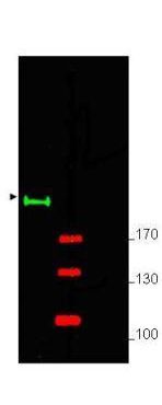
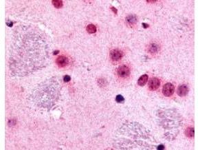
Supportive validation
- Submitted by
- GeneTex (provider)
- Main image
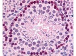
- Experimental details
- GeneTex's Affinity Purified anti-mouse Gli-2 antibody (GTX27195) was used at 10 ?g/ml to evaluate staining on several mouse tissues. Moderate to strong staining was seen on many tissues, with low background staining. This image shows Gli-2 staining of mouse testis. Tissue was formalin-fixed and paraffin embedded.
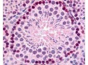
- Submitted by
- GeneTex (provider)
- Main image

- Experimental details
- GeneTex's Affinity Purified anti-mouse Gli-2 antibody (GTX27195) was used at 10 ?g/ml to evaluate staining on several mouse tissues. Moderate to strong staining was seen on many tissues with low background staining. This image shows Gli-2 staining of mouse brain. Tissue was formalin-fixed and paraffin embedded.