Explore
 Validate
 Learn
Learn Western blot
Antibody data
- Antibody Data
- Antigen structure
- References [3]
- Comments [0]
- Validations
- Western blot [1]
- Immunohistochemistry [2]
Submit
Validation data
Reference
Comment
Report error
- Product number
- PAB10270 - Provider product page

- Provider
- Abnova Corporation
- Proper citation
- Abnova Corporation Cat#PAB10270, RRID:AB_1674688
- Product name
- Gli2 polyclonal antibody
- Antibody type
- Polyclonal
- Description
- Rabbit polyclonal antibody raised against synthetic peptide of Gli2.
- Storage
- Store at 4°C. For long term storage store at -20°C.Aliquot to avoid repeated freezing and thawing.
Submitted references Molecular control of spinal accessory motor neuron/axon development in the mouse spinal cord.
Gli2 and Gli3 have redundant and context-dependent function in skeletal muscle formation.
Gli3 null mice display glandular overgrowth of the developing stomach.
Dillon AK, Fujita SC, Matise MP, Jarjour AA, Kennedy TE, Kollmus H, Arnold HH, Weiner JA, Sanes JR, Kaprielian Z
The Journal of neuroscience : the official journal of the Society for Neuroscience 2005 Nov 2;25(44):10119-30
The Journal of neuroscience : the official journal of the Society for Neuroscience 2005 Nov 2;25(44):10119-30
Gli2 and Gli3 have redundant and context-dependent function in skeletal muscle formation.
McDermott A, Gustafsson M, Elsam T, Hui CC, Emerson CP Jr, Borycki AG
Development (Cambridge, England) 2005 Jan;132(2):345-57
Development (Cambridge, England) 2005 Jan;132(2):345-57
Gli3 null mice display glandular overgrowth of the developing stomach.
Kim JH, Huang Z, Mo R
Developmental dynamics : an official publication of the American Association of Anatomists 2005 Dec;234(4):984-91
Developmental dynamics : an official publication of the American Association of Anatomists 2005 Dec;234(4):984-91
No comments: Submit comment
Supportive validation
- Submitted by
- Abnova Corporation (provider)
- Main image

- Experimental details
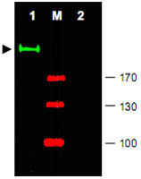
- Western blot using Gli2 polyclonal antibody (Cat # PAB10270) shows detection of a predominant band at ~190 KDa corresponding to Gli2 (arrowhead) in mouse brain tissue lysate (Lane 1).Pre-incubation of antibody with immunizing peptide completely blocks staining of this band (Lane 2).Approximately 25 ug of lysate was resolved on a 4-8% Tris-glycine gel by SDS-PAGE and transferred onto nitrocellulose.After blocking with 5% goat serum and 0.5% BLOTTO in PBS, the membrane was probed with the primary antibody diluted to 1 : 750.Incubation was at room temperature for 2 h followed by washes and reaction with a 1 : 10,000 dilution of IRDye® 800 conjugated Gt-a-Rabbit IgG (H&L) MX10 for 45 min at room temperature.Molecular weight markers are shown (M) using the 700 nm channel (red).IRDye® 800 fluorescence image was captured using the Odyssey® Infrared Imaging System developed by LI-COR.IRDye is a trademark of LI-COR, Inc.
Supportive validation
- Submitted by
- Abnova Corporation (provider)
- Main image

- Experimental details
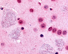
- Immunohistochemistry of Gli2 polyclonal antibody (Cat # PAB10270) was used at 10 ug/mL to evaluate staining on several mouse tissues.Moderate to strong staining was seen on many tissues with low background staining.This image shows Gli2 staining of mouse brain.Tissue was formalin-fixed and paraffin embedded.Personal Communication, Tina Roush, Life Span Biosciences, Seattle, WA.
- Validation comment
- Immunohistochemistry (Formalin/PFA-fixed paraffin-embedded sections)
- Submitted by
- Abnova Corporation (provider)
- Main image
- Experimental details
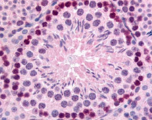
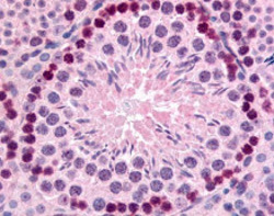
- Immunohistochemistry of Gli2 polyclonal antibody (Cat # PAB10270) was used at 10 ug/mL to evaluate staining on several mouse tissues.Moderate to strong staining was seen on many tissues, with low background staining.This image shows Gli2 staining of mouse testis.Tissue was formalin-fixed and paraffin embedded.Personal Communication, Tina Roush, Life Span Biosciences, Seattle, WA.
- Validation comment
- Immunohistochemistry (Formalin/PFA-fixed paraffin-embedded sections)