Explore
 Validate
 Learn
Learn Western blot
Antibody data
- Antibody Data
- Antigen structure
- References [4]
- Comments [0]
- Validations
- Western blot [1]
- Immunohistochemistry [1]
Submit
Validation data
Reference
Comment
Report error
- Product number
- PAB10051 - Provider product page

- Provider
- Abnova Corporation
- Proper citation
- Abnova Corporation Cat#PAB10051, RRID:AB_1674711
- Product name
- GLI2 polyclonal antibody
- Antibody type
- Polyclonal
- Description
- Rabbit polyclonal antibody raised against synthetic peptide of GLI2.
- Storage
- Store at 4°C. For long term storage store at -20°C.Aliquot to avoid repeated freezing and thawing.
Submitted references Inhibition of hedgehog signaling for the treatment of murine sclerodermatous chronic graft-versus-host disease.
A previously unidentified amino-terminal domain regulates transcriptional activity of wild-type and disease-associated human GLI2.
GLI2 is expressed in normal human epidermis and BCC and induces GLI1 expression by binding to its promoter.
The zinc-finger transcription factor GLI2 antagonizes contact inhibition and differentiation of human epidermal cells.
Zerr P, Palumbo-Zerr K, Distler A, Tomcik M, Vollath S, Munoz LE, Beyer C, Dees C, Egberts F, Tinazzi I, Del Galdo F, Distler O, Schett G, Spriewald BM, Distler JH
Blood 2012 Oct 4;120(14):2909-17
Blood 2012 Oct 4;120(14):2909-17
A previously unidentified amino-terminal domain regulates transcriptional activity of wild-type and disease-associated human GLI2.
Roessler E, Ermilov AN, Grange DK, Wang A, Grachtchouk M, Dlugosz AA, Muenke M
Human molecular genetics 2005 Aug 1;14(15):2181-8
Human molecular genetics 2005 Aug 1;14(15):2181-8
GLI2 is expressed in normal human epidermis and BCC and induces GLI1 expression by binding to its promoter.
Ikram MS, Neill GW, Regl G, Eichberger T, Frischauf AM, Aberger F, Quinn A, Philpott M
The Journal of investigative dermatology 2004 Jun;122(6):1503-9
The Journal of investigative dermatology 2004 Jun;122(6):1503-9
The zinc-finger transcription factor GLI2 antagonizes contact inhibition and differentiation of human epidermal cells.
Regl G, Kasper M, Schnidar H, Eichberger T, Neill GW, Ikram MS, Quinn AG, Philpott MP, Frischauf AM, Aberger F
Oncogene 2004 Feb 12;23(6):1263-74
Oncogene 2004 Feb 12;23(6):1263-74
No comments: Submit comment
Supportive validation
- Submitted by
- Abnova Corporation (provider)
- Main image
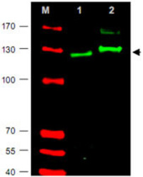
- Experimental details
- Western blot using GLI2 polyclonal antibody (Cat # PAB10051) shows detection of GLI2 protein in rat testes (Lane 1) and human HEK293 (Lane 2) whole cell lysates (arrowhead).See Ruppert et al for testing conditions.Each lane contains approximately 35 ug of lysate.Primary antibody was used at a 1:400 dilution in 5% BLOTTO in PBS overnight at 4°C.The membrane was washed and reacted with a 1:10,000 dilution of IRDye® 800 conjugated Gt-a-Rabbit IgG [H&L] MX10 for 45 min at room temperature (800 nm channel, green).Molecular weight estimation was made by comparison to prestained MW markers in lane M (700 nm channel, red).IRDye® 800 fluorescence image was captured using the Odyssey® Infrared Imaging System developed by LI-COR.IRDye is a trademark of LI-COR, Inc.

Supportive validation
- Submitted by
- Abnova Corporation (provider)
- Main image
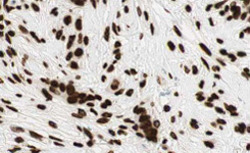
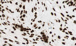
- Experimental details
- Immunohistochemistry of GLI2 polyclonal antibody (Cat # PAB10051) shows strong cytoplasmic and membranous staining of tumor cells in human breast tissue.Tissue was formalin-fixed and paraffin embedded.Brown color indicates presence of protein, blue color shows cell nuclei.Personal Communication, Kenneth Wester, www.proteinatlas.org, Uppsala, Sweden.
- Validation comment
- Immunohistochemistry (Formalin/PFA-fixed paraffin-embedded sections)