 Explore
 Validate
 Learn
Learn Western blot
 ELISA
ELISAAntibody data
- Antibody Data
- Antigen structure
- References [0]
- Comments [0]
- Validations
- Western blot [1]
- Immunohistochemistry [2]
Submit
Validation data
Reference
Comment
Report error
- Product number
- AP09261PU-N - Provider product page

- Provider
- Acris Antibodies GmbH
- Proper citation
- Acris Antibodies GmbH Cat#AP09261PU-N, RRID:AB_2035558
- Product name
- anti GLI2
- Antibody type
- Polyclonal
- Antigen
- Synthetic peptide corresponding to amino acids from an internal region of Mouse Gli-2
- Reactivity
- Mouse, Rat
- Host
- Rabbit
- Isotype
- IgG
- Vial size
- 0.1 mg
- Concentration
- 1.02 mg/ml (by UV absorbance at 280 nm)
No comments: Submit comment
Supportive validation
- Submitted by
- Acris Antibodies GmbH (provider)
- Main image

- Experimental details
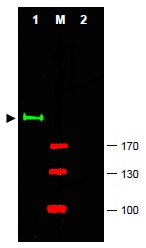
- Western blot using AP09261PU-N Gli2 antibody shows detection of a predominant band at ~190 kDa corresponding to Gli-2 (arrowhead) in Mouse brain whole cell lysate (Lane 1). Pre-incubation of antibody with immunizing peptide completely blocks staining of this band (Lane 2). ~ 25 μg of lysate was resolved on a 4-8% Tris-glycine gel by SDS-PAGE and transferred onto nitrocellulose. After blocking with 5% goat serum and 0.5% BLOTTO in PBS, the membrane was probed with the primary antibody diluted to 1/750. Incubation was at RT for 2 h followed by washes and reaction with a 1/10,000 dilution of IRDye® 800 conjugated Goat anti-Rabbit IgG (H&L) MX10 for 45 min at RT. Molecular weight markers are shown (M) using the 700 nm channel (red). IRDye® 800 fluorescence image was captured using the Odyssey® Infrared Imaging System developed by LI-COR. IRDye is a trademark of LI-COR, Inc. Other detection systems will yield similar results.
Supportive validation
- Submitted by
- Acris Antibodies GmbH (provider)
- Main image

- Experimental details
- AP09261PU-N Gli2 antibody staining of Formalin-Fixed, Paraffin Embedded of several Mouse Tesis tissues at 10 μg/ml . Moderate to strong staining was seen on many tissues, with low background staining.

- Submitted by
- Acris Antibodies GmbH (provider)
- Main image

- Experimental details
- AP09261PU-N Gli2 antibody staining of Formalin-Fixed, Paraffin Embedded of several Mouse Brain tissues at 10 μg/ml . Moderate to strong staining was seen on many tissues, with low background staining.
