 Explore
 Validate
 Learn
Learn Western blot
 ELISA
ELISAAntibody data
- Antibody Data
- Antigen structure
- References [0]
- Comments [0]
- Validations
- Western blot [1]
- Immunohistochemistry [1]
Submit
Validation data
Reference
Comment
Report error
- Product number
- AP09264PU-N - Provider product page

- Provider
- Acris Antibodies GmbH
- Proper citation
- Acris Antibodies GmbH Cat#AP09264PU-N, RRID:AB_2035559
- Product name
- anti GLI2 (40-60)
- Antibody type
- Polyclonal
- Antigen
- Synthetic peptide corresponding to amino acids 46-60 of Human Gli-2 (isoform alpha).
- Reactivity
- Human, Mouse, Rat, Bovine
- Host
- Rabbit
- Isotype
- IgG
- Vial size
- 0.1 mg
- Concentration
- 1.1 mg/ml (by UV absorbance at 280 nm)
No comments: Submit comment
Supportive validation
- Submitted by
- Acris Antibodies GmbH (provider)
- Main image
- Experimental details
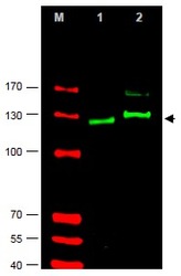
- Figure 1. Western blot using AP09264PU-N Gli-2 antibody shows detection of Gli-2 protein in Rat testes (Lane 1) and Human HEK293 (Lane 2)whole cell lysates (arrowhead). See Ruppert et al for testing conditions. Each lane contains approximately 35 μg of lysate. Primary antibody was used at a 1/400 dilution in 5% BLOTTO in PBS overnight at 4°C. The membrane was washed and reacted with a 1/10,000 dilution of IRDye® 800 conjugated Goat anti-Rabbit IgG [H&L] for 45 min at RT (800 nm channel, green). Molecular weight estimation was made by comparison to prestained MW markers in lane M (700 nm channel, red). IRDye® 800 fluorescence image was captured using the Odyssey® Infrared Imaging System developed by LI-COR. IRDye is a trademark of LI-COR, Inc. Other detection systems will yield similar results.

Supportive validation
- Submitted by
- Acris Antibodies GmbH (provider)
- Main image
- Experimental details
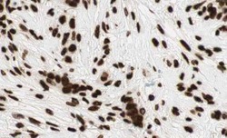
- Figure 2. AP09264PU-N Gli2 antibody staining of Formalin-Fixed, Paraffin Embedded Human breast tissue (strong cytoplasmic and membranous satining). Brown color indicates presence of protein, blue color shows cell nuclei. Personal Communication, Kenneth Wester, www.proteinatlas.org, Uppsala, Sweden.
