Explore
 Validate
 Learn
Learn Western blot
 ELISA
ELISAAntibody data
- Antibody Data
- Antigen structure
- References [0]
- Comments [0]
- Validations
- Western blot [6]
- Immunocytochemistry [4]
Submit
Validation data
Reference
Comment
Report error
- Product number
- MA1-23332 - Provider product page

- Provider
- Invitrogen Antibodies
- Product name
- PCNA Monoclonal Antibody (339)
- Antibody type
- Monoclonal
- Antigen
- Recombinant full-length protein
- Description
- MA1-23332 detects PCNA interdomain connector loop in Human samples.
- Reactivity
- Human, Mouse, Rat
- Host
- Mouse
- Isotype
- IgG
- Antibody clone number
- 339
- Vial size
- 100 µL
- Concentration
- 1 mg/mL
- Storage
- Store at 4°C short term. For long term storage, store at -20°C, avoiding freeze/thaw cycles.
No comments: Submit comment
Supportive validation
- Submitted by
- Invitrogen Antibodies (provider)
- Main image

- Experimental details
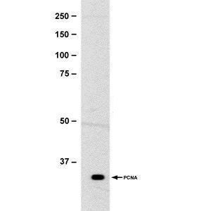
- Western blot analysis of PCNA interdomain connector loop using a PCNA interdomain connector loop monoclonal antibody (Product # MA1-23332).
- Submitted by
- Invitrogen Antibodies (provider)
- Main image

- Experimental details
- Western Blot using PCNA Monoclonal Antibody (339) (Product # MA1-23332). Sample (30 µg of whole cell lysate). Lane A: Jurkat. Lane B: Raji. Lane C: 293T. Lane D: A431. Lane E: HeLa. Lane F: HepG2. Lane G: H1299. Lane H: HCT116. I: MCF-7. J: NT2D1. K: PC-3. L: U87-MG. 12% SDS PAGE. PCNA Monoclonal Antibody (339) (Product # MA1-23332) diluted at 1:1,000.

- Submitted by
- Invitrogen Antibodies (provider)
- Main image

- Experimental details
- Western Blot using PCNA Monoclonal Antibody (339) (Product # MA1-23332). Sample (whole cell lysate). Lane A: 293T 20 µg. B: 293T 10 µg. Lane C: 293T 5 µg. 12% SDS PAGE. PCNA Monoclonal Antibody (339) (Product # MA1-23332) diluted at 1:1,000.

- Submitted by
- Invitrogen Antibodies (provider)
- Main image

- Experimental details
- Western Blot using PCNA Monoclonal Antibody (339) (Product # MA1-23332). Sample (30 µg of whole cell lysate). A: 293T. Lane B: NIH-3T3. C: PC-12. 12% SDS PAGE. PCNA Monoclonal Antibody (339) (Product # MA1-23332) diluted at 1:1,000.

- Submitted by
- Invitrogen Antibodies (provider)
- Main image
- Experimental details
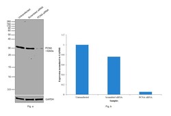
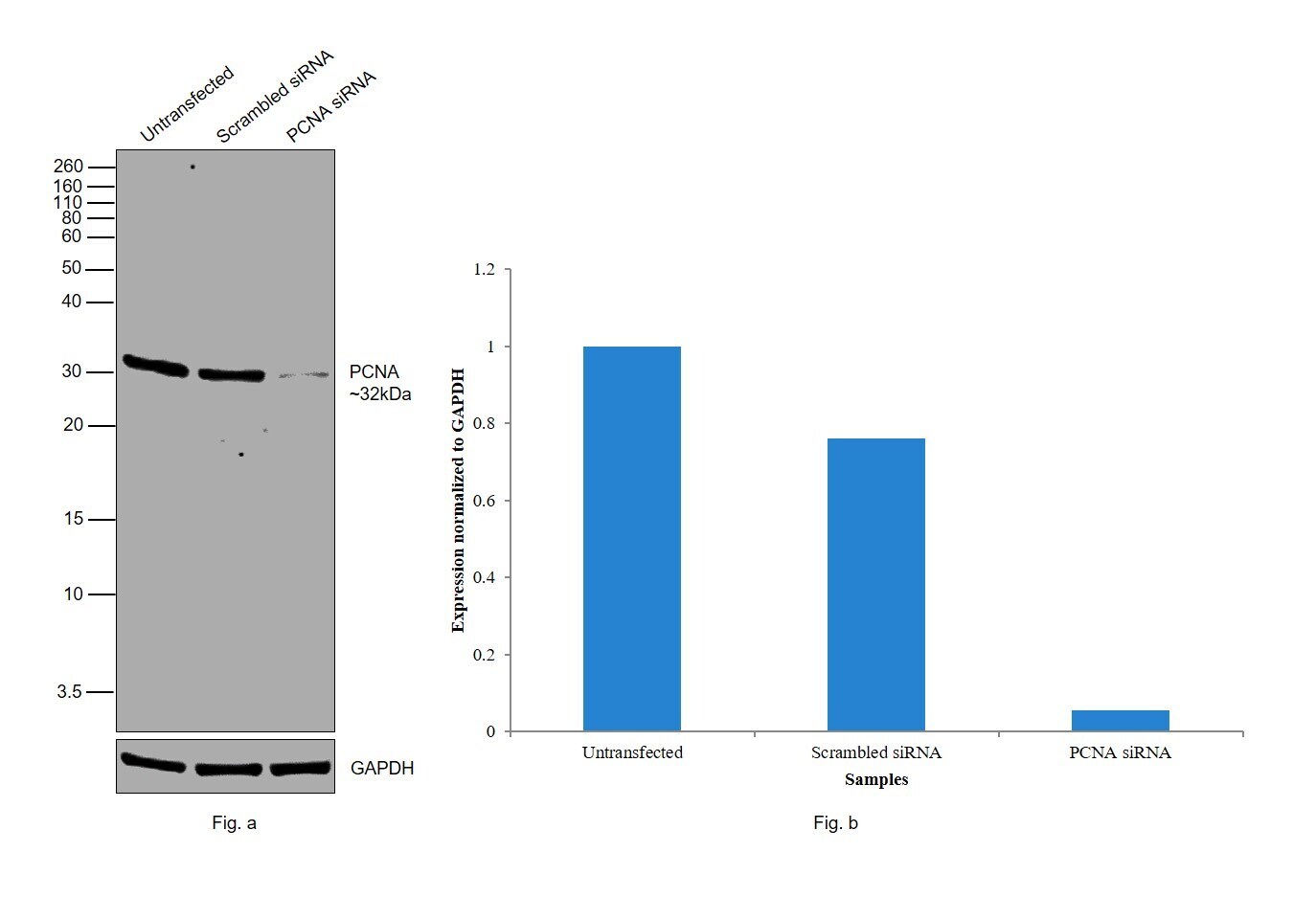
- Knockdown of PCNA was achieved by transfecting HeLa with PCNA specific siRNAs (Silencer® select Product # S10133, S10134). Western blot analysis (Fig. a) was performed using Nuclear enriched extracts from the PCNA knockdown cells (lane 3), non-targeting scrambled siRNA transfected cells (lane 2) and untransfected cells (lane 1). The blot was probed with PCNA Monoclonal Antibody (339) (Product # MA1-23332, 1:1000 ) and Goat anti-Mouse IgG (H+L) Superclonal™ Recombinant Secondary Antibody, HRP (Product # A28177, 1:4000). Densitometric analysis of this western blot is shown in histogram (Fig. b). Decrease in signal upon siRNA mediated knock down confirms that antibody is specific to PCNA.
- Submitted by
- Invitrogen Antibodies (provider)
- Main image
- Experimental details
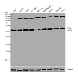
- Western blot was performed using Anti-PCNA Monoclonal Antibody (339)(Product # MA1-23332) and a 32kDa band corresponding to PCNA was observed across all tested cell lines. Nuclear enriched extracts (30 µg lysate) of HeLa (Lane 1), MCF7 (Lane 2), Jurkat (Lane 3), HEK-293 (Lane 4), NIH/3T3 (Lane 5), MDA-MB-231 (Lane 6), A549 (Lane 7), SH-SY5Y (Lane 8), PC-12 (Lane 9) were electrophoresed using NuPAGE™ 12% Bis-Tris Protein Gel (Product # NP0342BOX). Resolved proteins were then transferred onto a Nitrocellulose membrane (Product # IB23001) by iBlot® 2 Dry Blotting System (Product # IB21001). The blot was probed with the primary antibody (1:1000) and detected by chemiluminescence with Goat anti-Mouse IgG (H+L) Superclonal™ Recombinant Secondary Antibody, HRP (Product # A28177,1:4000) using the iBright FL 1000 (Product # A32752). Chemiluminescent detection was performed using SuperSignal™ West Dura Extended Duration Substrate (Product # 34076).

Supportive validation
- Submitted by
- Invitrogen Antibodies (provider)
- Main image

- Experimental details
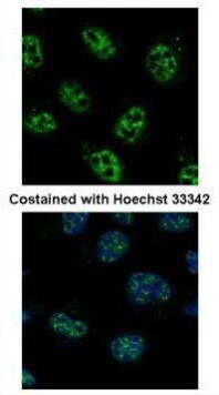
- Immunofluorescent analysis of PCNA interdomain connector loop in paraformaldehyde-fixed A431 cells using a PCNA interdomain connector loop monoclonal antibody (Product # MA1-23332) at a 1:500 dilution.
- Submitted by
- Invitrogen Antibodies (provider)
- Main image
- Experimental details
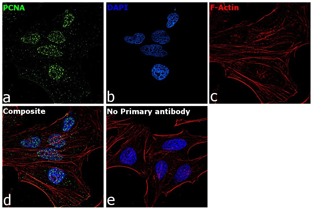
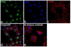
- Immunofluorescence analysis of PCNA was performed using 70% confluent log phase A549 cells. The cells were fixed with 4% paraformaldehyde for 10 minutes, permeabilized with 0.1% Triton™ X-100 for 15 minutes, and blocked with 2% BSA for 1 hour at room temperature. The cells were labeled with PCNA Monoclonal Antibody (339) (Product # MA1-23332, 1:100 dilution) in 0.1% BSA, incubated at 4 degree celsius overnight and then labeled with Donkey anti-Mouse IgG (H+L) Highly Cross-Adsorbed Secondary Antibody, Alexa Fluor Plus 488 (Product # A32766, 1:2000 dilution) for 45 minutes at room temperature (Panel a: Green). Nuclei (Panel b:Blue) were stained with Hoechst 33342 (Product # H1399). F-actin (Panel c: Red) was stained with Rhodamine Phalloidin (Product # R415, 1:300 dilution). Panel d represents the merged image showing nuclear localization. Panel e represents control cells with no primary antibody to assess background. The images were captured at 40X magnification in CellInsight CX7 LZR High-Content Screening (HCS) Platform (Product # CX7A1110LZR) and externally deconvoluted (D.Sage et al. / Methods 115 (2017) 28–41).

- Submitted by
- Invitrogen Antibodies (provider)
- Main image

- Experimental details
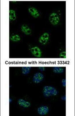
- Immunofluorescence analysis of paraformaldehyde-fixed A431, using (Product # MA1-23332) antibody at 1:500 dilution.
- Submitted by
- Invitrogen Antibodies (provider)
- Main image
- Experimental details
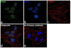
- Immunofluorescence analysis of PCNA was performed using 70 percent confluent log phase HeLa cells. The cells were fixed with 4% paraformaldehyde for 10 minutes, permeabilized with 0.1% Triton™ X-100 for 15 minutes, and blocked with 2% BSA for 45 minutes at room temperature. The cells were labeled with PCNA Monoclonal Antibody (339) (Product # MA1-23332) at 1:200 in 0.1% BSA, incubated at 4 degree celsius overnight and then labeled with Goat anti-Mouse IgG (H+L) Highly Cross-Adsorbed Secondary Antibody, Alexa Fluor Plus 488 (Product # A32723), (1:2000), for 45 minutes at room temperature (Panel a: Green). Nuclei (Panel b:Blue) were stained with ProLong™ Diamond Antifade Mountant with DAPI (Product # P36962). F-actin (Panel c: Red) was stained with Rhodamine Phalloidin (Product # R415, 1:300). Panel d represents the merged image showing nuclear localization. Panel e represents control cells with no primary antibody to assess background. The images were captured at 60X magnification.