Explore
 Validate
 Learn
Learn Western blot
 Immunohistochemistry
Antibody data
- Antibody Data
- Antigen structure
- References [2]
- Comments [0]
- Validations
- Immunohistochemistry [1]
Submit
Validation data
Reference
Comment
Report error
- Product number
- HPA030521 - Provider product page

- Provider
- Atlas Antibodies
- Proper citation
- Atlas Antibodies Cat#HPA030521, RRID:AB_10600218
- Product name
- Anti-PCNA
- Antibody type
- Polyclonal
- Description
- Polyclonal Antibody against Human PCNA, Gene description: proliferating cell nuclear antigen, Validated applications: IHC, WB, Uniprot ID: P12004, Storage: Store at +4°C for short term storage. Long time storage is recommended at -20°C.
- Reactivity
- Human
- Host
- Rabbit
- Conjugate
- Unconjugated
- Isotype
- IgG
- Vial size
- 100 µl
- Concentration
- 0.05 mg/ml
- Storage
- Store at +4°C for short term storage. Long time storage is recommended at -20°C.
- Handling
- The antibody solution should be gently mixed before use.
Submitted references TUG1-mediated R-loop resolution at microsatellite loci as a prerequisite for cancer cell proliferation
Histological Analysis of Gonadal Ridge Development and Sex Differentiation of Gonads in Three Gecko Species
Suzuki M, Iijima K, Ogami K, Shinjo K, Murofushi Y, Xie J, Wang X, Kitano Y, Mamiya A, Kibe Y, Nishimura T, Ohka F, Saito R, Sato S, Kobayashi J, Yao R, Miyata K, Kataoka K, Suzuki H, Kondo Y
Nature Communications 2023;14(1)
Nature Communications 2023;14(1)
Histological Analysis of Gonadal Ridge Development and Sex Differentiation of Gonads in Three Gecko Species
Rams-Pociecha I, Mizia P, Piprek R
Biology 2023;13(1):7
Biology 2023;13(1):7
No comments: Submit comment
Supportive validation
- Submitted by
- Atlas Antibodies (provider)
- Enhanced method
- Orthogonal validation
- Main image
- Experimental details
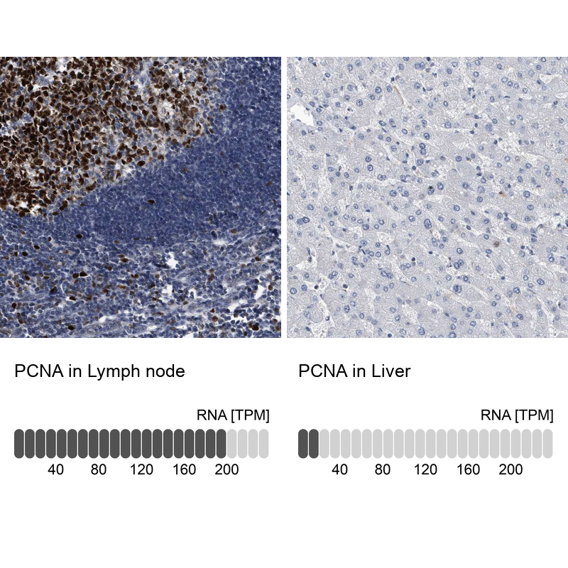
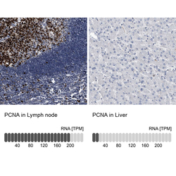
- Immunohistochemistry analysis in human lymph node and liver tissues using HPA030521 antibody. Corresponding PCNA RNA-seq data are presented for the same tissues.
- Sample type
- Human
- Protocol
- Protocol