 Explore
 Validate
 Learn
Learn Western blot
 Immunoprecipitation
Antibody data
- Antibody Data
- Antigen structure
- References [5]
- Comments [0]
- Validations
- Western blot [3]
- Flow cytometry [1]
- Other assay [1]
Submit
Validation data
Reference
Comment
Report error
- Product number
- 14-9910-82 - Provider product page

- Provider
- Invitrogen Antibodies
- Product name
- PCNA Monoclonal Antibody (PC10 (3F81)), eBioscience™
- Antibody type
- Monoclonal
- Antigen
- Other
- Description
- Description: The PC10 antibody recognizes the proliferating cell nuclear antigen (PCNA), a 36 kDa protein, also known as polymerase delta auxiliary protein. PC10 antibody reacts with human, mouse, and rat PCNA. The peak expression of PCNA occurs during the S-phase. Applications Reported: The PC10 (a.k.a. 3F81) antibody has been reported for use in flow cytometric analysis, immunoprecipitation, immunoblotting (WB), and immunohistochemical staining. Applications Tested: The PC10 (a.k.a. 3F81) antibody has been tested by flow cytometric analysis. This can be used at less than or equal to 0.25 µg per test. A test is defined as the amount (µg) of antibody that will stain a cell sample in a final volume of 100 µL. Cell number should be determined empirically but can range from 10^5 to 10^8 cells/test. The PC10 antibody has also been tested by immunocytochemistry of methanol-fixed MDCK cells and can be used at less than or equal to 5 µg/mL. It is recommended that the antibody be carefully titrated for optimal performance in the assay of interest. Purity: Greater than 90%, as determined by SDS-PAGE. Aggregation: Less than 10%, as determined by HPLC. Filtration: 0.2 µm post-manufacturing filtered.
- Reactivity
- Human, Mouse, Rat
- Host
- Mouse
- Isotype
- IgG
- Antibody clone number
- PC10 (3F81)
- Vial size
- 100 µg
- Concentration
- 0.5 mg/mL
- Storage
- 4° C
Submitted references Inosine pranobex enhances human NK cell cytotoxicity by inducing metabolic activation and NKG2D ligand expression.
The giant danio (D. aequipinnatus) as a model of cardiac remodeling and regeneration.
Antibodies to proliferating cell nuclear antigen as S-phase probes in flow cytometric cell cycle analysis.
Monoclonal antibody analysis of the proliferating cell nuclear antigen (PCNA). Structural conservation and the detection of a nucleolar form.
Proliferating cell nuclear antigen (PCNA) immunolocalization in paraffin sections: an index of cell proliferation with evidence of deregulated expression in some neoplasms.
McCarthy MT, Lin D, Soga T, Adam J, O'Callaghan CA
European journal of immunology 2020 Jan;50(1):130-137
European journal of immunology 2020 Jan;50(1):130-137
The giant danio (D. aequipinnatus) as a model of cardiac remodeling and regeneration.
Lafontant PJ, Burns AR, Grivas JA, Lesch MA, Lala TD, Reuter SP, Field LJ, Frounfelter TD
Anatomical record (Hoboken, N.J. : 2007) 2012 Feb;295(2):234-48
Anatomical record (Hoboken, N.J. : 2007) 2012 Feb;295(2):234-48
Antibodies to proliferating cell nuclear antigen as S-phase probes in flow cytometric cell cycle analysis.
Landberg G, Roos G
Cancer research 1991 Sep 1;51(17):4570-4
Cancer research 1991 Sep 1;51(17):4570-4
Monoclonal antibody analysis of the proliferating cell nuclear antigen (PCNA). Structural conservation and the detection of a nucleolar form.
Waseem NH, Lane DP
Journal of cell science 1990 May;96 ( Pt 1):121-9
Journal of cell science 1990 May;96 ( Pt 1):121-9
Proliferating cell nuclear antigen (PCNA) immunolocalization in paraffin sections: an index of cell proliferation with evidence of deregulated expression in some neoplasms.
Hall PA, Levison DA, Woods AL, Yu CC, Kellock DB, Watkins JA, Barnes DM, Gillett CE, Camplejohn R, Dover R
The Journal of pathology 1990 Dec;162(4):285-94
The Journal of pathology 1990 Dec;162(4):285-94
No comments: Submit comment
Supportive validation
- Submitted by
- Invitrogen Antibodies (provider)
- Main image

- Experimental details
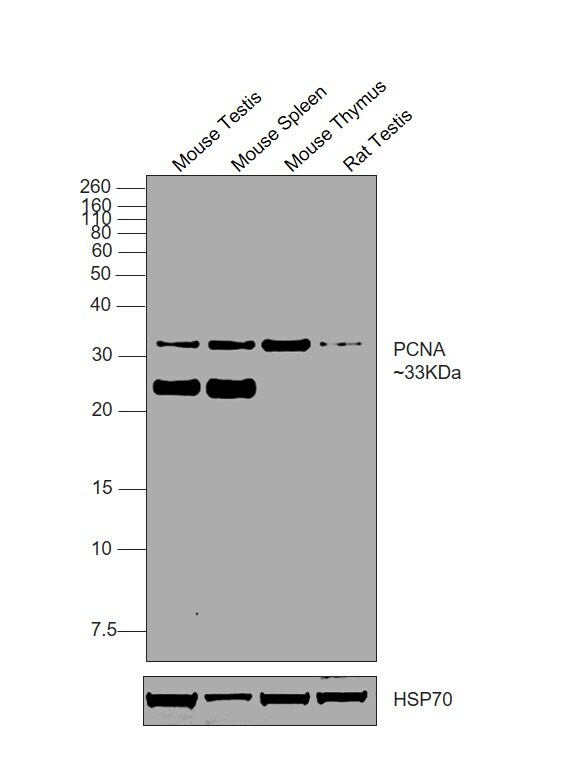
- Western blot was performed using PCNA Monoclonal Antibody (PC10 (3F81)), eBioscience (Product # 14-9910-82) and a 33 kDa band corresponding to PCNA was observed across in all tissues tested. Tissue extracts (30 µg lysate) of Mouse Testis (Lane 1), Mouse Spleen (Lane 2), Mouse Thymus (Lane 3), Rat Testis (Lane 4) were electrophoresed using NuPAGE™ 12% Bis-Tris Protein Gel (Product # NP0341BOX), 10 well. Resolved proteins were then transferred onto a nitrocellulose membrane (Product # IB23002) by iBlot® 2 Dry Blotting System (Product # IB21001). The blot was probed with the primary antibody (1 µg/mL) and detected by chemiluminescence with Goat anti-Mouse IgG (H+L) Superclonal™ Recombinant Secondary Antibody, HRP (Product # A28177, 1:10,000) using the iBright™ FL1500 Imaging System (Product # A44115). Chemiluminescent detection was performed using SuperSignal™ West Pico PLUS Chemiluminescent Substrate (Product # 34580).
- Submitted by
- Invitrogen Antibodies (provider)
- Main image

- Experimental details
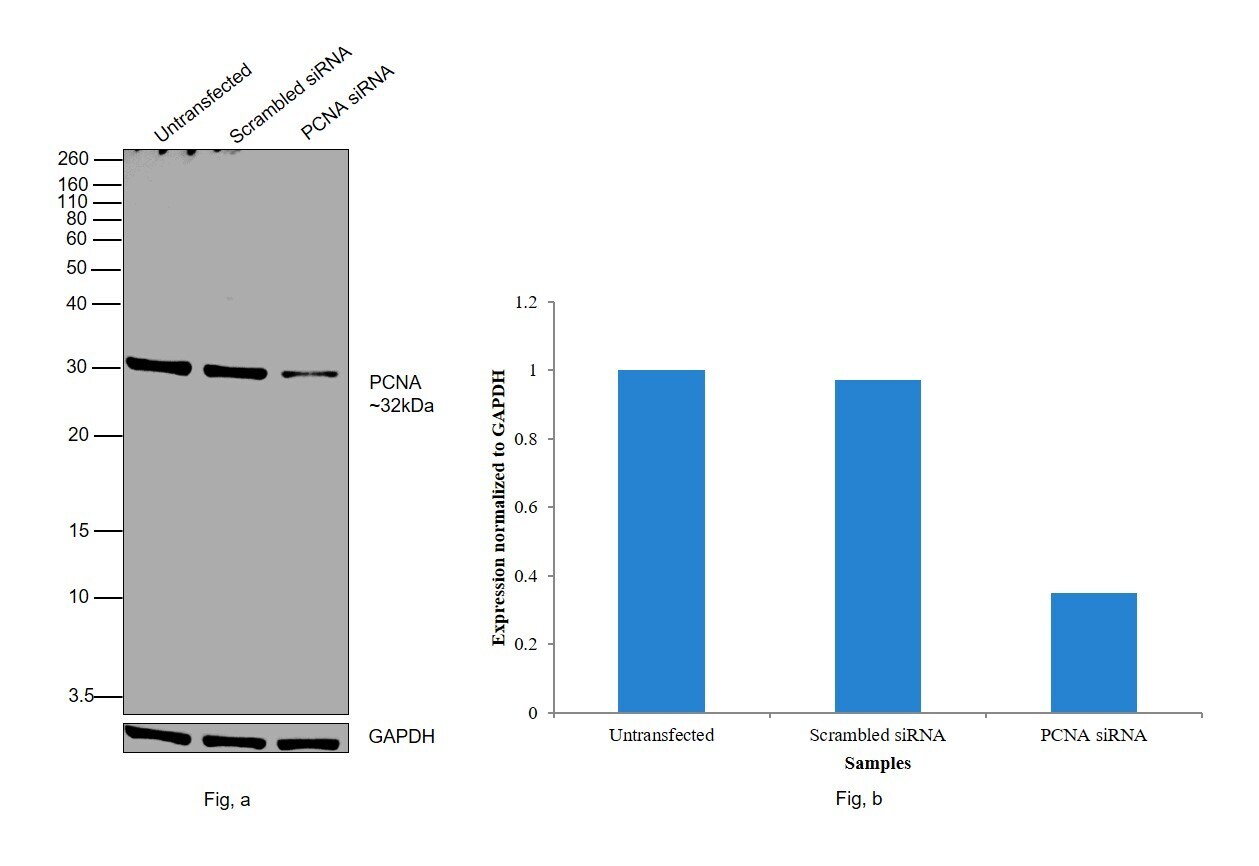
- Knockdown of PCNA was achieved by transfecting HeLa with PCNA specific siRNAs (Silencer® select Product # S10133, S10134). Western blot analysis (Fig. a) was performed using Nuclear enriched extracts from the PCNA knockdown cells (lane 3), non-targeting scrambled siRNA transfected cells (lane 2) and untransfected cells (lane 1). The blot was probed with PCNA Monoclonal Antibody (PC10 (3F81)), eBioscience™ (Product # 14-9910-82, 1 µg/mL) and Goat anti-Mouse IgG (H+L) Superclonal™ Recombinant Secondary Antibody, HRP (Product # A28177, 1:4000). Densitometric analysis of this western blot is shown in histogram (Fig. b). Decrease in signal upon siRNA mediated knock down confirms that antibody is specific to PCNA.
- Submitted by
- Invitrogen Antibodies (provider)
- Main image

- Experimental details
- Western blot was performed using Anti-PCNA Monoclonal Antibody (PC10 (3F81)), eBioscience™(Product # 14-9910-82) and a 32kDa band corresponding to PCNA was observed across all tested cell lines. Nuclear enriched extracts (30 µg lysate) of HeLa (Lane 1), MCF7 (Lane 2), Jurkat (Lane 3), HEK-293 (Lane 4), NIH/3T3 (Lane 5), MDA-MB-231 (Lane 6), A549 (Lane 7), SH-SY5Y (Lane 8), PC-12 (Lane 9) were electrophoresed using NuPAGE™ 12% Bis-Tris Protein Gel (Product # NP0342BOX). Resolved proteins were then transferred onto a Nitrocellulose membrane (Product # IB23001) by iBlot® 2 Dry Blotting System (Product # IB21001). The blot was probed with the primary antibody (1 µg/mL) and detected by chemiluminescence with Goat anti-Mouse IgG (H+L) Superclonal™ Recombinant Secondary Antibody, HRP (Product # A28177,1:4000) using the iBright FL 1000 (Product # A32752). Chemiluminescent detection was performed using Novex® ECL Chemiluminescent Substrate Reagent Kit (Product # WP20005).

Supportive validation
- Submitted by
- Invitrogen Antibodies (provider)
- Main image

- Experimental details
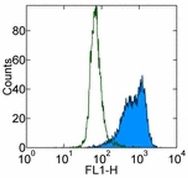
- Staining of Molt-4 cell line with 0.125 µg of Mouse IgG2a kappa Isotype Control Purified (Product # 14-4724-82) (open histogram) or 0.125 µg of Anti-Human PCNA Purified (filled histogram) followed by Anti-Mouse IgG FITC (Product # 11-4011-85).Total cells were used for analysis.
Supportive validation
- Submitted by
- Invitrogen Antibodies (provider)
- Main image

- Experimental details
- Figure 1 IP induces dose-dependent cell surface NKG2D ligand expression. (A) NKG2D ligands are not typically expressed on healthy quiescent cells. Stimuli including malignant transformation, viral infection, and proliferative lymphocyte activation are associated with NKG2D ligand induction. Expression can cause cytotoxicity, cytokine secretion, or costimulation through binding to the activating receptor, NKG2D. (B) HEK293T cells were cultured in 5 mM glucose with 0.25, 1, or 2 mM IP for 48 h, and cell surface expression of MICA (2C10) was measured by flow cytometry. A strong dose-dependent increase in MICA expression was observed. Isotype controls (dotted histogram), cells cultured in 5 mM glucose only (light grey shaded histogram) or in 25 mM glucose (dark grey shaded histogram) are also shown. (C) Cells were cultured in 5 or 25 mM glucose with IP in biological triplicates and MICA expression was measured by flow cytometry. In 5 mM glucose, IP produced a significant increase in cell surface MICA expression compared to untreated cells. In 25 mM glucose, a significant increase in MICA expression was observed at higher IP concentrations. (D) HEK293T cells, (E) HT1080 cells (human fibrosarcoma), and (F) HeLa cells (human cervical carcinoma) demonstrate dose-dependent MICA (2C10) expression when cultured with IP. (G) We tested whether IP influenced total cellular MICA levels by staining permeabilized and non-permeabilized cells in parallel. Permeabilized cells displayed the sa
