 Explore
 Validate
 Learn
Learn Western blot
Antibody data
- Antibody Data
- Antigen structure
- References [2]
- Comments [0]
- Validations
- Western blot [4]
- Immunocytochemistry [3]
- Immunohistochemistry [1]
- Flow cytometry [1]
- Other assay [1]
Submit
Validation data
Reference
Comment
Report error
- Product number
- PA5-16797 - Provider product page

- Provider
- Invitrogen Antibodies
- Product name
- PCNA Polyclonal Antibody
- Antibody type
- Polyclonal
- Antigen
- Recombinant full-length protein
- Description
- Methanol fixation is recommended for IF applications. Paraformaldehyde fixation for IF will result in mostly nuclear staining with some cytoplasmic staining.
- Reactivity
- Human, Mouse, Rat, Canine
- Host
- Rabbit
- Isotype
- IgG
- Vial size
- 500 µL
- Storage
- -20° C, Avoid Freeze/Thaw Cycles
Submitted references The Neurotoxicity of Vesicles Secreted by ALS Patient Myotubes Is Specific to Exosome-Like and Not Larger Subtypes.
PGC-1α Protects from Notch-Induced Kidney Fibrosis Development.
Anakor E, Milla V, Connolly O, Martinat C, Pradat PF, Dumonceaux J, Duddy W, Duguez S
Cells 2022 Mar 1;11(5)
Cells 2022 Mar 1;11(5)
PGC-1α Protects from Notch-Induced Kidney Fibrosis Development.
Han SH, Wu MY, Nam BY, Park JT, Yoo TH, Kang SW, Park J, Chinga F, Li SY, Susztak K
Journal of the American Society of Nephrology : JASN 2017 Nov;28(11):3312-3322
Journal of the American Society of Nephrology : JASN 2017 Nov;28(11):3312-3322
No comments: Submit comment
Supportive validation
- Submitted by
- Invitrogen Antibodies (provider)
- Main image

- Experimental details
- Western blot of PCNA (Proliferating Cell Nuclear Antigen) using PCNA (Proliferating Cell Nuclear Antigen) Polyclonal Antibody (Product # PA5-16797) on Raji Cells.

- Submitted by
- Invitrogen Antibodies (provider)
- Main image

- Experimental details
- Western blot analysis was performed on modified whole cell extracts (1% SDS) (30 µg lysate) of HeLa (Lane 1), MCF7 (Lane 2), Jurkat (Lane 3), HEK-293 (Lane 4), NIH/3T3 (Lane 5), MDA-MB-231 (Lane 6), A549 (Lane 7), SH-SY5Y (Lane 8) and Raji (Lane 9). The blot was probed with Anti- PCNA Polyclonal Antibody (Product # PA5-16797, 1 µg/mL) and detected by chemiluminescence using Goat anti-Rabbit IgG (H+L) Superclonal™ Secondary Antibody, HRP conjugate (Product # A27036, 0.25 µg/mL, 1:4000 dilution). A 34 kDa band corresponding to PCNA was observed across the cell lines tested.

- Submitted by
- Invitrogen Antibodies (provider)
- Main image

- Experimental details
- Western blot analysis of PCNA was performed by loading 50 µg of the indicated whole cell lysates per well, and 10 µL of PageRuler Plus Prestained Protein Ladder (Product # 26619) onto a 4-20% Tris-HCl polyacrylamide gel. Proteins were transferred to a PVDF membrane using the G2 Fast Blotter (Product # 62288), and blocked with StartingBlock T20 (TBS) Blocking Buffer (Product # 37543) for 1 hour at room temperature. PCNA was detected between 30-35 kDa using a PCNA polyclonal antibody (Product # PA5-16797) at a dilution of 1:50 in StartingBlock T20 (TBS) Blocking Buffer (Product # 37543) overnight at 4C on a rocking platform, followed by an HRP-conjugated goat anti-rabbit IgG secondary antibody (Product # 31460) at a dilution of 1:20,000 for 1 hour. Chemiluminescent detection was performed using SuperSignal West Pico (Product # 34080). Images were acquired on a Thermo Scientific myECL Imager (Product # 62236).

- Submitted by
- Invitrogen Antibodies (provider)
- Main image

- Experimental details
- Knockdown of PCNA was achieved by transfecting HeLa cells with PCNA specific siRNAs (Silencer® select Product # s10133, s10135). Western blot analysis (Fig. a) was performed using modified whole cell extracts (1% SDS) from PCNA knockdown cells (lane 3), non-specific scrambled siRNA transfected cells (lane 2) and untransfected cells (lane 1). The blots were probed with PCNA Polyclonal Antibody (Product # PA5-16797, 1:500 dilution) and Goat anti-Rabbit IgG (H+L) Superclonal™ Secondary Antibody, HRP conjugate (Product # A27036, 0.25 µg/mL, 1:4000 dilution). Densitometric analysis of this western blot is shown in histogram (Fig. b). Reduction of signal upon siRNA mediated knock down confirms that antibody is specific to PCNA.

Supportive validation
- Submitted by
- Invitrogen Antibodies (provider)
- Main image
- Experimental details
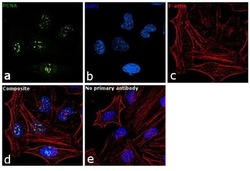
- Immunofluorescence analysis of PCNA was performed using 70% confluent log phase HeLa cells. The cells were fixed with 4% paraformaldehyde for 10 minutes, permeabilized with 0.1% Triton™ X-100 for 15 minutes, and blocked with 1% BSA for 1 hour at room temperature. The cells were labeled with PCNA Polyclonal Antibody (Product # PA5-16797) at 5 µg/mL in 0.1% BSA, incubated at 4 degree Celsius overnight and then labeled with Goat anti-Rabbit IgG (H+L) Superclonal™ Secondary Antibody, Alexa Fluor® 488 conjugate (Product # A27034) at a dilution of 1:2000 for 45 minutes at room temperature (Panel a: green). Nuclei (Panel b: blue) were stained with SlowFade® Gold Antifade Mountant with DAPI (Product # S36938). F-actin (Panel c: red) was stained with Rhodamine Phalloidin (Product # R415, 1:300). Panel d represents the merged image showing nuclear localization. Panel e represents control cells with no primary antibody to assess background. The images were captured at 60X magnification.

- Submitted by
- Invitrogen Antibodies (provider)
- Main image
- Experimental details
- Immunofluorescent analysis of PCNA (green) in U-2 OS cells. The cells were fixed with formaldehyde for 15 minutes, permeabilized with 0.1% Triton X-100 in TBS for 10 minutes, and blocked with 1% Blocker BSA in PBS (Product # 37525) for 15 minutes, all at room temperature. Cells were stained with a PCNA polyclonal antibody (Product # PA5-16797) at a dilution of 1:25 in 1% Blocker BSA in PBS (Product # 37525) for 1 hour at room temperature, and then incubated with a DyLight 488-conjugated goat anti-rabbit IgG secondary antibody (Product # 35552) at a dilution of 1:250 for 30 minutes at room temperature. F-Actin (red) was stained with DyLight-554 Phalloidin (Product # 21834) and nuclei (blue) were stained with Hoechst 33342 dye (Product # 62249). Images were taken on a Thermo Scientific ToxInsight Instrument at 20X magnification.

- Submitted by
- Invitrogen Antibodies (provider)
- Main image
- Experimental details
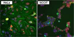
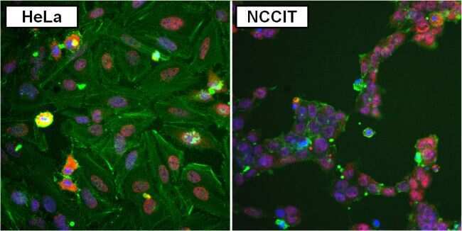
- Immunofluorescent analysis of PCNA (purple) in HeLa (left panel) and NCCIT (right panel) cells. The cells were fixed and permeabilized with ice-cold methanol for 15 minutes, and blocked with 1% Blocker BSA in PBS (Product # 37525) for 15 minutes at room temperature. Cells were stained with a PCNA polyclonal antibody (Product # PA5-16797) at a dilution of 1:50 in 1% Blocker BSA in PBS (Product # 37525) for 1 hour at room temperature, and then incubated with a DyLight 594-conjugated goat anti-rabbit IgG secondary antibody (Product # 35560) at a dilution of 1:250 for 30 minutes at room temperature. Actin (green) was stained with a DyLight 488-conjugated beta-actin monoclonal antibody (Product # MA5-15739-D488) and nuclei (blue) were stained with Hoechst 33342 dye (Product # 62249). Images were taken on a Thermo Scientific ToxInsight Instrument at 20X magnification.
Supportive validation
- Submitted by
- Invitrogen Antibodies (provider)
- Main image

- Experimental details
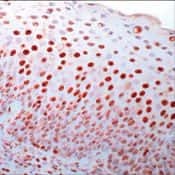
- Formalin-fixed, paraffin-embedded human tonsil stained with PCNA antibody using peroxidase-conjugate and AEC chromogen. Note nuclear staining of proliferating cells.
Supportive validation
- Submitted by
- Invitrogen Antibodies (provider)
- Main image

- Experimental details
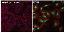
- Flow cytometry analysis of PCNA on HeLa cells. Cells were permeabilized with ice-cold methanol, and blocked with 1% Blocker BSA in PBS (Product # 37525) for 1 hour at room temperature. Cells were stained with a PCNA polyclonal antibody (Product # PA5-16797) at a dilution of 1:50 in 1% Blocker BSA (red histogram), or incubated in blocking buffer with no antibody as a negative control (black histogram). After incubation with blocking buffer alone or the primary antibody for 1 hour at room temperature, the cells were stained with a DyLight 488-conjugated goat anti-rabbit IgG secondary antibody (Product # 35552) for 30-minutes at room temperature. Approximately 15,000 cells were acquired for each sample.

Supportive validation
- Submitted by
- Invitrogen Antibodies (provider)
- Main image

- Experimental details
- Effects of MuVs or lmEVs on astrocytes. ( A ) Quantification of astrocyte survival following treatment with EVs secreted by the myotubes of ALS or healthy subjects. ( B ) Representative images of astrocyte shapes. Green: GFAP staining, blue: DAPI. ( C ) Distributions of astrocyte shapes when treated or not with MuVs (NT-M = non-treated, H-M = treated with Healthy MuVs, A-M = treated with ALS MuVs) or with lmEVs (NT-L = non-treated, H-L = treated with Healthy lmEVs, A-L = treated with ALS lmEVs), ( n = 4 per treatment). **, p < 0.01, ***, p < 0.001, significantly different from NT, ##, p < 0.01, significantly different from Healthy MuV values, ns: non-significant. Plots are shown for ( D ) percentage of Ki67 positive astrocytes, ( E ) measurement of GFAP intensity in astrocytes, and ( F ) quantification of ROS production in rat astrocytes, following treatment with MuVs (ALS or Healthy) or lmEVs (ALS or Healthy) ( n = 3-4 per treatment). Dotted lines indicate levels for untreated cells or in ( D ) for H 2 O 2 positive control and NAC (N-acetyl cysteine) negative control. Lower panels: representative Western blots showing ( D ) the low expression level of PCNA in astrocytes, and ( E ) the constant expression level of GFAP in astrocytes, regardless of treatment; ns--non-significant.
