 Explore
 Validate
 Learn
Learn Western blot
 Immunoprecipitation
Antibody data
- Antibody Data
- Antigen structure
- References [10]
- Comments [0]
- Validations
- Western blot [8]
- Immunocytochemistry [3]
- Immunohistochemistry [6]
- Other assay [6]
Submit
Validation data
Reference
Comment
Report error
- Product number
- PA5-27214 - Provider product page

- Provider
- Invitrogen Antibodies
- Product name
- PCNA Polyclonal Antibody
- Antibody type
- Polyclonal
- Antigen
- Recombinant protein fragment
- Description
- Recommended positive controls: 293T, A431, HeLa, HepG2, NIH-3T3, PC-12, A549, SiHa. Predicted reactivity: Mouse (97%), Rat (98%), Zebrafish (92%), Xenopus laevis (89%), Pig (100%), Chicken (96%), Rhesus Monkey (100%), Bovine (99%). Store product as a concentrated solution. Centrifuge briefly prior to opening the vial.
- Reactivity
- Human, Mouse, Rat, Hamster
- Host
- Rabbit
- Isotype
- IgG
- Vial size
- 100 µL
- Concentration
- 0.11 mg/mL
- Storage
- Store at 4°C short term. For long term storage, store at -20°C, avoiding freeze/thaw cycles.
Submitted references Cartilage Formation In Vivo Using High Concentration Collagen-Based Bioink with MSC and Decellularized ECM Granules.
Transmembrane protein ADAM29 facilitates cell proliferation, invasion and migration in clear cell renal cell carcinoma.
Sinoporphyrin sodium is a promising sensitizer for photodynamic and sonodynamic therapy in glioma.
Protective Effects of Dietary Grape on UVB-Mediated Cutaneous Damages and Skin Tumorigenesis in SKH-1 Mice.
Nephropathy induced by renal microembolism: a characterization of biochemical and histopathological changes in rats.
PCNA is recruited to irradiated chromatin in late S-phase and is most pronounced in G2 phase of the cell cycle.
STAT3 deficiency prevents hepatocarcinogenesis and promotes biliary proliferation in thioacetamide-induced liver injury.
Effects of the aryl hydrocarbon receptor agonist 3-methylcholanthrene on the 17β-estradiol regulated mRNA transcriptome of the rat uterus.
Geraniol attenuates 2-acetylaminofluorene induced oxidative stress, inflammation and apoptosis in the liver of wistar rats.
Therapeutic Touch Has Significant Effects on Mouse Breast Cancer Metastasis and Immune Responses but Not Primary Tumor Size.
Isaeva EV, Beketov EE, Demyashkin GA, Yakovleva ND, Arguchinskaya NV, Kisel AA, Lagoda TS, Malakhov EP, Smirnova AN, Petriev VM, Eremin PS, Osidak EO, Domogatsky SP, Ivanov SA, Shegay PV, Kaprin AD
International journal of molecular sciences 2022 Feb 28;23(5)
International journal of molecular sciences 2022 Feb 28;23(5)
Transmembrane protein ADAM29 facilitates cell proliferation, invasion and migration in clear cell renal cell carcinoma.
Li SL, Jiang TQ, Cao QW, Liu SM
Journal of chemotherapy (Florence, Italy) 2021 Feb;33(1):40-50
Journal of chemotherapy (Florence, Italy) 2021 Feb;33(1):40-50
Sinoporphyrin sodium is a promising sensitizer for photodynamic and sonodynamic therapy in glioma.
An YW, Liu HQ, Zhou ZQ, Wang JC, Jiang GY, Li ZW, Wang F, Jin HT
Oncology reports 2020 Oct;44(4):1596-1604
Oncology reports 2020 Oct;44(4):1596-1604
Protective Effects of Dietary Grape on UVB-Mediated Cutaneous Damages and Skin Tumorigenesis in SKH-1 Mice.
Mintie CA, Musarra AK, Singh CK, Ndiaye MA, Sullivan R, Eickhoff JC, Ahmad N
Cancers 2020 Jul 1;12(7)
Cancers 2020 Jul 1;12(7)
Nephropathy induced by renal microembolism: a characterization of biochemical and histopathological changes in rats.
Bersani-Amado LE, da Rocha BA, Schneider LCL, Ames FQ, Breithaupt Faloppa AC, Araújo GB, Dantas JA, Bersani-Amado CA, Cuman RKN
International journal of clinical and experimental pathology 2019;12(6):2311-2323
International journal of clinical and experimental pathology 2019;12(6):2311-2323
PCNA is recruited to irradiated chromatin in late S-phase and is most pronounced in G2 phase of the cell cycle.
Bártová E, Suchánková J, Legartová S, Malyšková B, Hornáček M, Skalníková M, Mašata M, Raška I, Kozubek S
Protoplasma 2017 Sep;254(5):2035-2043
Protoplasma 2017 Sep;254(5):2035-2043
STAT3 deficiency prevents hepatocarcinogenesis and promotes biliary proliferation in thioacetamide-induced liver injury.
Abe M, Yoshida T, Akiba J, Ikezono Y, Wada F, Masuda A, Sakaue T, Tanaka T, Iwamoto H, Nakamura T, Sata M, Koga H, Yoshimura A, Torimura T
World journal of gastroenterology 2017 Oct 7;23(37):6833-6844
World journal of gastroenterology 2017 Oct 7;23(37):6833-6844
Effects of the aryl hydrocarbon receptor agonist 3-methylcholanthrene on the 17β-estradiol regulated mRNA transcriptome of the rat uterus.
Helle J, Keiler AM, Zierau O, Dörfelt P, Vollmer G, Lehmann L, Chittur SV, Tenniswood M, Welsh J, Kretzschmar G
The Journal of steroid biochemistry and molecular biology 2017 Jul;171:133-143
The Journal of steroid biochemistry and molecular biology 2017 Jul;171:133-143
Geraniol attenuates 2-acetylaminofluorene induced oxidative stress, inflammation and apoptosis in the liver of wistar rats.
Hasan SK, Sultana S
Toxicology mechanisms and methods 2015;25(7):559-73
Toxicology mechanisms and methods 2015;25(7):559-73
Therapeutic Touch Has Significant Effects on Mouse Breast Cancer Metastasis and Immune Responses but Not Primary Tumor Size.
Gronowicz G, Secor ER Jr, Flynn JR, Jellison ER, Kuhn LT
Evidence-based complementary and alternative medicine : eCAM 2015;2015:926565
Evidence-based complementary and alternative medicine : eCAM 2015;2015:926565
No comments: Submit comment
Supportive validation
- Submitted by
- Invitrogen Antibodies (provider)
- Main image

- Experimental details
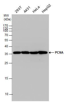
- Western blot analysis of PCNA using A) 30 µg 293T whole cell lysate (B) 30 µg A431 whole cell lysate (C) 30 µg HeLa whole cell lysate and D) 30 µg HepG2 whole cell lysate. Samples were loaded onto a 12% SDS-PAGE gel and probed with a PCNA polyclonal antibody (Product # PA5-27214) at a dilution of 1:2500.
- Submitted by
- Invitrogen Antibodies (provider)
- Main image

- Experimental details
- Western Blot analysis of PCNA was performed by separating 30 µg of various whole cell extracts by 12% SDS-PAGE. Proteins were transferred to a membrane and probed with a PCNA Polyclonal Antibody (Product # PA5-27214) at a dilution of 1:2500 and a HRP-conjugated anti-rabbit IgG secondary antibody.

- Submitted by
- Invitrogen Antibodies (provider)
- Main image

- Experimental details
- PCNA Polyclonal Antibody detects PCNA protein by Western blot analysis. Various whole cell extracts (30 µg) were separated by 12% SDS-PAGE, and the membrane was blotted with PCNA Polyclonal Antibody (Product # PA5-27214) diluted at 1:2,500.

- Submitted by
- Invitrogen Antibodies (provider)
- Main image

- Experimental details
- Western blot analysis was performed on modified whole cell extracts (1% SDS) (30 µg lysate) of HeLa (Lane 1), MCF7 (Lane 2), Jurkat (Lane 3), HEK-293 (Lane 4), NIH/3T3 (Lane 5), MDA-MB-231 (Lane 6), A549 (Lane 7), SH-SY5Y (Lane 8) and Raji (Lane 9). The blot was probed with Anti- PCNA Polyclonal Antibody (Product # PA5-27214, 1:1000 dilution) and detected by chemiluminescence using Goat anti-Rabbit IgG (H+L) Superclonal™ Secondary Antibody, HRP conjugate (Product # A27036, 0.25µg/mL, 1:4000 dilution). A 34 kDa band corresponding to PCNA was observed across the cell lines tested.

- Submitted by
- Invitrogen Antibodies (provider)
- Main image

- Experimental details
- Knockdown of PCNA was achieved by transfecting HeLa cells with PCNA specific siRNAs (Silencer® select Product # s10133, s10135). Western blot analysis (Fig. a) was performed using modified whole cell extracts (1% SDS) from the PCNA knockdown cells (lane 3), non-specific scrambled siRNA transfected cells (lane 2) and untransfected cells (lane 1). The blots were probed with PCNA Polyclonal Antibody (Product # PA5-27214, 1:1000 dilution) and Goat anti-Rabbit IgG (H+L) Superclonal™ Secondary Antibody, HRP conjugate (Product # A27036, 0.25µg/mL, 1:4000 dilution). Densitometric analysis of this western blot is shown in histogram (Fig. b). Decrease in signal upon siRNA mediated knock down confirms that antibody is specific to PCNA.

- Submitted by
- Invitrogen Antibodies (provider)
- Main image

- Experimental details
- PCNA Polyclonal Antibody detects PCNA protein by western blot analysis. A. 30 µg NIH-3T3 whole cell lysate/extract.12% SDS-PAGE. PCNA Polyclonal Antibody (Product # PA5-27214) dilution: 1:2,500. The HRP-conjugated anti-rabbit IgG antibody was used to detect the primary antibody.

- Submitted by
- Invitrogen Antibodies (provider)
- Main image

- Experimental details
- PCNA Polyclonal Antibody detects PCNA protein by western blot analysis. A. 30 µg PC-12 whole cell lysate/extract.12% SDS-PAGE. PCNA Polyclonal Antibody (Product # PA5-27214) dilution: 1:2,500. The HRP-conjugated anti-rabbit IgG antibody was used to detect the primary antibody.

- Submitted by
- Invitrogen Antibodies (provider)
- Main image

- Experimental details
- PCNA Polyclonal Antibody detects PCNA protein by Western blot analysis. Various whole cell extracts (30 µg) were separated by 12% SDS-PAGE, and the membrane was blotted with PCNA Polyclonal Antibody (Product # PA5-27214) diluted at 1:2,500.

Supportive validation
- Submitted by
- Invitrogen Antibodies (provider)
- Main image

- Experimental details
- Immunocytochemistry-Immunofluorescence analysis of PCNA was performed in HeLa cells fixed in ice-cold MeOH for 5 min. Green: PCNA Polyclonal Antibody (Product # PA5-27214) diluted at 1:1000. Blue: Hoechst 33342 staining. Scale bar = 10 µm.

- Submitted by
- Invitrogen Antibodies (provider)
- Main image

- Experimental details
- Immunofluorescence analysis of PCNA was performed using 70% confluent log phase A549 cells. The cells were fixed with 4% paraformaldehyde for 10 minutes, permeabilized with 0.1% Triton™ X-100 for 15 minutes, and blocked with 2% BSA for 1 hour at room temperature. The cells were labeled with PCNA Polyclonal Antibody (Product # PA5-27214, 1:100 dilution) in 0.1% BSA, incubated at 4 degree celsius overnight and then labeled with Donkey anti-Rabbit IgG (H+L) Highly Cross-Adsorbed Secondary Antibody, Alexa Fluor Plus 488 (Product # A32790, 1:2000 dilution), for 45 minutes at room temperature (Panel a: Green). Nuclei (Panel b:Blue) were stained with Hoechst 33342 (Product # H1399). F-actin (Panel c: Red) was stained with Rhodamine Phalloidin (Product # R415, 1:300 dilution). Panel d represents the merged image showing nuclear localization. Panel e represents control cells with no primary antibody to assess background. The images were captured at 40X magnification in CellInsight CX7 LZR High-Content Screening (HCS) Platform (Product # CX7A1110LZR) and externally deconvoluted (D.Sage et al. / Methods 115 (2017) 28–41).

- Submitted by
- Invitrogen Antibodies (provider)
- Main image

- Experimental details
- PCNA Polyclonal Antibody detects PCNA protein at nucleus by immunofluorescent analysis. Sample: HeLa cells were fixed in 4% paraformaldehyde at RT for 15 min. Green: PCNA protein stained by PCNA Polyclonal Antibody (Product # PA5-27214) diluted at 1:500. Red: Phalloidin, a cytoskeleton marker, diluted at 1:200. Scale bar = 10 µm.

Supportive validation
- Submitted by
- Invitrogen Antibodies (provider)
- Main image
- Experimental details
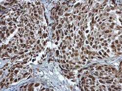
- Immunohistochemistry (Paraffin) analysis of PCNA was performed in paraffin-embedded human cervical carcinoma tissue using PCNA Polyclonal Antibody (Product # PA5-27214) at a dilution of 1:500.

- Submitted by
- Invitrogen Antibodies (provider)
- Main image
- Experimental details
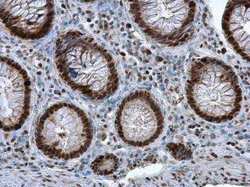
- Immunohistochemistry (Paraffin) analysis of PCNA was performed in paraffin-embedded human colon tissue using PCNA Polyclonal Antibody (Product # PA5-27214) at a dilution of 1:500.

- Submitted by
- Invitrogen Antibodies (provider)
- Main image

- Experimental details
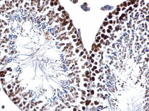
- PCNA Polyclonal Antibody detects PCNA protein at nucleus on mouse testis by immunohistochemical analysis. Sample: Paraffin-embedded mouse testis. PCNA Polyclonal Antibody (Product # PA5-27214) dilution: 1:500. Antigen Retrieval: EDTA based buffer, pH 8.0, 15 min.
- Submitted by
- Invitrogen Antibodies (provider)
- Main image
- Experimental details
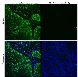
- Immunohistochemical analysis of PCNA was performed using formalin-fixed paraffin-embedded human alveolar ridge mucosa tissue sections. To expose the target protein, heat-induced epitope retrieval was performed on de-paraffinized sections using eBioscience™ IHC Antigen Retrieval Solution - Low pH (10X) (Product # 00-4955-58) diluted to 1X solution in water in a decloaking chamber at 110 degree Celsius for 15 minutes. Following antigen retrieval, the sections were blocked with 2% normal goat serum in 1X PBS for 45 minutes at room temperature and then probed with or without PCNA Polyclonal Antibody (Product # PA5-27214) at 1:100 dilution in 0.1% normal goat serum overnight at 4 degree Celsius in a humidified chamber. Detection was performed using Goat anti-Rabbit IgG (H+L) Highly Cross-Adsorbed Secondary Antibody, Alexa Fluor Plus 488 (Product # A32731) at a dilution of 1:2000 in 0.1% normal goat serum for 45 minutes at room temperature. ReadyProbes™ Tissue Autofluorescence Quenching Kit (Product # R37630) was used to quench autofluorescence from the tissues. Nuclei were stained with DAPI (Product # D1306) and the sections were mounted using ProLong™ Glass Antifade Mountant (Product # P36984). The images were captured on EVOS™ M7000 Imaging System (Product # AMF7000) at 20X magnification.

- Submitted by
- Invitrogen Antibodies (provider)
- Main image
- Experimental details
- PCNA Polyclonal Antibody detects PCNA protein at nucleus on mouse testis by immunohistochemical analysis. Sample: Paraffin-embedded mouse testis. PCNA Polyclonal Antibody (Product # PA5-27214) dilution: 1:500. Antigen Retrieval: EDTA based buffer, pH 8.0, 15 min.
- Submitted by
- Invitrogen Antibodies (provider)
- Main image
- Experimental details
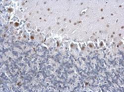
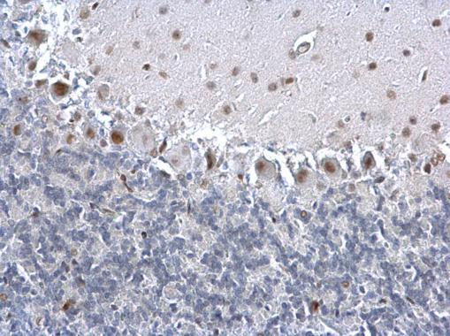
- PCNA Polyclonal Antibody detects PCNA protein at nucleus on rat hind brain by immunohistochemical analysis. Sample: Paraffin-embedded rat hind brain. PCNA Polyclonal Antibody (Product # PA5-27214) dilution: 1:500. Antigen Retrieval: EDTA based buffer, pH 8.0, 15 min.
Supportive validation
- Submitted by
- Invitrogen Antibodies (provider)
- Main image

- Experimental details
- Figure 1 Proliferation and apoptosis in the primary tumor. In (a) the normal morphology of the foot is seen in a PBS-treated mouse (PBS). Panels (b) and (c) demonstrate at successively higher magnifications the change in the foot injected with 66cl4 cells from a mouse breast carcinoma (CA group). Mostly undifferentiated tumor tissue is found. In panel (d) PCNA-stained cells are seen in the TT-treated tumor (arrows), and the graph (e) shows no significant differences in proliferation between CA and TT groups. Apoptosis was assessed by immunohistochemistry (arrows) of the TT-treated tumor (f) and quantified in the graph (g) showing no significant differences between mice from the CA and TT groups. Bar = 250 mu m in (a) and (b). Bar = 50 mu m in (c), (d), and (f).

- Submitted by
- Invitrogen Antibodies (provider)
- Main image

- Experimental details
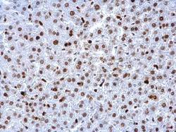
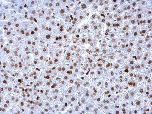
- Figure 2 Hepatic STAT3 deficiency reduced hepatocyte proliferation and inhibited the development of hepatocellular carcinoma. A: Representative immunohistochemical staining for PCNA in livers of control and STAT3 Deltahep mice with TAA treatment for 16 wk. PCNA positive hepatocytes were decreased in STAT3 Deltahep mice. Original magnification x 200. B: The number of PCNA positive hepatocytes. The ratio of PCNA positive hepatocytes to all hepatocytes was quantified in three different samples per a group. Values represent means +- standard error of the mean. b P < 0.01 ( P = 0.000232) vs control. C: Gross liver appearance of control and STAT3 Deltahep mice with TAA treatment for 30 wk. Tumor formation with TAA treatment for 30 wk (upper panel). HE staining of representative sections of hepatocellular carcinoma. Original magnification x 100 (lower left panel) and x 400 (lower right panel). D: The number of HCC in control and STAT3 Deltahep mice. Values represent means +- SE of the mean ( n = 6). a P < 0.05 ( P = 0.0219) vs control. TAA: Thioacetamide.

- Submitted by
- Invitrogen Antibodies (provider)
- Main image

- Experimental details
- Figure 1 Dietary GP consumption results in UVB-mediated cutaneous hyperplasia and mast cell infiltrations in SKH-1 hairless mice. ( a ) Timeline of short-term UVB-mediated cutaneous damage study. ( b ) GP consumption significantly reduced epidermal thickness as determined by measuring epidermal thickness from the base of the stratum basale to the base of the stratum corneum on H&E-stained tissue sections. ( c ) Mast cell infiltration shown by toluidine blue staining. At least five images were taken at 10x magnification across five different skin sections of each mouse. Mast cells (violet metachromatic cytoplasmic granules) were counted using ImageJ, and then averaged per mouse. ( d ) Expression of proliferative markers PCNA and Ki67 was assessed using immunohistochemical staining techniques. Representative sections are shown. All images are represented at 10x magnification, with inset at 40x magnification. The data represent the mean +- SEM of all five animals per group. A two-way ANOVA with Tukey's multiple comparison test was performed (* p

- Submitted by
- Invitrogen Antibodies (provider)
- Main image

- Experimental details
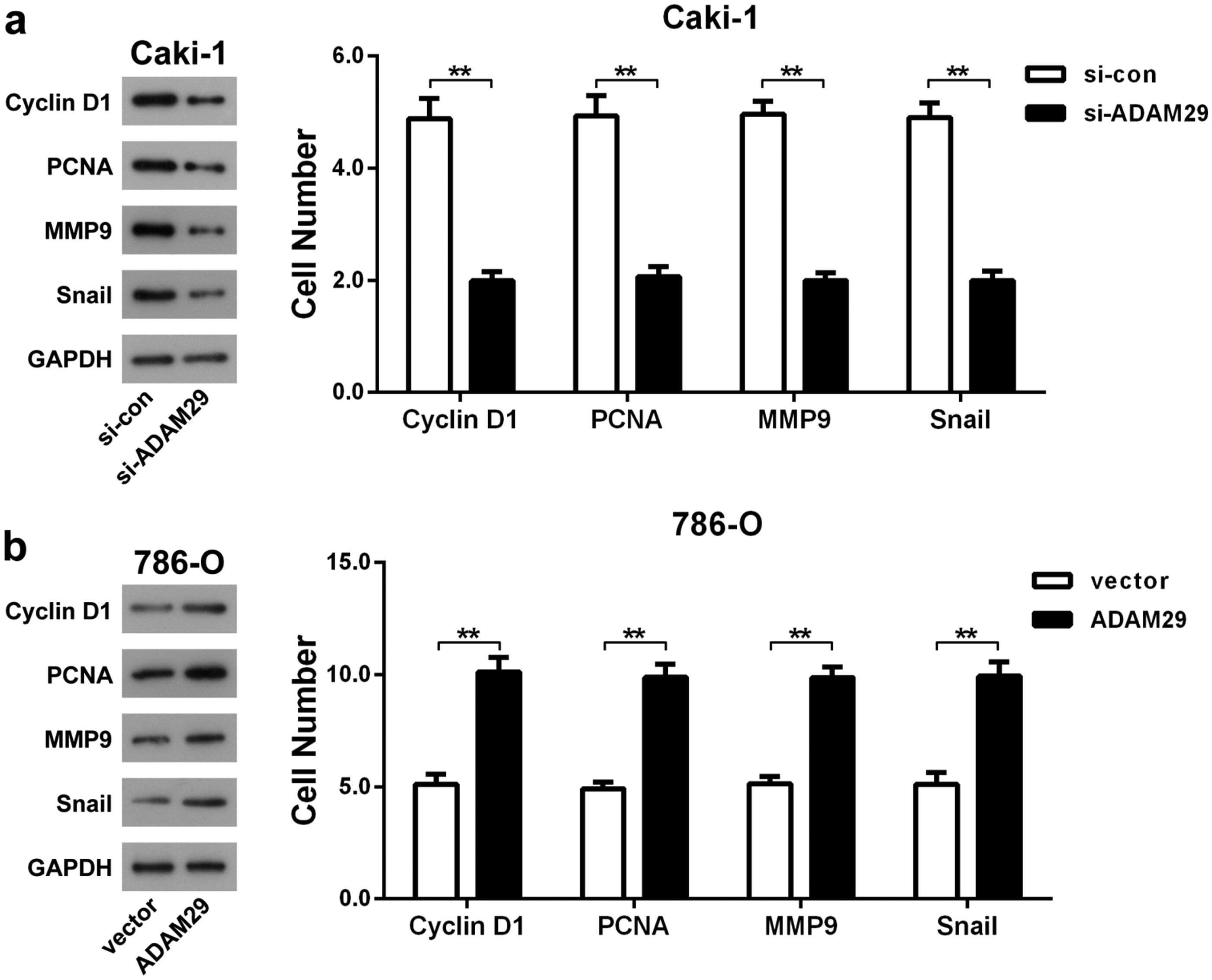
- Figure 5. Effects of knockdown or overexpression of ADAM29 on proliferation- and motion-related proteins. Western blot assay was adopted to determine the effect of ADAM29 knockdown (a) or overexpression (b) on the level of Cyclin D1, PCNA, MMP9 and Snail. ** p < 0.01 vs. si-con group or vector group.
- Submitted by
- Invitrogen Antibodies (provider)
- Main image

- Experimental details
- Figure 7 1st group (dECM only), 1 week. PCNA-positive multinucleated cells resorb around the perimeter of the scaffold in the connective tissue capsule (arrows). Immunohistochemical staining for PCNA, objective lens x40, scale bar--50 mum.

- Submitted by
- Invitrogen Antibodies (provider)
- Main image

- Experimental details
- Figure 21 3rd group (dECM and MSC), 2 weeks. Immunohistochemical staining (PCNA) of multinucleated cells of cartilage tissue resorption (arrows), objective lens x40, scale bar--50 mum.
