Explore
 Validate
 Learn
Learn Western blot
Antibody data
- Antibody Data
- Antigen structure
- References [6]
- Comments [0]
- Validations
- Western blot [4]
- Immunocytochemistry [2]
- Immunohistochemistry [1]
Submit
Validation data
Reference
Comment
Report error
- Product number
- PA5-32541 - Provider product page

- Provider
- Invitrogen Antibodies
- Product name
- PCNA Polyclonal Antibody
- Antibody type
- Polyclonal
- Antigen
- Recombinant full-length protein
- Description
- A suggested positive control is tonsil, lymph node or small intestine tissue.
- Reactivity
- Human, Mouse
- Host
- Rabbit
- Isotype
- IgG
- Vial size
- 500 µL
- Concentration
- 0.14 mg/mL
- Storage
- Store at 4°C short term. For long term storage, store at -20°C, avoiding freeze/thaw cycles.
Submitted references Flavonoids from each of the six structural groups reactivate BRM, a possible cofactor for the anticancer effects of flavonoids.
Two drugs with paradoxical effects on liver regeneration through antiangiogenesis and antifibrosis: Losartan and Spironolactone: a pharmacologic dilemma on hepatocyte proliferation.
Metastatic pancreatic cancer is dependent on oncogenic Kras in mice.
The protective effects of Ginkgo biloba extract (EGb-761) on radiation-induced dermatitis: an experimental study.
Transgenic expression of interferon-γ in mouse stomach leads to inflammation, metaplasia, and dysplasia.
Essential role of ribosomal protein L11 in mediating growth inhibition-induced p53 activation.
Kahali B, Marquez SB, Thompson KW, Yu J, Gramling SJ, Lu L, Aponick A, Reisman D
Carcinogenesis 2014 Oct;35(10):2183-93
Carcinogenesis 2014 Oct;35(10):2183-93
Two drugs with paradoxical effects on liver regeneration through antiangiogenesis and antifibrosis: Losartan and Spironolactone: a pharmacologic dilemma on hepatocyte proliferation.
Parlakgumus A, Colakoglu T, Kayaselcuk F, Colakoglu S, Ezer A, Calıskan K, Karakaya J, Yildirim S
The Journal of surgical research 2013 Jan;179(1):60-5
The Journal of surgical research 2013 Jan;179(1):60-5
Metastatic pancreatic cancer is dependent on oncogenic Kras in mice.
Collins MA, Brisset JC, Zhang Y, Bednar F, Pierre J, Heist KA, Galbán CJ, Galbán S, di Magliano MP
PloS one 2012;7(12):e49707
PloS one 2012;7(12):e49707
The protective effects of Ginkgo biloba extract (EGb-761) on radiation-induced dermatitis: an experimental study.
Yirmibesoglu E, Karahacioglu E, Kilic D, Lortlar N, Akbulut G, Omeroglu S
Clinical and experimental dermatology 2012 Jun;37(4):387-94
Clinical and experimental dermatology 2012 Jun;37(4):387-94
Transgenic expression of interferon-γ in mouse stomach leads to inflammation, metaplasia, and dysplasia.
Syu LJ, El-Zaatari M, Eaton KA, Liu Z, Tetarbe M, Keeley TM, Pero J, Ferris J, Wilbert D, Kaatz A, Zheng X, Qiao X, Grachtchouk M, Gumucio DL, Merchant JL, Samuelson LC, Dlugosz AA
The American journal of pathology 2012 Dec;181(6):2114-25
The American journal of pathology 2012 Dec;181(6):2114-25
Essential role of ribosomal protein L11 in mediating growth inhibition-induced p53 activation.
Bhat KP, Itahana K, Jin A, Zhang Y
The EMBO journal 2004 Jun 16;23(12):2402-12
The EMBO journal 2004 Jun 16;23(12):2402-12
No comments: Submit comment
Supportive validation
- Submitted by
- Invitrogen Antibodies (provider)
- Main image
- Experimental details
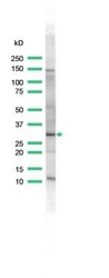
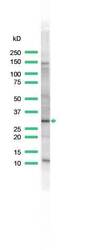
- Western blot analysis of 293T Cells using anti-PCNA Polyclonal Antibody (Product # PA5-32541). The recommened dilution for this antibody in western blot applications is 1:25.
- Submitted by
- Invitrogen Antibodies (provider)
- Main image

- Experimental details
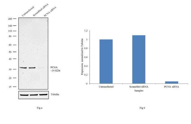
- Knockdown of PCNA was achieved by transfecting HeLa cells with PCNA specific siRNAs (Silencer® select Product # s10133, s10135). Western blot analysis (Fig. a) was performed using modified whole cell extracts (1% SDS) from the PCNA knockdown cells (lane 3), non-specific scrambled siRNA transfected cells (lane 2) and untransfected cells (lane 1). The blots were probed with PCNA Polyclonal Antibody (Product # PA5-32541, 1:500 dilution) and Goat anti-Rabbit IgG (H+L) Superclonal™ Secondary Antibody, HRP conjugate (Product # A27036, 0.25µg/mL, 1:4000 dilution). Densitometric analysis of this western blot is shown in histogram (Fig. b). Decrease in signal upon siRNA mediated knock down confirms that antibody is specific to PCNA.
- Submitted by
- Invitrogen Antibodies (provider)
- Main image

- Experimental details
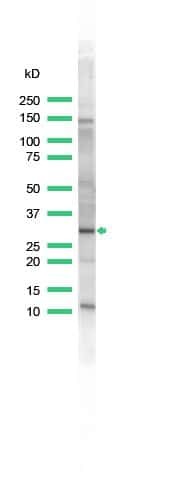
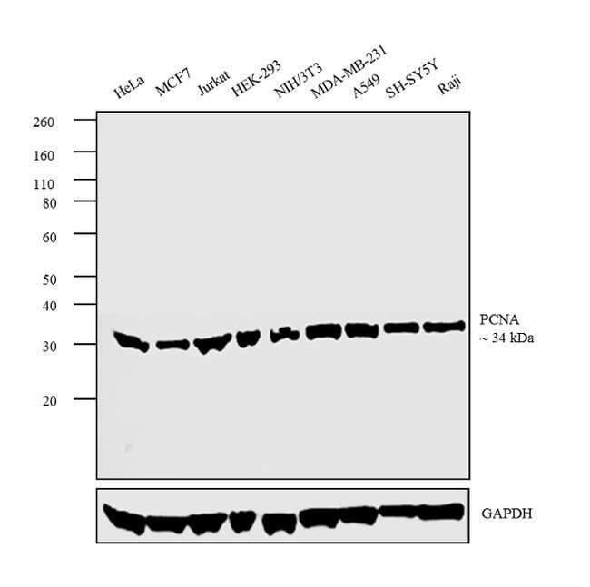
- Western blot analysis was performed on modified whole cell extracts (1% SDS) (30 µg lysate) of HeLa (Lane 1), MCF7 (Lane 2), Jurkat (Lane 3), HEK-293 (Lane 4), NIH/3T3 (Lane 5), MDA-MB-231 (Lane 6), A549 (Lane 7), SH-SY5Y (Lane 8) and Raji (Lane 9). The blot was probed with Anti- PCNA Polyclonal Antibody (Product # PA5-32541, 1:500 dilution) and detected by chemiluminescence using Goat anti-Rabbit IgG (H+L) Superclonal™ Secondary Antibody, HRP conjugate (Product # A27036, 0.25µg/mL, 1:4000 dilution). A 34 kDa band corresponding to PCNA was observed across the cell lines tested.
- Submitted by
- Invitrogen Antibodies (provider)
- Main image
- Experimental details
- Western blot analysis of 293T Cells using anti-PCNA Polyclonal Antibody (Product # PA5-32541). The recommened dilution for this antibody in western blot applications is 1:25.

Supportive validation
- Submitted by
- Invitrogen Antibodies (provider)
- Main image

- Experimental details
- Immunofluorescence analysis of PCNA was performed using 70% confluent log phase HeLa cells. The cells were fixed with 4% paraformaldehyde for 10 minutes, permeabilized with 0.1% Triton™ X-100 for 10 minutes, and blocked with 1% BSA for 1 hour at room temperature. The cells were labeled with PCNA Polyclonal Antibody (Product # PA1-38424) at 1:100 dilution in 0.1% BSA and incubated overnight at 4 degree and then labeled with Goat anti-Rabbit IgG (H+L) Superclonal™ Secondary Antibody, Alexa Fluor® 488 conjugate (Product # A27034) at a dilution of 1:2000 for 45 minutes at room temperature (Panel a: green). Nuclei (Panel b: blue) were stained with ProLong™ Diamond Antifade Mountant with DAPI (Product # P36962). F-actin (Panel c: red) was stained with Rhodamine Phalloidin (Product # R415, 1:300). Panel d represents the merged image showing Nuclear localization. Panel e represents control cells with no primary antibody to assess background. The images were captured at 60X magnification.

- Submitted by
- Invitrogen Antibodies (provider)
- Main image

- Experimental details
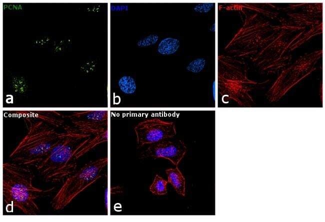
- Immunofluorescence analysis of PCNA was performed using 70% confluent log phase HeLa cells. The cells were fixed with 4% paraformaldehyde for 10 minutes, permeabilized with 0.1% Triton™ X-100 for 10 minutes, and blocked with 1% BSA for 1 hour at room temperature. The cells were labeled with PCNA Polyclonal Antibody (Product # PA5-32541) at 1:100 dilution in 0.1% BSA and incubated overnight at 4 degree and then labeled with Goat anti-Rabbit IgG (H+L) Superclonal™ Secondary Antibody, Alexa Fluor® 488 conjugate (Product # A27034) at a dilution of 1:2000 for 45 minutes at room temperature (Panel a: green). Nuclei (Panel b: blue) were stained with ProLong™ Diamond Antifade Mountant with DAPI (Product # P36962). F-actin (Panel c: red) was stained with Rhodamine Phalloidin (Product # R415, 1:300). Panel d represents the merged image showing Nuclear localization. Panel e represents control cells with no primary antibody to assess background. The images were captured at 60X magnification.
Supportive validation
- Submitted by
- Invitrogen Antibodies (provider)
- Main image

- Experimental details
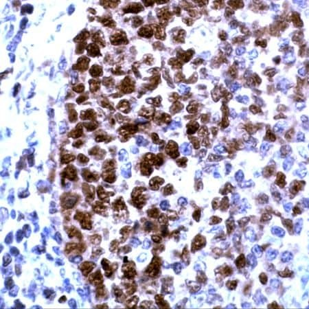
- Immunohistochemical analysis of PCNA using anti-PCNA Polyclonal Antibody (Product # PA5-32541) in Tonsil Tissue. The recommened dilution for this antibody in immunohistochemistry applications is 1:100.