Explore
 Validate
 Learn
Learn Western blot
 Immunohistochemistry
Antibody data
- Antibody Data
- Antigen structure
- References [3]
- Comments [0]
- Validations
- Immunohistochemistry [1]
- Other assay [3]
Submit
Validation data
Reference
Comment
Report error
- Product number
- PA5-27349 - Provider product page

- Provider
- Invitrogen Antibodies
- Product name
- alpha Galactosidase Polyclonal Antibody
- Antibody type
- Polyclonal
- Antigen
- Recombinant full-length protein
- Description
- Recommended positive controls: 293T, HeLa, Rat Lung. Predicted reactivity: Rat (82%), Pig (84%), Bovine (80%). Store product as a concentrated solution. Centrifuge briefly prior to opening the vial.
- Reactivity
- Human, Rat
- Host
- Rabbit
- Isotype
- IgG
- Vial size
- 100 μL
- Concentration
- 0.157 mg/mL
- Storage
- Store at 4°C short term. For long term storage, store at -20°C, avoiding freeze/thaw cycles.
Submitted references Drug Repositioning for Fabry Disease: Acetylsalicylic Acid Potentiates the Stabilization of Lysosomal Alpha-Galactosidase by Pharmacological Chaperones.
Characterization of Mono- and Bi-Transgenic Pig-Derived Epidermal Keratinocytes Expressing Human FUT2 and GLA Genes-In Vitro Studies.
Trichostatin A-Assisted Epigenomic Modulation Affects the Expression Profiles of Not Only Recombinant Human α1,2-Fucosyltransferase and α-Galactosidase A Enzymes But Also Galα1→3Gal Epitopes in Porcine Bi-Transgenic Adult Cutaneous Fibroblast Cells.
Monticelli M, Liguori L, Allocca M, Bosso A, Andreotti G, Lukas J, Monti MC, Morretta E, Cubellis MV, Hay Mele B
International journal of molecular sciences 2022 May 4;23(9)
International journal of molecular sciences 2022 May 4;23(9)
Characterization of Mono- and Bi-Transgenic Pig-Derived Epidermal Keratinocytes Expressing Human FUT2 and GLA Genes-In Vitro Studies.
Wiater J, Samiec M, Wartalski K, Smorąg Z, Jura J, Słomski R, Skrzyszowska M, Romek M
International journal of molecular sciences 2021 Sep 7;22(18)
International journal of molecular sciences 2021 Sep 7;22(18)
Trichostatin A-Assisted Epigenomic Modulation Affects the Expression Profiles of Not Only Recombinant Human α1,2-Fucosyltransferase and α-Galactosidase A Enzymes But Also Galα1→3Gal Epitopes in Porcine Bi-Transgenic Adult Cutaneous Fibroblast Cells.
Wiater J, Samiec M, Skrzyszowska M, Lipiński D
International journal of molecular sciences 2021 Jan 30;22(3)
International journal of molecular sciences 2021 Jan 30;22(3)
No comments: Submit comment
Supportive validation
- Submitted by
- Invitrogen Antibodies (provider)
- Main image
- Experimental details
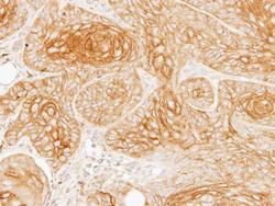
- Immunohistochemical analysis of paraffin-embedded Cal27 xenograft, using Galactosidase alpha (Product # PA5-27349) antibody at 1:100 dilution. Antigen Retrieval: EDTA based buffer, pH 8.0, 15 min.
Supportive validation
- Submitted by
- Invitrogen Antibodies (provider)
- Main image
- Experimental details
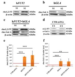
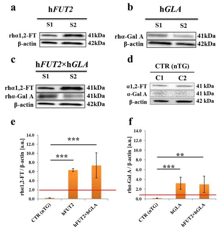
- Figure 3 Western blot-mediated estimation of the semiquantitative profiles noticed for expression of recombinant human alpha1,2-fucosyltransferase (rhalpha1,2-FT) and alpha-galactosidase A (rhalpha-Gal A) enzymes in total protein samples extracted from the ex vivo-expanded control nontransgenic (CTR nTG) PEKs and their cell counterparts stemming from h FUT2 and h GLA single-transgenic pigs and h FUT2 xh GLA double-transgenic specimens. Representative blots corresponding to expression of rhalpha1,2-FT and rhalpha-Gal A enzymes in total protein samples that were isolated from PEKs stemming from h FUT2 mono-transgenic ( a ), h GLA mono-transgenic ( b ), h FUT2 xh GLA bi-transgenic ( c ) and CTR nTG pigs ( d ). beta-Actin provides a loading control for all analyzed protein samples. Results of the semiquantitatively analyzing the relative abundances (RAs) determined for rhalpha1,2-FT and rhalpha-Gal A enzymes (in arbitrary units) were presented in panels ( e , f ), respectively. Bar graphs show mean +- standard error of mean (SEM) of relative optical density (ROD) descended from three separate analyses of three animals for each variant. Red line is taken as the cut-off value 1.0. Statistics: one-way analysis of variance (ANOVA) followed by Tukey's honestly significant difference (HSD) post hoc test. Values that are denoted as ** and *** indicate incidence of statistically significant differences between experimental groups with a probability of occurring random errors at the level
- Submitted by
- Invitrogen Antibodies (provider)
- Main image
- Experimental details
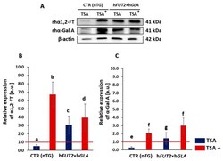
- Figure 1 Western blot analysis of the relative expression of human alpha1,2-fucosyltransferase (alpha1,2-FT) and alpha-galactosidase A (alpha-Gal A) proteins in porcine bi-transgenic and non-transgenic adult cutaneous fibroblast cells (ACFCs) undergoing or not undergoing treatment with trichostatin A (TSA + and TSA - , respectively). Representative blots of the expression of alpha1,2-FT and alpha-Gal A proteins in the ACFC samples derived from epigenetically modulated (TSA + ) and non-modulated (TSA - ) cells. The samples of non-transgenic animals served as a control group (CTR nTG--panel ( A )). beta-Actin served as a loading control for all analyzed samples. The results of relative expression (in arbitrary units) of alpha1,2-FT and alpha-Gal A are shown in panels ( B , C ), respectively. Relative optical density (ROD) from three separate analyses of at least three animals for each variant is expressed as mean. Bar graphs show mean +- standard error of the mean (SEM). The red line is taken as the cut-off value 1.0. Statistics: One-way ANOVA and Tukey's honestly significant difference (HSD) post hoc test. The bars marked with different letters differ significantly; values denoted as a-b, a-d, b-c, b-d, e-f: p < 0.01; a-c, c-d, e-g, f-g: p < 0.05.

- Submitted by
- Invitrogen Antibodies (provider)
- Main image
- Experimental details
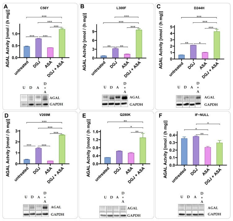
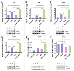
- Acetylsalicylic acid is a PC-enhancer. IF-GLA-MUTs, specifically C56Y ( A ), L300F ( B ), D244H ( C ), V269M ( D ), and Q280K ( E ), were treated for 72 h with the following drugs: i. untreated; ii. 10 muM DGJ; iii. 4 mM acetylsalicylic acid (ASA); iv. 10 muM DGJ + 4 mM ASA. IF- was used as a control ( F ). AGAL specific activity measured on protein extracts is shown. Tukey's HSD was used to evaluate significative differences among treatments (***: p < 0.001; **: p < 0.01; *: p < 0.05; n = 2). The effects of combined treatment (DGJ + ASA) are significantly higher than those of DGJ monotherapy. Immunoblots confirmed the results (U = untreated; D = DGJ 10 muM; A = ASA 4 mM; D + A = DGJ 10 muM + ASA 4 mM). Each panel includes specific activity and an immunoblot for a cell line.