 Explore
 Validate
 Learn
Learn Immunocytochemistry
 Immunohistochemistry
Antibody data
- Antibody Data
- Antigen structure
- References [2]
- Comments [0]
- Validations
- Immunocytochemistry [1]
Submit
Validation data
Reference
Comment
Report error
- Product number
- AMAb91297 - Provider product page

- Provider
- Atlas Antibodies
- Proper citation
- Atlas Antibodies Cat#AMAb91297, RRID:AB_2665884
- Product name
- Anti-SOX10
- Antibody type
- Monoclonal
- Description
- Monoclonal Antibody against Human SOX10, Clone ID: CL4455, Gene description: SRY (sex determining region Y)-box 10, Validated applications: IHC, ICC, Uniprot ID: P56693, Storage: Store at +4°C for short term storage. Long time storage is recommended at -20°C.
- Reactivity
- Human, Mouse
- Host
- Mouse
- Conjugate
- Unconjugated
- Isotype
- IgG
- Antibody clone number
- CL4455
- Vial size
- 100 µl
- Concentration
- 1.0 mg/ml
- Storage
- Store at +4°C for short term storage. Long time storage is recommended at -20°C.
- Handling
- The antibody solution should be gently mixed before use.
Submitted references Astrocyte-oligodendrocyte interaction regulates central nervous system regeneration
Stage-dependent differential gene expression profiles of cranial neural crest-like cells derived from mouse-induced pluripotent stem cells.
Molina-Gonzalez I, Holloway R, Jiwaji Z, Dando O, Kent S, Emelianova K, Lloyd A, Forbes L, Mahmood A, Skripuletz T, Gudi V, Febery J, Johnson J, Fowler J, Kuhlmann T, Williams A, Chandran S, Stangel M, Howden A, Hardingham G, Miron V
Nature Communications 2023;14(1)
Nature Communications 2023;14(1)
Stage-dependent differential gene expression profiles of cranial neural crest-like cells derived from mouse-induced pluripotent stem cells.
Odashima A, Onodera S, Saito A, Ogihara Y, Ichinohe T, Azuma T
Medical molecular morphology 2020 Mar;53(1):28-41
Medical molecular morphology 2020 Mar;53(1):28-41
No comments: Submit comment
Supportive validation
- Submitted by
- Atlas Antibodies (provider)
- Main image
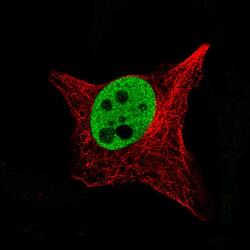
- Experimental details
- Immunofluorescence staining of SK-MEL-30 cells using the Anti-SOX10 monoclonal antibody, showing specific staining in the nucleoplasm in green. Microtubule- and nuclear probes are visualized in red and blue, respectively (where available).
- Sample type
- Human
