 Explore
 Validate
 Learn
Learn Western blot
 ELISA
ELISAAntibody data
- Antibody Data
- Antigen structure
- References [0]
- Comments [0]
- Validations
- Western blot [3]
Submit
Validation data
Reference
Comment
Report error
- Product number
- LS-C19061 - Provider product page

- Provider
- LSBio
- Product name
- AKT2 Antibody (aa450-475) LS-C19061
- Antibody type
- Polyclonal
- Description
- Affinity purified
- Reactivity
- Human
- Host
- Rabbit
- Isotype
- IgG
- Storage
- Store vial at -20°C prior to opening. Centrifuge product if not completely clear after standing at room temperature. Dilute only prior to immediate use. For extended storage aliquot contents and freeze at -20°C or below.
No comments: Submit comment
Supportive validation
- Submitted by
- LSBio (provider)
- Enhanced method
- Genetic validation
- Main image
- Experimental details
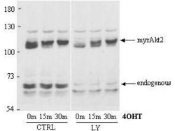
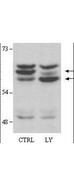
- Anti-AKT2 Antibody - Western Blot. Anti-AKT2 Antibody in Western blot shows detection of AKT2. A lysate from a stable HEK293 cell line expressing an inducible, myristoylated form of Akt2 (MyrAkt2-ER) was loaded for SDS- PAGE, separated and then transferred to nitrocellulose. The blot was used at 1:1000 and reacted with Anti-AKT2 Antibody for 1h at room temperature. In response to 4OHT (tamoxifen), the Akt2 is recruited to the plasma membrane via its myristoylation sequence, and becomes phosphorylated and activated. The blot (right panel) shows 0 m, 15 m, and 30 m of 4OHT treatment. This treatment has no effect on endogenous Akt2, but causes a band shift upwards in the MyrAkt2. Endogenous Akt2 runs at about 60kD whereas the myristoylated construct runs at around 110kD. Antibody to AKT2 detects both unphosphorylated and phosphorylated AKT2. As before approximately 20 mg/lane of crude HEK293 lysate was loaded for SDS-PAGE, separated and then transferred to nitrocellulose. The right lane contains lysate pretreated-for-15min-with 25 uM LY294002 which affects the phosphorylation of endogenous Akt2 but has no effect on phosphorylation of the myristoylated construct. This antibody clearly detects both the phosphorylated (top arrow) and the non-phosphorylated (bottom arrow) forms of endogenous Akt2.

- Submitted by
- LSBio (provider)
- Enhanced method
- Genetic validation
- Main image

- Experimental details
- Anti-AKT2 Antibody - Western Blot. Antibody to AKT2 detects both unphosphorylated and phosphorylated AKT2 in Western blot. As before approximately 20 mg/lane of crude HEK293 lysate was loaded for SDS-PAGE, separated and then transferred to nitrocellulose. The right lane contains lysate pretreated for 15min with 25 uM LY294002 which affects the phosphorylation of endogenous Akt2 but has no effect on phosphorylation of the myristoylated construct. This antibody clearly detects both the phosphorylated (top arrow) and the non-phosphorylated (bottom arrow) forms of endogenous Akt2.
- Submitted by
- LSBio (provider)
- Enhanced method
- Genetic validation
- Main image
- Experimental details
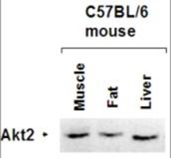
- Anti-AKT2 Antibody - Western Blot. Affinity Purified antibody to AKT2 was used at a 1:1000 dilution to detect AKT2 by Western blot of lysates from mouse tissues. The antibody is shown to react with mouse Akt2 in mouse liver, skeletal muscle and fat using standard western blotting methods.
