Explore
 Validate
 Learn
Learn Western blot
Antibody data
- Antibody Data
- Antigen structure
- References [1]
- Comments [0]
- Validations
- Western blot [2]
- Immunohistochemistry [3]
Submit
Validation data
Reference
Comment
Report error
- Product number
- AF23151 - Provider product page

- Provider
- Novus Biologicals
- Product name
- Goat Polyclonal AKT2 Antibody
- Antibody type
- Polyclonal
- Description
- Antigen Affinity-purified. Detects human, mouse, and rat Akt2 in Western blots. Also detects recombinant human Akt2 but not recombinant Akt1 or Akt3 in Western blots.
- Reactivity
- Human, Mouse, Rat
- Host
- Goat
- Conjugate
- Unconjugated
- Isotype
- IgG
- Vial size
- 100 ug
- Concentration
- LYOPH
- Storage
- Use a manual defrost freezer and avoid repeated freeze-thaw cycles. 12 months from date of receipt, -20 to -70 degreesC as supplied. 1 month, 2 to 8 degreesC under sterile conditions after reconstitution. 6 months, -20 to -70 degreesC under sterile conditions after reconstitution.
Submitted references Involvement of the protein kinase Akt2 in insulin-stimulated Rac1 activation leading to glucose uptake in mouse skeletal muscle.
Takenaka N, Araki N, Satoh T
PloS one 2019;14(2):e0212219
PloS one 2019;14(2):e0212219
No comments: Submit comment
Supportive validation
- Submitted by
- Novus Biologicals (provider)
- Main image
- Experimental details
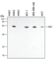
- Detection of Human/Mouse/Rat Akt2 by Western Blot. Western blot shows lysates of MCF-7 human breast cancer cell line, MDA-MB-468 human breast cancer cell line, and U937 human histiocytic lymphoma cell line. PVDF membrane was probed with 0.5 µg/mL Goat Anti-Human/Mouse/Rat Akt2 Antigen Affinity-purified Polyclonal Antibody (Catalog # AF23151) followed by HRP-conjugated Anti-Goat IgG Secondary Antibody (Catalog # HAF109). For additional reference, Recombinant Human Active Akt1 (Catalog # 1775-KS), recombinant human Akt2, and recombinant human Akt3 (5 ng/lane) were included. A specific band for Akt2 was detected at approximately 60 kDa (as indicated). This experiment was conducted under reducing conditions and using Immunoblot Buffer Group 1.
- Submitted by
- Novus Biologicals (provider)
- Main image
- Experimental details
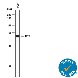
- Detection of Human Akt2 by Simple WesternTM. Simple Western lane view shows lysates of MCF-7 human breast cancer cell line, loaded at 0.2 mg/mL. A specific band was detected for Akt2 at approximately 60 kDa (as indicated) using 5 µg/mL of Goat Anti-Human/Mouse/Rat Akt2 Antigen Affinity-purified Polyclonal Antibody (Catalog # AF23151) followed by 1:50 dilution of HRP-conjugated Anti-Goat IgG Secondary Antibody (Catalog # HAF109). This experiment was conducted under reducing conditions and using the 12-230 kDa separation system.

Supportive validation
- Submitted by
- Novus Biologicals (provider)
- Main image

- Experimental details
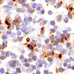
- Akt2 in Human Kidney Cancer Tissue. Akt2 was detected in immersion fixed paraffin-embedded sections of human kidney cancer tissue using 15 µg/mL Goat Anti-Human/Mouse/Rat Akt2 Antigen Affinity-purified Polyclonal Antibody (Catalog # AF23151) overnight at 4 °C. Tissue was stained with the Anti-Goat HRP-DAB Cell & Tissue Staining Kit (brown; Catalog # CTS008) and counterstained with hematoxylin (blue). Specific labeling was localized to epithelial cells in tubules. View our protocol for Chromogenic IHC Staining of Paraffin-embedded Tissue Sections.

- Submitted by
- Novus Biologicals (provider)
- Main image

- Experimental details
- Akt2 in Rat Testis. Akt2 was detected in perfusion fixed frozen sections of rat testis using Goat Anti-Human/Mouse/Rat Akt2 Antigen Affinity-purified Polyclonal Antibody (Catalog # AF23151) at 3 µg/mL for 1 hour at room temperature followed by incubation with the Anti-Goat IgG VisUCyte™ HRP Polymer Antibody (Catalog # VC004). Tissue was stained using DAB (brown) and counterstained with hematoxylin (blue). Specific staining was localized to cytoplasm and nuclei. View our protocol for IHC Staining with VisUCyte HRP Polymer Detection Reagents.

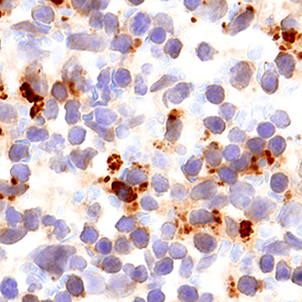
- Submitted by
- Novus Biologicals (provider)
- Main image
- Experimental details
- Akt2 in Mouse Spleen. Akt2 was detected in perfusion fixed frozen sections of mouse spleen using Goat Anti-Human/Mouse/Rat Akt2 Antigen Affinity-purified Polyclonal Antibody (Catalog # AF23151) at 3 µg/mL for 1 hour at room temperature followed by incubation with the Anti-Goat IgG VisUCyte™ HRP Polymer Antibody (Catalog # VC004). Tissue was stained using DAB (brown) and counterstained with hematoxylin (blue). Specific staining was localized to cytoplasm and nuclei. View our protocol for IHC Staining with VisUCyte HRP Polymer Detection Reagents.