Explore
 Validate
 Learn
Learn Western blot
 ELISA
ELISAAntibody data
- Antibody Data
- Antigen structure
- References [2]
- Comments [0]
- Validations
- Western blot [1]
- Immunohistochemistry [1]
- Other assay [1]
Submit
Validation data
Reference
Comment
Report error
- Product number
- 39-3900 - Provider product page

- Provider
- Invitrogen Antibodies
- Product name
- AKT2 Monoclonal Antibody (ZA006)
- Antibody type
- Monoclonal
- Antigen
- Synthetic peptide
- Reactivity
- Human, Rat
- Host
- Mouse
- Isotype
- IgG
- Antibody clone number
- ZA006
- Vial size
- 100 µg
- Concentration
- 0.5 mg/mL
- Storage
- -20°C
Submitted references Reduced FHIT expression is associated with mismatch repair deficient and high CpG island methylator phenotype colorectal cancer.
Proximity ligation assays for isoform-specific Akt activation in breast cancer identify activated Akt1 as a driver of progression.
Al-Temaimi RA, Jacob S, Al-Ali W, Thomas DA, Al-Mulla F
The journal of histochemistry and cytochemistry : official journal of the Histochemistry Society 2013 Sep;61(9):627-38
The journal of histochemistry and cytochemistry : official journal of the Histochemistry Society 2013 Sep;61(9):627-38
Proximity ligation assays for isoform-specific Akt activation in breast cancer identify activated Akt1 as a driver of progression.
Spears M, Cunningham CA, Taylor KJ, Mallon EA, Thomas JS, Kerr GR, Jack WJ, Kunkler IH, Cameron DA, Chetty U, Bartlett JM
The Journal of pathology 2012 Aug;227(4):481-9
The Journal of pathology 2012 Aug;227(4):481-9
No comments: Submit comment
Supportive validation
- Submitted by
- Invitrogen Antibodies (provider)
- Main image
- Experimental details
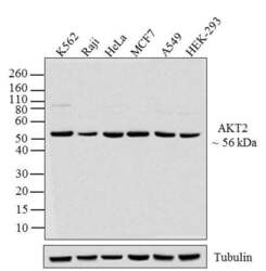
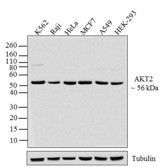
- Western blot analysis of AKT2 was performed by loading 20 µg of K562 (lane1), Raji (lane2), HeLa (lane3), MCF7 (lane4), A549 (lane5) and HEK-293 (lane6) cell lysates using Novex® NuPAGE® 4-12 % Bis-Tris gel (Product # NP0321BOX), XCell SureLock Electrophoresis System (Product # EI0002), Novex® Sharp Pre-Stained Protein Standard (LC5800), and iBlot® Dry Blotting System (IB21001). Proteins were transferred to a nitrocellulose membrane and blocked with 5 % skim milk for 1 hour at room temperature. AKT2 was detected at ~60 kDa using AKT2 Mouse Monoclonal Antibody (Product # 39-3900) at 0.5-1 µg/mL in 2.5 % skim milk at 4°C overnight on a rocking platform. Goat Anti-Mouse IgG - HRP Secondary Antibody (Product # 62-6520) at 1:4000 dilution was used and chemiluminescent detection was performed using Pierce™ ECL Western Blotting Substrate (Product # 32106).
Supportive validation
- Submitted by
- Invitrogen Antibodies (provider)
- Main image
- Experimental details
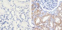
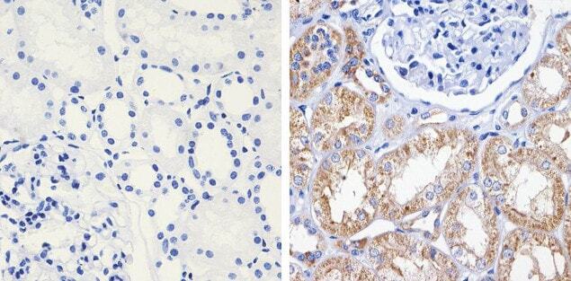
- Immunohistochemistry analysis of AKT2 showing staining in the cytoplasm of paraffin-embedded human kidney tissue (right) compared to a negative control without primary antibody (left). To expose target proteins, antigen retrieval was performed using 10 mM sodium citrate (pH 6.0), microwaved for 8-15 min. Following antigen retrieval, tissues were blocked in 3% H2O2-methanol for 15 min at room temperature, washed with ddH2O and PBS, and then probed with AKT2 monoclonal antibody (Product # 39-3900) diluted in 3% BSA-PBS at a dilution of 1:200 overnight at 4°C in a humidified chamber. Tissues were washed extensively in PBST and detection was performed using a HRP-conjugated secondary antibody followed by colorimetric detection using a DAB kit. Tissues were counterstained with hematoxylin and dehydrated with ethanol and xylene to prep for mounting.
Supportive validation
- Submitted by
- Invitrogen Antibodies (provider)
- Main image
- Experimental details
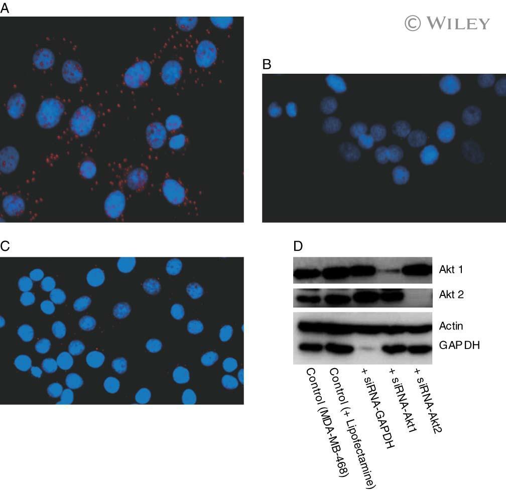
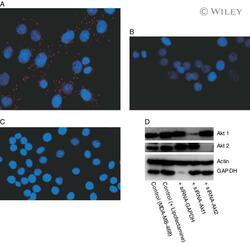
- 1 pAkt:Akt1 and pAkt:Akt2 activation in MDA-MB-468 cells as visualized by the PLA technique. Control MDA-MB-468 cells (A) and MDA-MB-468 cells after treatment with Akt1 siRNA (B) or Akt2 siRNA (C). Western blot analysis of proteins extracted from MDA-MB-468 after transfection with Akt1 siRNA and Akt2 siRNA (D); control cells were untreated, or with transfection reagents only or with siRNA targeting GAPDH; actin was used as a loading control