Explore
 Validate
 Learn
Learn Western blot
 ELISA
ELISAAntibody data
- Antibody Data
- Antigen structure
- References [0]
- Comments [0]
- Validations
- Western blot [4]
- Immunocytochemistry [1]
- Immunohistochemistry [1]
Submit
Validation data
Reference
Comment
Report error
- Product number
- 100-401-401S - Provider product page

- Provider
- Invitrogen Antibodies
- Product name
- AKT Polyclonal Antibody
- Antibody type
- Polyclonal
- Antigen
- Synthetic peptide
- Reactivity
- Human, Mouse, Rat, Chicken/Avian
- Host
- Rabbit
- Isotype
- IgG
- Vial size
- 25 µL
- Concentration
- 85 mg/mL
- Storage
- -20° C, Avoid Freeze/Thaw Cycles
No comments: Submit comment
Supportive validation
- Submitted by
- Invitrogen Antibodies (provider)
- Main image

- Experimental details
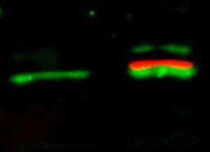
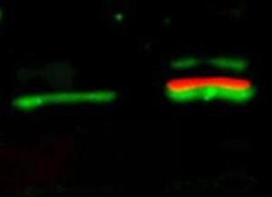
- Western Blot of simultaneous detection of unphosphorylated and phosphorylated Rabbit Anti-AKT antibody. Lane 1: unstimulated NIH/3T3 lysates contain inactive unphosphorylated Akt1, green band. Lane 2: PDGF stimulated NIH/3T3 lysate contains both inactive (green band) and activated phosphorylated Akt1 (red band). Load: 35 µg per lane. Primary antibody: rabbit anti-Akt (pan) and mouse anti-Akt pS473 specific antibodies at 1:1000 for overnight at 4°C. Secondary antibody: DyLight™ 549 conjugated anti-rabbit IgG (green) and DyLight™ 649 conjugated anti-mouse IgG (red) secondary antibodies at 1:10,000 for 45 min at RT. Block: 5% BLOTTO overnight at 4°C.
- Submitted by
- Invitrogen Antibodies (provider)
- Main image
- Experimental details
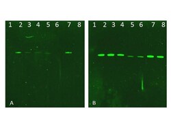
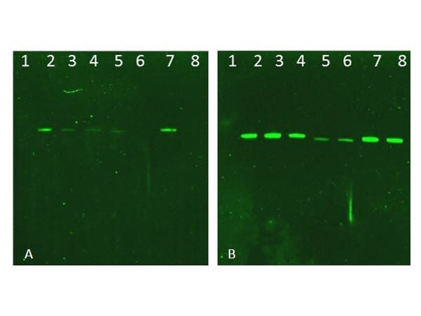
- Western Blot of Rabbit AKT Antibody. Lane 1: NIR MW protein ladder. Lane 2: AKT1, recombinant. Lane 3: AKT1, phosphatase-treated. Lane 4: AKT1, mutant T308A/S473A. Lane 5: AKT2, recombinant. Lane 6: AKT2, phosphatase-treated. Lane 7: AKT3, recombinant. Lane 8: AKT3, phosphatase-treated. Load: 50ng per lane. Blot A: 600-401-269 Anti-Akt pT308 used at 1:2270, Blot B: 100-401-401 Anti-Akt used 1:1000.
- Submitted by
- Invitrogen Antibodies (provider)
- Main image

- Experimental details
- Western Blot of simultaneous detection of unphosphorylated and phosphorylated Rabbit Anti-AKT antibody. Lane 1: unstimulated NIH/3T3 lysates contain inactive unphosphorylated Akt1, green band. Lane 2: PDGF stimulated NIH/3T3 lysate contains both inactive (green band) and activated phosphorylated Akt1 (red band). Load: 35 µg per lane. Primary antibody: rabbit anti-Akt (pan) and mouse anti-Akt pS473 specific antibodies at 1:1000 for overnight at 4°C. Secondary antibody: DyLight™ 549 conjugated anti-rabbit IgG (green) and DyLight™ 649 conjugated anti-mouse IgG (red) secondary antibodies at 1:10,000 for 45 min at RT. Block: 5% BLOTTO overnight at 4°C.
- Submitted by
- Invitrogen Antibodies (provider)
- Main image
- Experimental details
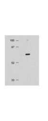
- Western Blot of Rabbit Anti-AKT antibody. Lane 1: Molecular Weight. Lane 2: NIH/3T3 whole cell lysate. Load: 20 µg lysate per lane. Primary antibody: Anti-AKT antibody at 1:500 for overnight at 4°C. Secondary antibody: HRP conjugated GT-a-Rabbit IgG (611-103-122) at 1:10,000 preceded color development using Pierce Chemicals SuperSignal™ substrate. Block: MOPS buffer overnight at 4°C. Predicted/Observed size: 56 kDa, 56 kDa for AKT. Other band(s): none.

Supportive validation
- Submitted by
- Invitrogen Antibodies (provider)
- Main image
- Experimental details
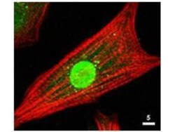
- Immunofluorescence Microscopy of Rabbit Anti-AKT Antibody. Tissue: neonatal rat cardiomyocytes. Fixation: 0.5% PFA. Antigen retrieval: not required. Primary antibody: AKT antibody at 1:80 dilution for 1 h at RT. Secondary antibody: Texas-red™ conjugated rabbit secondary antibody at 1:10,000 for 45 min at RT. Localization: AKT is nuclear. Staining: Anti-AKT staining appears green. Actin filaments are labeled red using a Texas-red™ conjugated phalloidin.

Supportive validation
- Submitted by
- Invitrogen Antibodies (provider)
- Main image
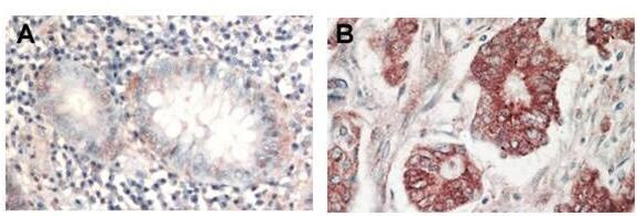
- Experimental details
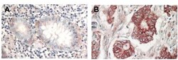
- Immunohistochemistry of Rabbit Anti-AKT antibody. Tissue: (A) normal colon tissue, (B) colon tumor tissue. Fixation: formalin fixed paraffin embedded. Antigen retrieval: not required. Primary antibody: AKT antibody at 1:1,000 dilution for 1 h at RT. Secondary antibody: Peroxidase rabbit secondary antibody at 1:10,000 for 45 min at RT. Localization: AKT is nuclear. Staining: AKT as precipitated red signal with hematoxylin purple nuclear counterstain.