 Explore
 Validate
 Learn
Learn60203-2-IG
antibody from Invitrogen Antibodies
Targeting: AKT2
PKBβ
 Western blot Immunocytochemistry
 Immunoprecipitation Immunohistochemistry Flow cytometry Other assay
Antibody data
- Antibody Data
- Antigen structure
- References [0]
- Comments [0]
- Validations
- Western blot [9]
- Immunocytochemistry [1]
- Immunohistochemistry [8]
- Flow cytometry [1]
- Other assay [1]
Submit
Validation data
Reference
Comment
Report error
- Product number
- 60203-2-IG - Provider product page

- Provider
- Invitrogen Antibodies
- Product name
- AKT Monoclonal Antibody (2C5D1)
- Antibody type
- Monoclonal
- Antigen
- Other
- Reactivity
- Human, Mouse, Rat
- Host
- Mouse
- Isotype
- IgG
- Antibody clone number
- 2C5D1
- Vial size
- 150 µL
- Concentration
- 1 mg/mL
- Storage
- -20°C
No comments: Submit comment
Supportive validation
- Submitted by
- Invitrogen Antibodies (provider)
- Main image

- Experimental details
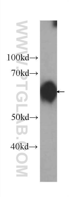
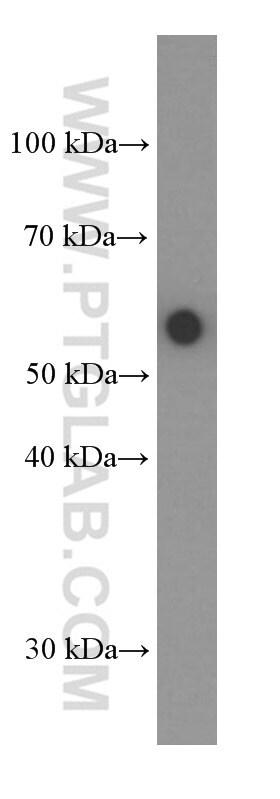
- Rat liver tissue were subjected to SDS PAGE followed by western blot with 60203-2-Ig (AKT Antibody) at dilution of 1:500 incubated at room temperature for 1.5 hours.
- Submitted by
- Invitrogen Antibodies (provider)
- Main image
- Experimental details
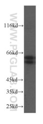
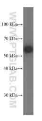
- HeLa cells were subjected to SDS PAGE followed by western blot with 60203-2-Ig (AKT antibody) at dilution of 1:500 incubated at room temperature for 1.5 hours.

- Submitted by
- Invitrogen Antibodies (provider)
- Main image
- Experimental details
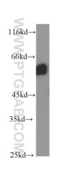
- HeLa cells were subjected to SDS PAGE followed by western blot with 60203-2-Ig (AKT antibody) at dilution of 1:1000 incubated at room temperature for 1.5 hours.

- Submitted by
- Invitrogen Antibodies (provider)
- Main image

- Experimental details
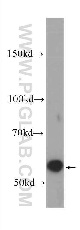
- Mouse brain tissue were subjected to SDS PAGE followed by western blot with 60203-2-Ig (AKT Antibody) at dilution of 1:1000 incubated at room temperature for 1.5 hours.
- Submitted by
- Invitrogen Antibodies (provider)
- Main image

- Experimental details
- NIH/3T3 cells were subjected to SDS PAGE followed by western blot with 60203-2-Ig (AKT Antibody) at dilution of 1:2000 incubated at room temperature for 1.5 hours.
- Submitted by
- Invitrogen Antibodies (provider)
- Main image
- Experimental details
- ROS1728 cells were subjected to SDS PAGE followed by western blot with 60203-2-Ig (AKT Antibody) at dilution of 1:1000 incubated at room temperature for 1.5 hours.

- Submitted by
- Invitrogen Antibodies (provider)
- Main image
- Experimental details
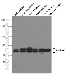
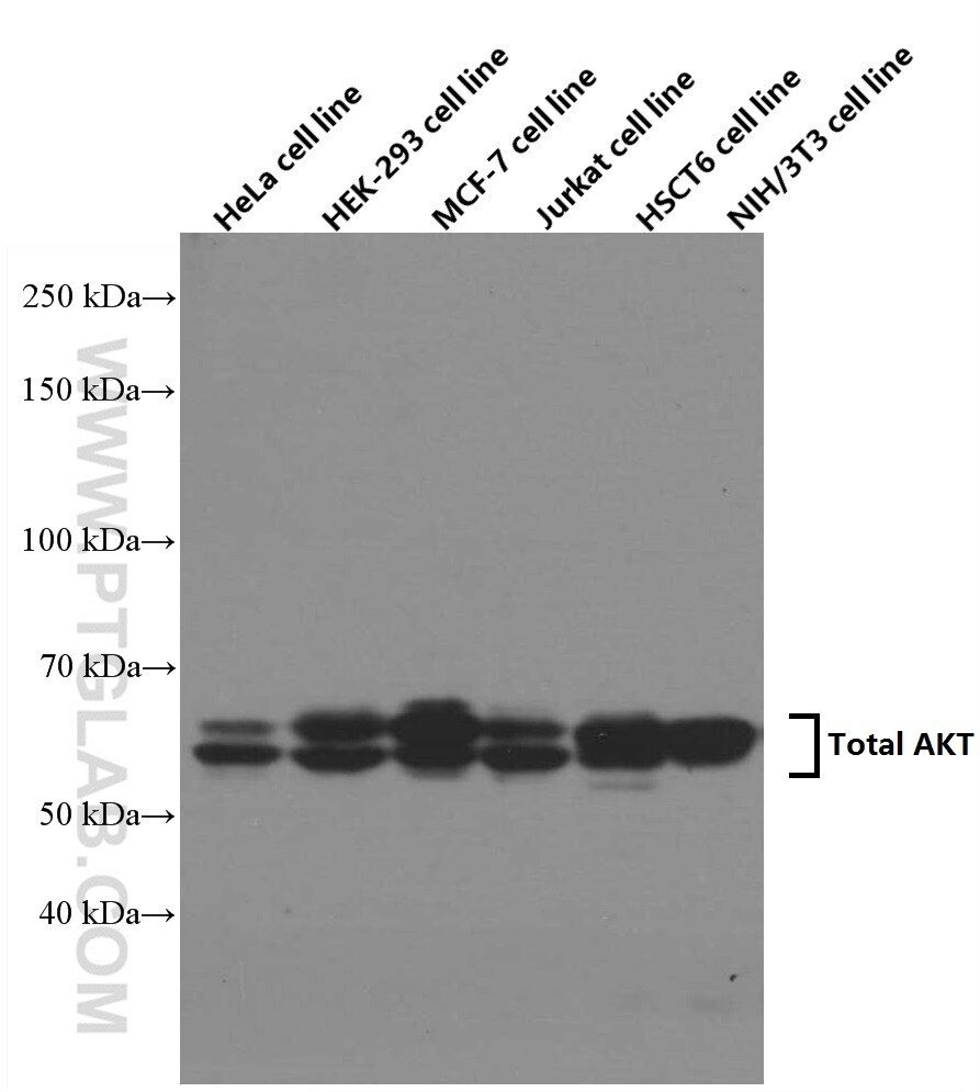
- Various lysates were subjected to SDS PAGE followed by western blot with 60203-2-IG (AKT antibody) at dilution of 1:5000 incubated at room temperature for 1.5 hours.
- Submitted by
- Invitrogen Antibodies (provider)
- Main image
- Experimental details
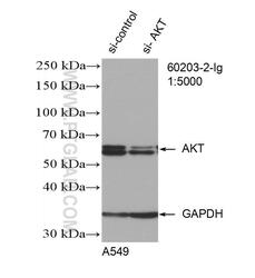
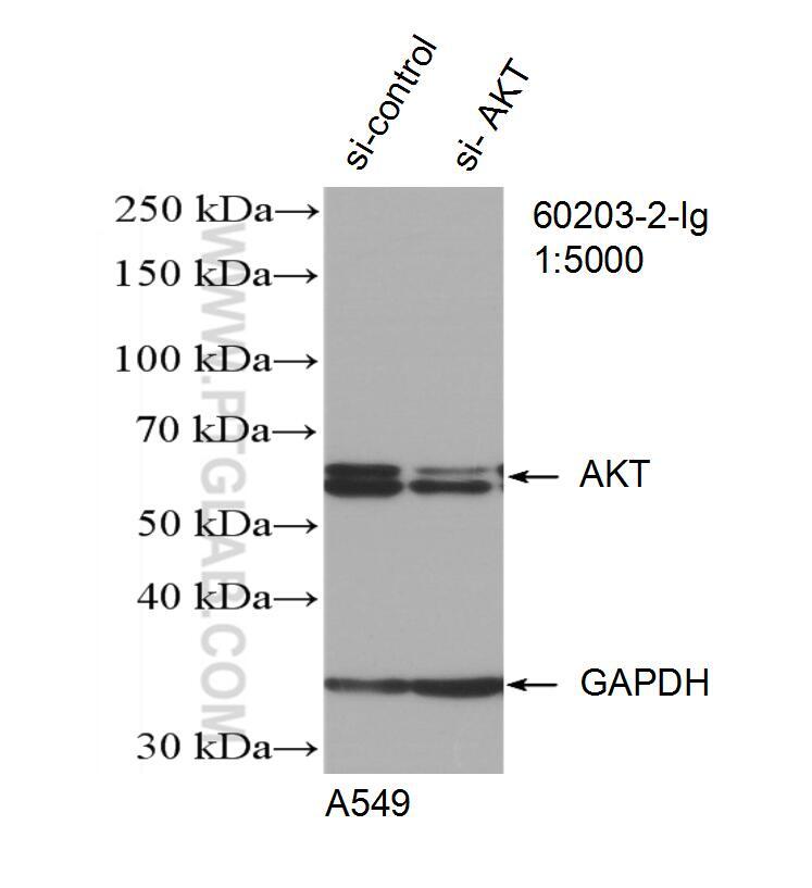
- WB result of AKT antibody (60203-2-IG; 1:5000; incubated at room temperature for 1.5 hours) with sh-Control and sh-AKT transfected A549 cells.
- Submitted by
- Invitrogen Antibodies (provider)
- Main image
- Experimental details
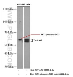
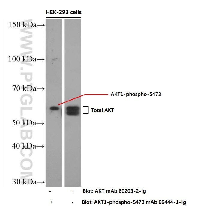
- HEK-293 cells were subjected to SDS PAGE followed by western blot with 60203-2-IG ( AKT Antibody) and 66444-1-IG (AKT1-phospho-S473 Antibody) at dilution of 1:4000 incubated at room temperature for 1.5 hours.
Supportive validation
- Submitted by
- Invitrogen Antibodies (provider)
- Main image

- Experimental details
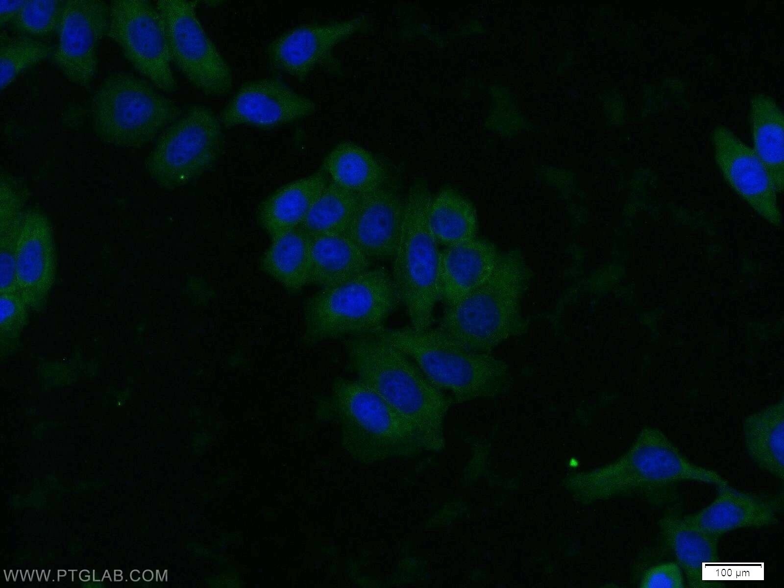
- Immunofluorescent analysis of HeLa cells using 60203-2-IG (AKT antibody) at dilution of 1:25 and Alexa Fluor 488-conjugated AffiniPure Goat Anti-Mouse IGG (H+L).
Supportive validation
- Submitted by
- Invitrogen Antibodies (provider)
- Main image
- Experimental details
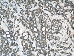
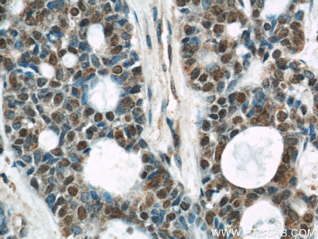
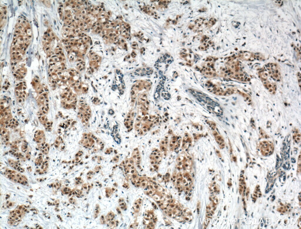
- Immunohistochemistry of paraffin-embedded human cervical cancer using 60203-2-Ig (AKT antibody) at dilution of 1:50 (under 10x lens).
- Submitted by
- Invitrogen Antibodies (provider)
- Main image
- Experimental details
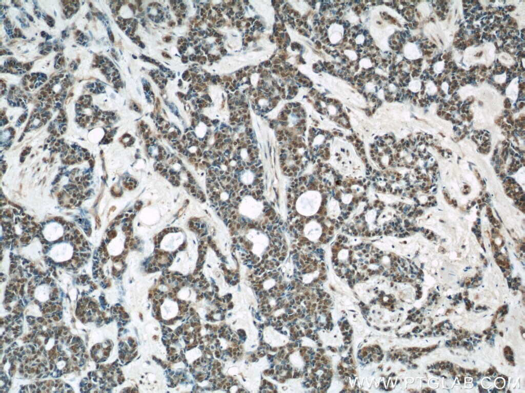
- Immunohistochemistry of paraffin-embedded human cervical cancer using 60203-2-Ig (AKT antibody) at dilution of 1:50 (under 40x lens).
- Submitted by
- Invitrogen Antibodies (provider)
- Main image
- Experimental details
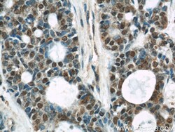
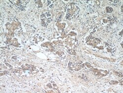
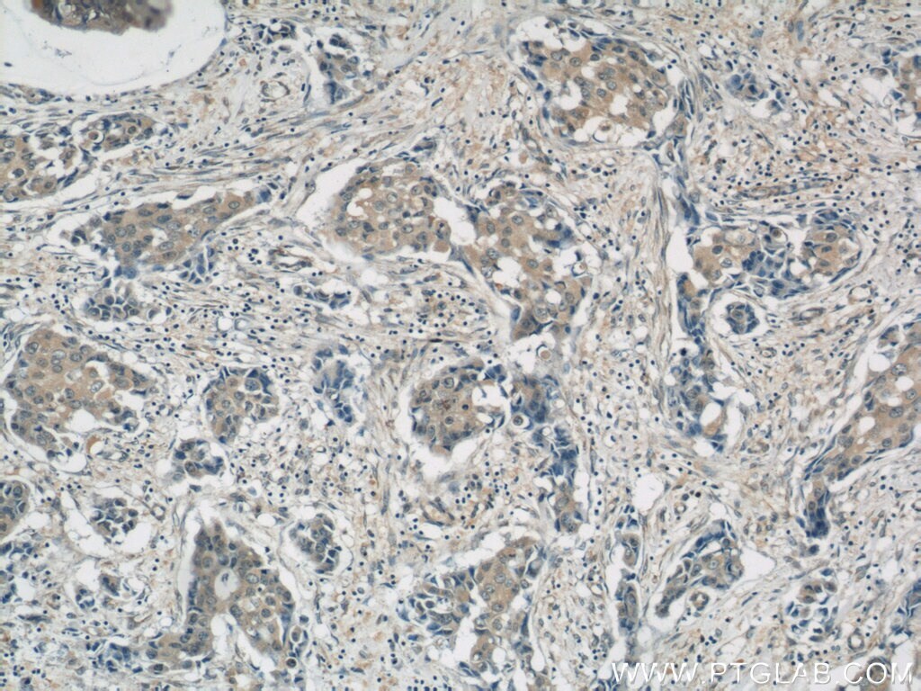
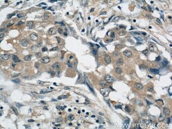
- Immunohistochemistry of paraffin-embedded human breast cancer using 60203-2-Ig (AKT antibody) at dilution of 1:50 (under 10x lens).

- Submitted by
- Invitrogen Antibodies (provider)
- Main image
- Experimental details
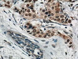
- Immunohistochemistry of paraffin-embedded human breast cancer using 60203-2-Ig (AKT antibody) at dilution of 1:50 (under 40x lens).
- Submitted by
- Invitrogen Antibodies (provider)
- Main image

- Experimental details
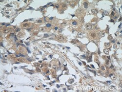
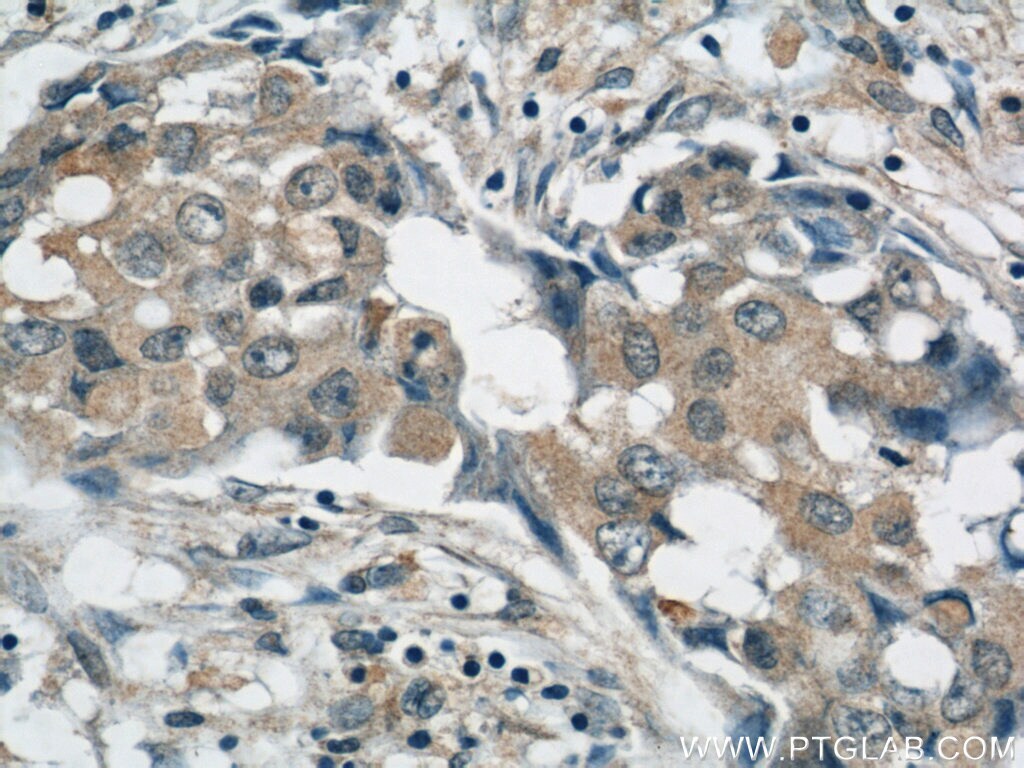
- Immunohistochemistry of paraffin-embedded human breast cancer using 60203-2-Ig (AKT antibody) at dilution of 1:50 (under 10x lens).
- Submitted by
- Invitrogen Antibodies (provider)
- Main image
- Experimental details
- Immunohistochemistry of paraffin-embedded human breast cancer using 60203-2-Ig (AKT antibody) at dilution of 1:50 (under 40x lens).
- Submitted by
- Invitrogen Antibodies (provider)
- Main image
- Experimental details
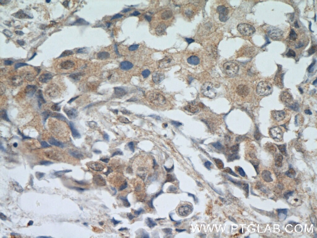
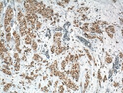
- Immunohistochemistry of paraffin-embedded human breast cancer tissue slide using 60203-2-IG ( AKT Antibody) at dilution of 1:200 (under 10x lens). heat mediated antigen retrieved with Tris-EDTA buffer (pH 9).
- Submitted by
- Invitrogen Antibodies (provider)
- Main image
- Experimental details
- Immunohistochemistry of paraffin-embedded human breast cancer tissue slide using 60203-2-IG ( AKT Antibody) at dilution of 1:200 (under 40x lens). heat mediated antigen retrieved with Tris-EDTA buffer (pH 9).

Supportive validation
- Submitted by
- Invitrogen Antibodies (provider)
- Main image

- Experimental details
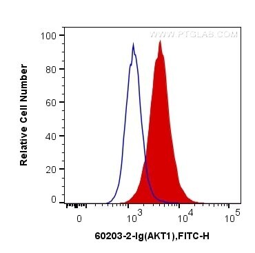
- 1X10^6 Jurkat cells were intracellularly stained with 0.2 µg Anti-Human AKT (Product # 60203-2-IG, Clone:2C5D1) and CoraLite®488-Conjugated AffiniPure Goat Anti-Mouse IgG(H+L) at dilution 1:1,000 (red), or 0.2 µg Mouse IgG1 Isotype Control (66360-1-IG, Clone: T1F8D3F10) (blue). Cells were fixed with 4% PFA and permeabilized with Flow Cytometry Perm Buffer (PF00011-C).
Supportive validation
- Submitted by
- Invitrogen Antibodies (provider)
- Main image
- Experimental details
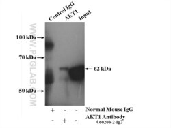
- IP result of anti-AKT (IP:60203-2-IG, 5ug; Detection:60203-2-IG 1:1000) with mouse brain tissue lysate 4000ug.
