Explore
 Validate
 Learn
Learn Western blot
 Immunocytochemistry
Antibody data
- Antibody Data
- Antigen structure
- References [18]
- Comments [0]
- Validations
- Immunocytochemistry [3]
- Immunohistochemistry [1]
- Flow cytometry [3]
Submit
Validation data
Reference
Comment
Report error
- Product number
- MA5-14000 - Provider product page

- Provider
- Invitrogen Antibodies
- Product name
- Anti-Bax Monoclonal Antibody (6A7), Biotin
- Antibody type
- Monoclonal
- Antigen
- Synthetic peptide
- Description
- MA5-14003 targets Bax in Western blot, FACS, immunohistochemistry (paraffin) and ICC/IF applications and shows reactivity with Human, mouse, and Rat samples. This antibody is not recommended for Jurkat cells in Western blot applications. The MA5-14003 immunogen is a Synthetic peptide, aa 12-24 (Cys-GPTSSEQIMKTGA), of human Bax protein. This amino acid sequence is shared by human, mouse and rat Bax protein.
- Reactivity
- Human, Mouse, Rat
- Host
- Mouse
- Conjugate
- Biotin
- Isotype
- IgG
- Antibody clone number
- 6A7
- Vial size
- 500 µL
- Concentration
- 0.2 mg/ml
- Storage
- 4° C
Submitted references TNF-α induces vascular endothelial cells apoptosis through overexpressing pregnancy induced noncoding RNA in Kawasaki disease model.
Phosphorylated nerve growth factor-induced clone B (NGFI-B) translocates from the nucleus to mitochondria of stressed rat cardiomyocytes and induces apoptosis.
Protective effects of trimetazidine on bone marrow mesenchymal stem cells viability in an ex vivo model of hypoxia and in vivo model of locally myocardial ischemia.
Prenatal stress induces long-term effects in cell turnover in the hippocampus-hypothalamus-pituitary axis in adult male rats.
Combined treatment with the Cox-2 inhibitor niflumic acid and PPARγ ligand ciglitazone induces ER stress/caspase-8-mediated apoptosis in human lung cancer cells.
Sequential caspase-2 and caspase-8 activation is essential for saikosaponin a-induced apoptosis of human colon carcinoma cell lines.
Cell cycle arrest and cytochrome c-mediated apoptotic induction by MCS-5A is associated with up-regulation of p16(INK4a) in HL-60 cells.
Heme oxygenase-1 prevents hyperthyroidism induced hepatic damage via an antioxidant and antiapoptotic pathway.
Notch-activated signaling cascade interacts with mitochondrial remodeling proteins to regulate cell survival.
Growth hormone-releasing peptide 6 protection of hypothalamic neurons from glutamate excitotoxicity is caspase independent and not mediated by insulin-like growth factor I.
N,N-dimethyl phytosphingosine induces caspase-8-dependent cytochrome c release and apoptosis through ROS generation in human leukemia cells.
Death of hypothalamic astrocytes in poorly controlled diabetic rats is associated with nuclear translocation of apoptosis inducing factor.
Desferrioxamine (DFX) induces apoptosis through the p38-caspase8-Bid-Bax pathway in PHA-stimulated human lymphocytes.
Preventive cardioprotection of erythropoietin against doxorubicin-induced cardiomyopathy.
Influence of glutathione on the induction of chromosome aberrations, delay in cell cycle kinetics and cell cycle regulator proteins in irradiated mouse bone marrow cells.
The N-terminus and alpha-5, alpha-6 helices of the pro-apoptotic protein Bax, modulate functional interactions with the anti-apoptotic protein Bcl-xL.
Iptakalim protects PC12 cell against H2O2-induced oxidative injury via opening mitochondrial ATP-sensitive potassium channel.
(+/-)-huprine Y, (-)-huperzine A and tacrine do not show neuroprotective properties in an apoptotic model of neuronal cytoskeletal alteration.
Jiang C, Fang X, Jiang Y, Shen F, Hu Z, Li X, Huang X
The international journal of biochemistry & cell biology 2016 Mar;72:118-124
The international journal of biochemistry & cell biology 2016 Mar;72:118-124
Phosphorylated nerve growth factor-induced clone B (NGFI-B) translocates from the nucleus to mitochondria of stressed rat cardiomyocytes and induces apoptosis.
Xinxing W, Hong F, Rui Z, Yun Z, Jingbo G, Lingjia Q
Stress (Amsterdam, Netherlands) 2012 Sep;15(5):545-53
Stress (Amsterdam, Netherlands) 2012 Sep;15(5):545-53
Protective effects of trimetazidine on bone marrow mesenchymal stem cells viability in an ex vivo model of hypoxia and in vivo model of locally myocardial ischemia.
Xu H, Zhu G, Tian Y
Journal of Huazhong University of Science and Technology. Medical sciences = Hua zhong ke ji da xue xue bao. Yi xue Ying De wen ban = Huazhong keji daxue xuebao. Yixue Yingdewen ban 2012 Feb;32(1):36-41
Journal of Huazhong University of Science and Technology. Medical sciences = Hua zhong ke ji da xue xue bao. Yi xue Ying De wen ban = Huazhong keji daxue xuebao. Yixue Yingdewen ban 2012 Feb;32(1):36-41
Prenatal stress induces long-term effects in cell turnover in the hippocampus-hypothalamus-pituitary axis in adult male rats.
Baquedano E, García-Cáceres C, Diz-Chaves Y, Lagunas N, Calmarza-Font I, Azcoitia I, Garcia-Segura LM, Argente J, Chowen JA, Frago LM
PloS one 2011;6(11):e27549
PloS one 2011;6(11):e27549
Combined treatment with the Cox-2 inhibitor niflumic acid and PPARγ ligand ciglitazone induces ER stress/caspase-8-mediated apoptosis in human lung cancer cells.
Kim BM, Maeng K, Lee KH, Hong SH
Cancer letters 2011 Jan 28;300(2):134-44
Cancer letters 2011 Jan 28;300(2):134-44
Sequential caspase-2 and caspase-8 activation is essential for saikosaponin a-induced apoptosis of human colon carcinoma cell lines.
Kim BM, Hong SH
Apoptosis : an international journal on programmed cell death 2011 Feb;16(2):184-97
Apoptosis : an international journal on programmed cell death 2011 Feb;16(2):184-97
Cell cycle arrest and cytochrome c-mediated apoptotic induction by MCS-5A is associated with up-regulation of p16(INK4a) in HL-60 cells.
Choi BY, Lee CH
Bioorganic & medicinal chemistry letters 2010 Jul 1;20(13):3880-4
Bioorganic & medicinal chemistry letters 2010 Jul 1;20(13):3880-4
Heme oxygenase-1 prevents hyperthyroidism induced hepatic damage via an antioxidant and antiapoptotic pathway.
Giriş M, Erbil Y, Depboylu B, Mete O, Türkoğlu U, Abbasoğlu SD, Uysal M
The Journal of surgical research 2010 Dec;164(2):266-75
The Journal of surgical research 2010 Dec;164(2):266-75
Notch-activated signaling cascade interacts with mitochondrial remodeling proteins to regulate cell survival.
Perumalsamy LR, Nagala M, Sarin A
Proceedings of the National Academy of Sciences of the United States of America 2010 Apr 13;107(15):6882-7
Proceedings of the National Academy of Sciences of the United States of America 2010 Apr 13;107(15):6882-7
Growth hormone-releasing peptide 6 protection of hypothalamic neurons from glutamate excitotoxicity is caspase independent and not mediated by insulin-like growth factor I.
Delgado-Rubín A, Chowen JA, Argente J, Frago LM
The European journal of neuroscience 2009 Jun;29(11):2115-24
The European journal of neuroscience 2009 Jun;29(11):2115-24
N,N-dimethyl phytosphingosine induces caspase-8-dependent cytochrome c release and apoptosis through ROS generation in human leukemia cells.
Kim BM, Choi YJ, Han Y, Yun YS, Hong SH
Toxicology and applied pharmacology 2009 Aug 15;239(1):87-97
Toxicology and applied pharmacology 2009 Aug 15;239(1):87-97
Death of hypothalamic astrocytes in poorly controlled diabetic rats is associated with nuclear translocation of apoptosis inducing factor.
García-Cáceres C, Lechuga-Sancho A, Argente J, Frago LM, Chowen JA
Journal of neuroendocrinology 2008 Dec;20(12):1348-60
Journal of neuroendocrinology 2008 Dec;20(12):1348-60
Desferrioxamine (DFX) induces apoptosis through the p38-caspase8-Bid-Bax pathway in PHA-stimulated human lymphocytes.
Kim BM, Chung HW
Toxicology and applied pharmacology 2008 Apr 1;228(1):24-31
Toxicology and applied pharmacology 2008 Apr 1;228(1):24-31
Preventive cardioprotection of erythropoietin against doxorubicin-induced cardiomyopathy.
Chen X, Chen Y, Bi Y, Fu N, Shan C, Wang S, Aslam S, Wang PW, Xu J
Cardiovascular drugs and therapy 2007 Oct;21(5):367-74
Cardiovascular drugs and therapy 2007 Oct;21(5):367-74
Influence of glutathione on the induction of chromosome aberrations, delay in cell cycle kinetics and cell cycle regulator proteins in irradiated mouse bone marrow cells.
Ray S, Chatterjee A
International journal of radiation biology 2007 May;83(5):347-54
International journal of radiation biology 2007 May;83(5):347-54
The N-terminus and alpha-5, alpha-6 helices of the pro-apoptotic protein Bax, modulate functional interactions with the anti-apoptotic protein Bcl-xL.
Parikh N, Koshy C, Dhayabaran V, Perumalsamy LR, Sowdhamini R, Sarin A
BMC cell biology 2007 May 23;8:16
BMC cell biology 2007 May 23;8:16
Iptakalim protects PC12 cell against H2O2-induced oxidative injury via opening mitochondrial ATP-sensitive potassium channel.
Chai Y, Niu L, Sun XL, Ding JH, Hu G
Biochemical and biophysical research communications 2006 Nov 17;350(2):307-14
Biochemical and biophysical research communications 2006 Nov 17;350(2):307-14
(+/-)-huprine Y, (-)-huperzine A and tacrine do not show neuroprotective properties in an apoptotic model of neuronal cytoskeletal alteration.
Jordá EG, Verdaguer E, Jiménez A, Canudas AM, Rimbau V, Camps P, Muñoz-Torrero D, Camins A, Pallàs M
Journal of Alzheimer's disease : JAD 2004 Dec;6(6):577-83; discussion 673-81
Journal of Alzheimer's disease : JAD 2004 Dec;6(6):577-83; discussion 673-81
No comments: Submit comment
Supportive validation
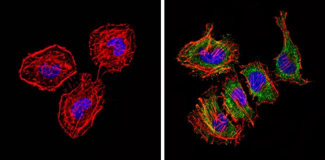
- Submitted by
- Invitrogen Antibodies (provider)
- Main image
- Experimental details
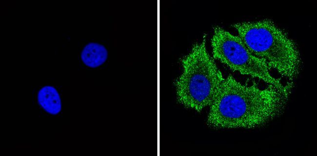
- Immunofluorescent analysis of Bax (green) showing staining in the cytoplasm of Hela cells (right) compared to a negative control without primary antibody (left). Formalin-fixed cells were permeabilized with 0.1% Triton X-100 in TBS for 5-10 minutes and blocked with 3% BSA-PBS for 30 minutes at room temperature. Cells were probed with a Bax monoclonal antibody (Product # MA5-14003) in 3% BSA-PBS at a dilution of 1:50 and incubated overnight at 4 ºC in a humidified chamber. Cells were washed with PBST and incubated with a DyLight-conjugated secondary antibody in PBS at room temperature in the dark. F-actin (red) was stained with a flourescent red phalloidin and nuclei (blue) were stained with Hoechst or DAPI. Images were taken at a magnification of 60x.
- Conjugate
- Biotin
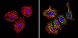
- Submitted by
- Invitrogen Antibodies (provider)
- Main image
- Experimental details
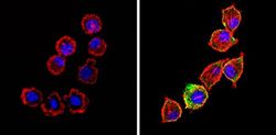
- Immunofluorescent analysis of Bax (green) showing staining in the cytoplasm of MCF-7 cells (right) compared to a negative control without primary antibody (left). Formalin-fixed cells were permeabilized with 0.1% Triton X-100 in TBS for 5-10 minutes and blocked with 3% BSA-PBS for 30 minutes at room temperature. Cells were probed with a Bax monoclonal antibody (Product # MA5-14003) in 3% BSA-PBS at a dilution of 1:50 and incubated overnight at 4 ºC in a humidified chamber. Cells were washed with PBST and incubated with a DyLight-conjugated secondary antibody in PBS at room temperature in the dark. F-actin (red) was stained with a flourescent red phalloidin and nuclei (blue) were stained with Hoechst or DAPI. Images were taken at a magnification of 60x.
- Conjugate
- Biotin
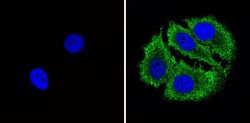
- Submitted by
- Invitrogen Antibodies (provider)
- Main image
- Experimental details
- Immunofluorescent analysis of Bax (green) showing staining in the cytoplasm of L929 cells (right) compared to a negative control without primary antibody (left). Formalin-fixed cells were permeabilized with 0.1% Triton X-100 in TBS for 5-10 minutes and blocked with 3% BSA-PBS for 30 minutes at room temperature. Cells were probed with a Bax monoclonal antibody (Product # MA5-14003) in 3% BSA-PBS at a dilution of 1:50 and incubated overnight at 4 ºC in a humidified chamber. Cells were washed with PBST and incubated with a DyLight-conjugated secondary antibody in PBS at room temperature in the dark. F-actin (red) was stained with a flourescent red phalloidin and nuclei (blue) were stained with Hoechst or DAPI. Images were taken at a magnification of 60x.
- Conjugate
- Biotin

Supportive validation
- Submitted by
- Invitrogen Antibodies (provider)
- Main image

- Experimental details
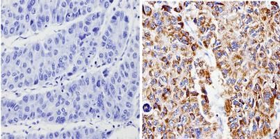
- Immunohistochemistry analysis of Bax showing positive staining in the cytoplasm of paraffin-treated Human hepatocarcinoma (right) compared with a negative control in the absence of primary antibody (left). To expose target proteins, antigen retrieval method was performed using 10mM sodium citrate (pH 6.0) microwaved for 8-15 min. Following antigen retrieval, tissues were blocked in 3% H2O2-methanol for 15 min at room temperature, washed with ddH2O and PBS, and then probed with a Bax monoclonal antibody (Product # MA5-14003) diluted by 3% BSA-PBS at a dilution of 1:100 overnight at 4°C in a humidified chamber. Tissues were washed extensively PBST and detection was performed using an HRP-conjugated secondary antibody followed by colorimetric detection using a DAB kit. Tissues were counterstained with hematoxylin and dehydrated with ethanol and xylene to prep for mounting.
- Conjugate
- Biotin
Supportive validation
- Submitted by
- Invitrogen Antibodies (provider)
- Main image

- Experimental details
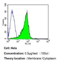
- Flow cytometry analysis of Bax in Hela cells compared to an isotype control (blue). Cells were harvested, adjusted to a concentration of 1-5x10^6 cells/ml, fixed with 2% paraformaldehyde, washed with PBS, and incubated with Bax monoclonal antibody (Product # MA5-14003) at a dilution of 0.5 ug/test for 60 min at room temperature. Cells were then blocked in a solution of 2% BSA-PBS for 30 min at room temperature, incubated for 40 min at room temperature in the dark using a Dylight 488-conjugated goat anti-mouse IgG (H+L) secondary antibody, and re-suspended in PBS for FACS analysis.
- Conjugate
- Biotin
- Submitted by
- Invitrogen Antibodies (provider)
- Main image

- Experimental details
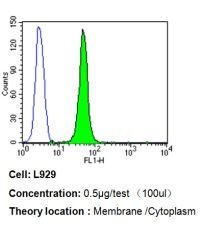
- Flow cytometry analysis of Bax in L929 cells compared to an isotype control (blue). Cells were harvested, adjusted to a concentration of 1-5x10^6 cells/ml, fixed with 2% paraformaldehyde, washed with PBS, and incubated with Bax monoclonal antibody (Product # MA5-14003) at a dilution of 0.5 ug/test for 60 min at room temperature. Cells were then blocked in a solution of 2% BSA-PBS for 30 min at room temperature, incubated for 40 min at room temperature in the dark using a Dylight 488-conjugated goat anti-mouse IgG (H+L) secondary antibody, and re-suspended in PBS for FACS analysis.
- Conjugate
- Biotin
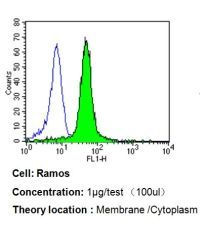
- Submitted by
- Invitrogen Antibodies (provider)
- Main image

- Experimental details
- Flow cytometry analysis of Bax in Ramos cells compared to an isotype control (blue). Cells were harvested, adjusted to a concentration of 1-5x10^6 cells/ml, fixed with 2% paraformaldehyde, washed with PBS, and incubated with Bax monoclonal antibody (Product # MA5-14003) at a dilution of 1 ug/test for 60 min at room temperature. Cells were then blocked in a solution of 2% BSA-PBS for 30 min at room temperature, incubated for 40 min at room temperature in the dark using a Dylight 488-conjugated goat anti-mouse IgG (H+L) secondary antibody, and re-suspended in PBS for FACS analysis.
- Conjugate
- Biotin