Explore
 Validate
 Learn
Learn Western blot
 Immunocytochemistry
Antibody data
- Antibody Data
- Antigen structure
- References [51]
- Comments [0]
- Validations
- Immunocytochemistry [6]
- Immunohistochemistry [1]
- Flow cytometry [6]
- Other assay [18]
Submit
Validation data
Reference
Comment
Report error
- Product number
- MA5-14003 - Provider product page

- Provider
- Invitrogen Antibodies
- Product name
- Bax Monoclonal Antibody (6A7)
- Antibody type
- Monoclonal
- Antigen
- Synthetic peptide
- Description
- MA5-14003 targets Bax in Western blot, FACS, immunohistochemistry (paraffin) and ICC/IF applications and shows reactivity with Human, mouse, and Rat samples. This antibody is not recommended for Jurkat cells in Western blot applications. The MA5-14003 immunogen is a Synthetic peptide, aa 12-24 (Cys-GPTSSEQIMKTGA), of human Bax protein. This amino acid sequence is shared by human, mouse and rat Bax protein.
- Reactivity
- Human, Mouse, Rat
- Host
- Mouse
- Isotype
- IgG
- Antibody clone number
- 6A7
- Vial size
- 500 μL
- Concentration
- 0.2 mg/mL
- Storage
- 4°C
Submitted references Preventive Effect of Limosilactobacillus fermentum SCHY34 on Lead Acetate-Induced Neurological Damage in SD Rats.
Size-Dependent Cytoprotective Effects of Selenium Nanoparticles during Oxygen-Glucose Deprivation in Brain Cortical Cells.
Infarct-preconditioning exosomes of umbilical cord mesenchymal stem cells promoted vascular remodeling and neurological recovery after stroke in rats.
AntiGan: An Epinutraceutical Bioproduct with Antitumor Properties in Cultured Cell Lines.
Astragaloside IV protects against ischemia/reperfusion (I/R)-induced kidney injury based on the Keap1-Nrf2/ARE signaling pathway.
DRP1 interacts directly with BAX to induce its activation and apoptosis.
LncRNA ASAP1-IT1 enhances cancer cell stemness via regulating miR-509-3p/YAP1 axis in NSCLC.
Human Adipose-derived mesenchymal stem cells promote lymphocyte apoptosis and alleviate atherosclerosis via miR-125b-1-3p/BCL11B signal axis.
Urine Sample-Derived Cerebral Organoids Suitable for Studying Neurodevelopment and Pharmacological Responses.
MiR-203-3p inhibits the oxidative stress, inflammatory responses and apoptosis of mice podocytes induced by high glucose through regulating Sema3A expression.
Desoxyrhapontigenin attenuates neuronal apoptosis in an isoflurane-induced neuronal injury model by modulating the TLR-4/cyclin B1/Sirt-1 pathway.
Elaeagnus angustifolia Plant Extract Inhibits Epithelial-Mesenchymal Transition and Induces Apoptosis via HER2 Inactivation and JNK Pathway in HER2-Positive Breast Cancer Cells.
Pulsed focused ultrasound enhances the therapeutic effect of mesenchymal stromal cell-derived extracellular vesicles in acute kidney injury.
Enhancement of Cisplatin Cytotoxicity by Cu(II)-Mn(II) Schiff Base Tetradentate Complex in Human Oral Squamous Cell Carcinoma.
Antifungal itraconazole ameliorates experimental autoimmune encephalomyelitis through a novel mechanism of action.
High fat diet consumption results in mitochondrial dysfunction, oxidative stress, and oligodendrocyte loss in the central nervous system.
MicroRNA-26a/b have protective roles in oral lichen planus.
Treatment-Induced Tumor Dormancy through YAP-Mediated Transcriptional Reprogramming of the Apoptotic Pathway.
Serine-70 phosphorylated Bcl-2 prevents oxidative stress-induced DNA damage by modulating the mitochondrial redox metabolism.
Photodynamic Therapy Using a Novel Phosphorus Tetraphenylporphyrin Induces an Anticancer Effect via Bax/Bcl-xL-related Mitochondrial Apoptosis in Biliary Cancer Cells.
Activation of Piezo1 sensitizes cells to TRAIL-mediated apoptosis through mitochondrial outer membrane permeability.
Diallyl sulfide ameliorates carbon tetrachloride-induced hepatotoxicity in rats via suppressing stress-activated MAPK signaling pathways.
The transcription factor ETS1 promotes apoptosis resistance of senescent cholangiocytes by epigenetically up-regulating the apoptosis suppressor BCL2L1.
The protective effects of total paeony glycoside on ischemia/reperfusion injury in H9C2 cells via inhibition of the PI3K/Akt signaling pathway.
Combined effects of octreotide and cisplatin on the proliferation of side population cells from anaplastic thyroid cancer cell lines.
Lactobacillus casei Strain Shirota Enhances the In Vitro Antiproliferative Effect of Geniposide in Human Oral Squamous Carcinoma HSC-3 Cells.
Diazoxide inhibits of ER stress‑mediated apoptosis during oxygen‑glucose deprivation in vitro and cerebral ischemia‑reperfusion in vivo.
Autophagic cell death is dependent on lysosomal membrane permeability through Bax and Bak.
Apoptosis induced by Ginkgo biloba (EGb761) in melanoma cells is Mcl-1-dependent.
Neuroprotective effect of resveratrol in diabetic cerebral ischemic-reperfused rats through regulation of inflammatory and apoptotic events.
Phosphorylated nerve growth factor-induced clone B (NGFI-B) translocates from the nucleus to mitochondria of stressed rat cardiomyocytes and induces apoptosis.
Protective effects of trimetazidine on bone marrow mesenchymal stem cells viability in an ex vivo model of hypoxia and in vivo model of locally myocardial ischemia.
Prenatal stress induces long-term effects in cell turnover in the hippocampus-hypothalamus-pituitary axis in adult male rats.
Combined treatment with the Cox-2 inhibitor niflumic acid and PPARγ ligand ciglitazone induces ER stress/caspase-8-mediated apoptosis in human lung cancer cells.
Sequential caspase-2 and caspase-8 activation is essential for saikosaponin a-induced apoptosis of human colon carcinoma cell lines.
Endosome-mitochondria juxtaposition during apoptosis induced by H. pylori VacA.
Cell cycle arrest and cytochrome c-mediated apoptotic induction by MCS-5A is associated with up-regulation of p16(INK4a) in HL-60 cells.
Heme oxygenase-1 prevents hyperthyroidism induced hepatic damage via an antioxidant and antiapoptotic pathway.
Notch-activated signaling cascade interacts with mitochondrial remodeling proteins to regulate cell survival.
Growth hormone-releasing peptide 6 protection of hypothalamic neurons from glutamate excitotoxicity is caspase independent and not mediated by insulin-like growth factor I.
N,N-dimethyl phytosphingosine induces caspase-8-dependent cytochrome c release and apoptosis through ROS generation in human leukemia cells.
Immunohistochemical expression of apoptosis regulating proteins and sex hormone receptors in meningiomas.
Death of hypothalamic astrocytes in poorly controlled diabetic rats is associated with nuclear translocation of apoptosis inducing factor.
Desferrioxamine (DFX) induces apoptosis through the p38-caspase8-Bid-Bax pathway in PHA-stimulated human lymphocytes.
Preventive cardioprotection of erythropoietin against doxorubicin-induced cardiomyopathy.
Hypoxia/reoxygenation induces apoptosis through a ROS-mediated caspase-8/Bid/Bax pathway in human lymphocytes.
Influence of glutathione on the induction of chromosome aberrations, delay in cell cycle kinetics and cell cycle regulator proteins in irradiated mouse bone marrow cells.
The N-terminus and alpha-5, alpha-6 helices of the pro-apoptotic protein Bax, modulate functional interactions with the anti-apoptotic protein Bcl-xL.
Iptakalim protects PC12 cell against H2O2-induced oxidative injury via opening mitochondrial ATP-sensitive potassium channel.
(+/-)-huprine Y, (-)-huperzine A and tacrine do not show neuroprotective properties in an apoptotic model of neuronal cytoskeletal alteration.
(+/-)-huprine Y, (-)-huperzine A and tacrine do not show neuroprotective properties in an apoptotic model of neuronal cytoskeletal alteration.
Long X, Wu H, Zhou Y, Wan Y, Kan X, Gong J, Zhao X
Frontiers in nutrition 2022;9:852012
Frontiers in nutrition 2022;9:852012
Size-Dependent Cytoprotective Effects of Selenium Nanoparticles during Oxygen-Glucose Deprivation in Brain Cortical Cells.
Varlamova EG, Gudkov SV, Plotnikov EY, Turovsky EA
International journal of molecular sciences 2022 Jul 5;23(13)
International journal of molecular sciences 2022 Jul 5;23(13)
Infarct-preconditioning exosomes of umbilical cord mesenchymal stem cells promoted vascular remodeling and neurological recovery after stroke in rats.
Ye YC, Chang ZH, Wang P, Wang YW, Liang J, Chen C, Wang JJ, Sun HT, Wang Y, Li XH
Stem cell research & therapy 2022 Jul 28;13(1):378
Stem cell research & therapy 2022 Jul 28;13(1):378
AntiGan: An Epinutraceutical Bioproduct with Antitumor Properties in Cultured Cell Lines.
Martínez-Iglesias O, Carrera I, Naidoo V, Cacabelos R
Life (Basel, Switzerland) 2022 Jan 10;12(1)
Life (Basel, Switzerland) 2022 Jan 10;12(1)
Astragaloside IV protects against ischemia/reperfusion (I/R)-induced kidney injury based on the Keap1-Nrf2/ARE signaling pathway.
Su Y, Xu J, Chen S, Feng J, Li J, Lei Z, Qiao L, Wang Y, Zeng D
Translational andrology and urology 2022 Aug;11(8):1177-1188
Translational andrology and urology 2022 Aug;11(8):1177-1188
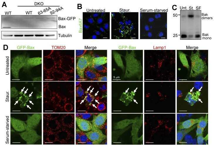
DRP1 interacts directly with BAX to induce its activation and apoptosis.
Jenner A, Peña-Blanco A, Salvador-Gallego R, Ugarte-Uribe B, Zollo C, Ganief T, Bierlmeier J, Mund M, Lee JE, Ries J, Schwarzer D, Macek B, Garcia-Saez AJ
The EMBO journal 2022 Apr 19;41(8):e108587
The EMBO journal 2022 Apr 19;41(8):e108587
LncRNA ASAP1-IT1 enhances cancer cell stemness via regulating miR-509-3p/YAP1 axis in NSCLC.
Liu Y, Yang Y, Zhang L, Lin J, Li B, Yang M, Li H, Chen K, Zhao W
Cancer cell international 2021 Oct 29;21(1):572
Cancer cell international 2021 Oct 29;21(1):572
Human Adipose-derived mesenchymal stem cells promote lymphocyte apoptosis and alleviate atherosclerosis via miR-125b-1-3p/BCL11B signal axis.
Yu C, Tang W, Lu R, Tao Y, Ren T, Gao Y
Annals of palliative medicine 2021 Feb;10(2):2123-2133
Annals of palliative medicine 2021 Feb;10(2):2123-2133
Urine Sample-Derived Cerebral Organoids Suitable for Studying Neurodevelopment and Pharmacological Responses.
Lin VJT, Hu J, Zolekar A, Yan LJ, Wang YC
Frontiers in cell and developmental biology 2020;8:304
Frontiers in cell and developmental biology 2020;8:304
MiR-203-3p inhibits the oxidative stress, inflammatory responses and apoptosis of mice podocytes induced by high glucose through regulating Sema3A expression.
Chen J, Xu Q, Zhang W, Zhen Y, Cheng F, Hua G, Lan J, Tu C
Open life sciences 2020;15(1):939-950
Open life sciences 2020;15(1):939-950
Desoxyrhapontigenin attenuates neuronal apoptosis in an isoflurane-induced neuronal injury model by modulating the TLR-4/cyclin B1/Sirt-1 pathway.
Liang F, Fu X, Li Y, Han F
AMB Express 2020 Sep 30;10(1):175
AMB Express 2020 Sep 30;10(1):175
Elaeagnus angustifolia Plant Extract Inhibits Epithelial-Mesenchymal Transition and Induces Apoptosis via HER2 Inactivation and JNK Pathway in HER2-Positive Breast Cancer Cells.
Jabeen A, Sharma A, Gupta I, Kheraldine H, Vranic S, Al Moustafa AE, Al Farsi HF
Molecules (Basel, Switzerland) 2020 Sep 16;25(18)
Molecules (Basel, Switzerland) 2020 Sep 16;25(18)
Pulsed focused ultrasound enhances the therapeutic effect of mesenchymal stromal cell-derived extracellular vesicles in acute kidney injury.
Ullah M, Liu DD, Rai S, Razavi M, Concepcion W, Thakor AS
Stem cell research & therapy 2020 Sep 14;11(1):398
Stem cell research & therapy 2020 Sep 14;11(1):398
Enhancement of Cisplatin Cytotoxicity by Cu(II)-Mn(II) Schiff Base Tetradentate Complex in Human Oral Squamous Cell Carcinoma.
Al-Serwi RH, Othman G, Attia MA, Enan ET, El-Sherbiny M, Mahmoud S, Elsherbiny N
Molecules (Basel, Switzerland) 2020 Oct 14;25(20)
Molecules (Basel, Switzerland) 2020 Oct 14;25(20)
Antifungal itraconazole ameliorates experimental autoimmune encephalomyelitis through a novel mechanism of action.
Huang H, Tian X, Peng X, Huang L, Mei L, Zhan Y, Chen S, Wu H, Wei G, Cai X
Advances in clinical and experimental medicine : official organ Wroclaw Medical University 2020 May;29(5):535-545
Advances in clinical and experimental medicine : official organ Wroclaw Medical University 2020 May;29(5):535-545
High fat diet consumption results in mitochondrial dysfunction, oxidative stress, and oligodendrocyte loss in the central nervous system.
Langley MR, Yoon H, Kim HN, Choi CI, Simon W, Kleppe L, Lanza IR, LeBrasseur NK, Matveyenko A, Scarisbrick IA
Biochimica et biophysica acta. Molecular basis of disease 2020 Mar 1;1866(3):165630
Biochimica et biophysica acta. Molecular basis of disease 2020 Mar 1;1866(3):165630
MicroRNA-26a/b have protective roles in oral lichen planus.
Du J, Gao R, Wang Y, Nguyen T, Yang F, Shi Y, Liu T, Liao W, Li R, Zhang F, Ge X, Zhao B
Cell death & disease 2020 Jan 6;11(1):15
Cell death & disease 2020 Jan 6;11(1):15
Treatment-Induced Tumor Dormancy through YAP-Mediated Transcriptional Reprogramming of the Apoptotic Pathway.
Kurppa KJ, Liu Y, To C, Zhang T, Fan M, Vajdi A, Knelson EH, Xie Y, Lim K, Cejas P, Portell A, Lizotte PH, Ficarro SB, Li S, Chen T, Haikala HM, Wang H, Bahcall M, Gao Y, Shalhout S, Boettcher S, Shin BH, Thai T, Wilkens MK, Tillgren ML, Mushajiang M, Xu M, Choi J, Bertram AA, Ebert BL, Beroukhim R, Bandopadhayay P, Awad MM, Gokhale PC, Kirschmeier PT, Marto JA, Camargo FD, Haq R, Paweletz CP, Wong KK, Barbie DA, Long HW, Gray NS, Jänne PA
Cancer cell 2020 Jan 13;37(1):104-122.e12
Cancer cell 2020 Jan 13;37(1):104-122.e12
Serine-70 phosphorylated Bcl-2 prevents oxidative stress-induced DNA damage by modulating the mitochondrial redox metabolism.
Chong SJF, Iskandar K, Lai JXH, Qu J, Raman D, Valentin R, Herbaux C, Collins M, Low ICC, Loh T, Davids M, Pervaiz S
Nucleic acids research 2020 Dec 16;48(22):12727-12745
Nucleic acids research 2020 Dec 16;48(22):12727-12745
Photodynamic Therapy Using a Novel Phosphorus Tetraphenylporphyrin Induces an Anticancer Effect via Bax/Bcl-xL-related Mitochondrial Apoptosis in Biliary Cancer Cells.
Mai NNH, Yamaguchi Y, Choijookhuu N, Matsumoto J, Nanashima A, Takagi H, Sato K, Tuan LQ, Hishikawa Y
Acta histochemica et cytochemica 2020 Aug 26;53(4):61-72
Acta histochemica et cytochemica 2020 Aug 26;53(4):61-72
Activation of Piezo1 sensitizes cells to TRAIL-mediated apoptosis through mitochondrial outer membrane permeability.
Hope JM, Lopez-Cavestany M, Wang W, Reinhart-King CA, King MR
Cell death & disease 2019 Nov 4;10(11):837
Cell death & disease 2019 Nov 4;10(11):837
Diallyl sulfide ameliorates carbon tetrachloride-induced hepatotoxicity in rats via suppressing stress-activated MAPK signaling pathways.
Elkhoely A
Journal of biochemical and molecular toxicology 2019 Jun;33(6):e22307
Journal of biochemical and molecular toxicology 2019 Jun;33(6):e22307
The transcription factor ETS1 promotes apoptosis resistance of senescent cholangiocytes by epigenetically up-regulating the apoptosis suppressor BCL2L1.
O'Hara SP, Splinter PL, Trussoni CE, Guicciardi ME, Splinter NP, Al Suraih MS, Nasser-Ghodsi N, Stollenwerk D, Gores GJ, LaRusso NF
The Journal of biological chemistry 2019 Dec 6;294(49):18698-18713
The Journal of biological chemistry 2019 Dec 6;294(49):18698-18713
The protective effects of total paeony glycoside on ischemia/reperfusion injury in H9C2 cells via inhibition of the PI3K/Akt signaling pathway.
Shen P, Chen J, Pan M
Molecular medicine reports 2018 Sep;18(3):3332-3340
Molecular medicine reports 2018 Sep;18(3):3332-3340
Combined effects of octreotide and cisplatin on the proliferation of side population cells from anaplastic thyroid cancer cell lines.
Li Z, Jiang X, Chen P, Wu X, Duan A, Qin Y
Oncology letters 2018 Sep;16(3):4033-4042
Oncology letters 2018 Sep;16(3):4033-4042
Lactobacillus casei Strain Shirota Enhances the In Vitro Antiproliferative Effect of Geniposide in Human Oral Squamous Carcinoma HSC-3 Cells.
Qian Y, Song JL, Sun P, Yi R, Liu H, Feng X, Park KY, Zhao X
Molecules (Basel, Switzerland) 2018 May 3;23(5)
Molecules (Basel, Switzerland) 2018 May 3;23(5)
Diazoxide inhibits of ER stress‑mediated apoptosis during oxygen‑glucose deprivation in vitro and cerebral ischemia‑reperfusion in vivo.
Lei X, Lei L, Zhang Z, Cheng Y
Molecular medicine reports 2018 Jun;17(6):8039-8046
Molecular medicine reports 2018 Jun;17(6):8039-8046
Autophagic cell death is dependent on lysosomal membrane permeability through Bax and Bak.
Karch J, Schips TG, Maliken BD, Brody MJ, Sargent MA, Kanisicak O, Molkentin JD
eLife 2017 Nov 17;6
eLife 2017 Nov 17;6
Apoptosis induced by Ginkgo biloba (EGb761) in melanoma cells is Mcl-1-dependent.
Wang Y, Lv J, Cheng Y, Du J, Chen D, Li C, Zhang J
PloS one 2015;10(4):e0124812
PloS one 2015;10(4):e0124812
Neuroprotective effect of resveratrol in diabetic cerebral ischemic-reperfused rats through regulation of inflammatory and apoptotic events.
Mohamed HE, El-Swefy SE, Hasan RA, Hasan AA
Diabetology & metabolic syndrome 2014;6(1):88
Diabetology & metabolic syndrome 2014;6(1):88
Phosphorylated nerve growth factor-induced clone B (NGFI-B) translocates from the nucleus to mitochondria of stressed rat cardiomyocytes and induces apoptosis.
Xinxing W, Hong F, Rui Z, Yun Z, Jingbo G, Lingjia Q
Stress (Amsterdam, Netherlands) 2012 Sep;15(5):545-53
Stress (Amsterdam, Netherlands) 2012 Sep;15(5):545-53
Protective effects of trimetazidine on bone marrow mesenchymal stem cells viability in an ex vivo model of hypoxia and in vivo model of locally myocardial ischemia.
Xu H, Zhu G, Tian Y
Journal of Huazhong University of Science and Technology. Medical sciences = Hua zhong ke ji da xue xue bao. Yi xue Ying De wen ban = Huazhong keji daxue xuebao. Yixue Yingdewen ban 2012 Feb;32(1):36-41
Journal of Huazhong University of Science and Technology. Medical sciences = Hua zhong ke ji da xue xue bao. Yi xue Ying De wen ban = Huazhong keji daxue xuebao. Yixue Yingdewen ban 2012 Feb;32(1):36-41
Prenatal stress induces long-term effects in cell turnover in the hippocampus-hypothalamus-pituitary axis in adult male rats.
Baquedano E, García-Cáceres C, Diz-Chaves Y, Lagunas N, Calmarza-Font I, Azcoitia I, Garcia-Segura LM, Argente J, Chowen JA, Frago LM
PloS one 2011;6(11):e27549
PloS one 2011;6(11):e27549
Combined treatment with the Cox-2 inhibitor niflumic acid and PPARγ ligand ciglitazone induces ER stress/caspase-8-mediated apoptosis in human lung cancer cells.
Kim BM, Maeng K, Lee KH, Hong SH
Cancer letters 2011 Jan 28;300(2):134-44
Cancer letters 2011 Jan 28;300(2):134-44
Sequential caspase-2 and caspase-8 activation is essential for saikosaponin a-induced apoptosis of human colon carcinoma cell lines.
Kim BM, Hong SH
Apoptosis : an international journal on programmed cell death 2011 Feb;16(2):184-97
Apoptosis : an international journal on programmed cell death 2011 Feb;16(2):184-97
Endosome-mitochondria juxtaposition during apoptosis induced by H. pylori VacA.
Calore F, Genisset C, Casellato A, Rossato M, Codolo G, Esposti MD, Scorrano L, de Bernard M
Cell death and differentiation 2010 Nov;17(11):1707-16
Cell death and differentiation 2010 Nov;17(11):1707-16
Cell cycle arrest and cytochrome c-mediated apoptotic induction by MCS-5A is associated with up-regulation of p16(INK4a) in HL-60 cells.
Choi BY, Lee CH
Bioorganic & medicinal chemistry letters 2010 Jul 1;20(13):3880-4
Bioorganic & medicinal chemistry letters 2010 Jul 1;20(13):3880-4
Heme oxygenase-1 prevents hyperthyroidism induced hepatic damage via an antioxidant and antiapoptotic pathway.
Giriş M, Erbil Y, Depboylu B, Mete O, Türkoğlu U, Abbasoğlu SD, Uysal M
The Journal of surgical research 2010 Dec;164(2):266-75
The Journal of surgical research 2010 Dec;164(2):266-75
Notch-activated signaling cascade interacts with mitochondrial remodeling proteins to regulate cell survival.
Perumalsamy LR, Nagala M, Sarin A
Proceedings of the National Academy of Sciences of the United States of America 2010 Apr 13;107(15):6882-7
Proceedings of the National Academy of Sciences of the United States of America 2010 Apr 13;107(15):6882-7
Growth hormone-releasing peptide 6 protection of hypothalamic neurons from glutamate excitotoxicity is caspase independent and not mediated by insulin-like growth factor I.
Delgado-Rubín A, Chowen JA, Argente J, Frago LM
The European journal of neuroscience 2009 Jun;29(11):2115-24
The European journal of neuroscience 2009 Jun;29(11):2115-24
N,N-dimethyl phytosphingosine induces caspase-8-dependent cytochrome c release and apoptosis through ROS generation in human leukemia cells.
Kim BM, Choi YJ, Han Y, Yun YS, Hong SH
Toxicology and applied pharmacology 2009 Aug 15;239(1):87-97
Toxicology and applied pharmacology 2009 Aug 15;239(1):87-97
Immunohistochemical expression of apoptosis regulating proteins and sex hormone receptors in meningiomas.
Takei H, Buckleair LW, Powell SZ
Neuropathology : official journal of the Japanese Society of Neuropathology 2008 Feb;28(1):62-8
Neuropathology : official journal of the Japanese Society of Neuropathology 2008 Feb;28(1):62-8
Death of hypothalamic astrocytes in poorly controlled diabetic rats is associated with nuclear translocation of apoptosis inducing factor.
García-Cáceres C, Lechuga-Sancho A, Argente J, Frago LM, Chowen JA
Journal of neuroendocrinology 2008 Dec;20(12):1348-60
Journal of neuroendocrinology 2008 Dec;20(12):1348-60
Desferrioxamine (DFX) induces apoptosis through the p38-caspase8-Bid-Bax pathway in PHA-stimulated human lymphocytes.
Kim BM, Chung HW
Toxicology and applied pharmacology 2008 Apr 1;228(1):24-31
Toxicology and applied pharmacology 2008 Apr 1;228(1):24-31
Preventive cardioprotection of erythropoietin against doxorubicin-induced cardiomyopathy.
Chen X, Chen Y, Bi Y, Fu N, Shan C, Wang S, Aslam S, Wang PW, Xu J
Cardiovascular drugs and therapy 2007 Oct;21(5):367-74
Cardiovascular drugs and therapy 2007 Oct;21(5):367-74
Hypoxia/reoxygenation induces apoptosis through a ROS-mediated caspase-8/Bid/Bax pathway in human lymphocytes.
Kim BM, Chung HW
Biochemical and biophysical research communications 2007 Nov 23;363(3):745-50
Biochemical and biophysical research communications 2007 Nov 23;363(3):745-50
Influence of glutathione on the induction of chromosome aberrations, delay in cell cycle kinetics and cell cycle regulator proteins in irradiated mouse bone marrow cells.
Ray S, Chatterjee A
International journal of radiation biology 2007 May;83(5):347-54
International journal of radiation biology 2007 May;83(5):347-54
The N-terminus and alpha-5, alpha-6 helices of the pro-apoptotic protein Bax, modulate functional interactions with the anti-apoptotic protein Bcl-xL.
Parikh N, Koshy C, Dhayabaran V, Perumalsamy LR, Sowdhamini R, Sarin A
BMC cell biology 2007 May 23;8:16
BMC cell biology 2007 May 23;8:16
Iptakalim protects PC12 cell against H2O2-induced oxidative injury via opening mitochondrial ATP-sensitive potassium channel.
Chai Y, Niu L, Sun XL, Ding JH, Hu G
Biochemical and biophysical research communications 2006 Nov 17;350(2):307-14
Biochemical and biophysical research communications 2006 Nov 17;350(2):307-14
(+/-)-huprine Y, (-)-huperzine A and tacrine do not show neuroprotective properties in an apoptotic model of neuronal cytoskeletal alteration.
Jordá EG, Verdaguer E, Jiménez A, Canudas AM, Rimbau V, Camps P, Muñoz-Torrero D, Camins A, Pallàs M
Journal of Alzheimer's disease : JAD 2004 Dec;6(6):577-83; discussion 673-81
Journal of Alzheimer's disease : JAD 2004 Dec;6(6):577-83; discussion 673-81
No comments: Submit comment
Supportive validation
- Submitted by
- Invitrogen Antibodies (provider)
- Main image
- Experimental details
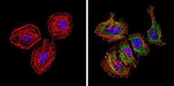
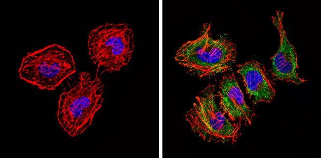
- Immunofluorescent analysis of Bax (green) showing staining in the cytoplasm of Hela cells (right) compared to a negative control without primary antibody (left). Formalin-fixed cells were permeabilized with 0.1% Triton X-100 in TBS for 5-10 minutes and blocked with 3% BSA-PBS for 30 minutes at room temperature. Cells were probed with a Bax monoclonal antibody (Product # MA5-14003) in 3% BSA-PBS at a dilution of 1:50 and incubated overnight at 4 ºC in a humidified chamber. Cells were washed with PBST and incubated with a DyLight-conjugated secondary antibody in PBS at room temperature in the dark. F-actin (red) was stained with a fluorescent red phalloidin and nuclei (blue) were stained with Hoechst or DAPI. Images were taken at a magnification of 60x.

- Submitted by
- Invitrogen Antibodies (provider)
- Main image
- Experimental details
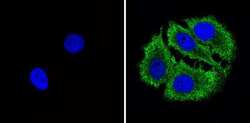
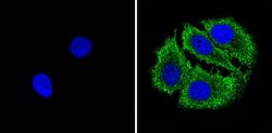
- Immunofluorescent analysis of Bax (green) showing staining in the cytoplasm of MCF-7 cells (right) compared to a negative control without primary antibody (left). Formalin-fixed cells were permeabilized with 0.1% Triton X-100 in TBS for 5-10 minutes and blocked with 3% BSA-PBS for 30 minutes at room temperature. Cells were probed with a Bax monoclonal antibody (Product # MA5-14003) in 3% BSA-PBS at a dilution of 1:50 and incubated overnight at 4 ºC in a humidified chamber. Cells were washed with PBST and incubated with a DyLight-conjugated secondary antibody in PBS at room temperature in the dark. F-actin (red) was stained with a fluorescent red phalloidin and nuclei (blue) were stained with Hoechst or DAPI. Images were taken at a magnification of 60x.

- Submitted by
- Invitrogen Antibodies (provider)
- Main image
- Experimental details
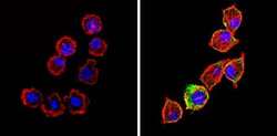
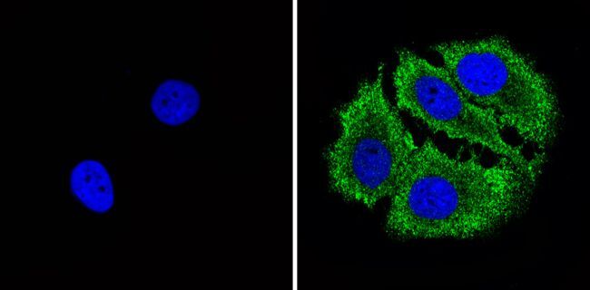
- Immunofluorescent analysis of Bax (green) showing staining in the cytoplasm of L929 cells (right) compared to a negative control without primary antibody (left). Formalin-fixed cells were permeabilized with 0.1% Triton X-100 in TBS for 5-10 minutes and blocked with 3% BSA-PBS for 30 minutes at room temperature. Cells were probed with a Bax monoclonal antibody (Product # MA5-14003) in 3% BSA-PBS at a dilution of 1:50 and incubated overnight at 4 ºC in a humidified chamber. Cells were washed with PBST and incubated with a DyLight-conjugated secondary antibody in PBS at room temperature in the dark. F-actin (red) was stained with a fluorescent red phalloidin and nuclei (blue) were stained with Hoechst or DAPI. Images were taken at a magnification of 60x.

- Submitted by
- Invitrogen Antibodies (provider)
- Main image

- Experimental details
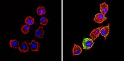
- Immunofluorescent analysis of Bax (green) showing staining in the cytoplasm of Hela cells (right) compared to a negative control without primary antibody (left). Formalin-fixed cells were permeabilized with 0.1% Triton X-100 in TBS for 5-10 minutes and blocked with 3% BSA-PBS for 30 minutes at room temperature. Cells were probed with a Bax monoclonal antibody (Product # MA5-14003) in 3% BSA-PBS at a dilution of 1:50 and incubated overnight at 4 ºC in a humidified chamber. Cells were washed with PBST and incubated with a DyLight-conjugated secondary antibody in PBS at room temperature in the dark. F-actin (red) was stained with a fluorescent red phalloidin and nuclei (blue) were stained with Hoechst or DAPI. Images were taken at a magnification of 60x.
- Submitted by
- Invitrogen Antibodies (provider)
- Main image
- Experimental details
- Immunofluorescent analysis of Bax (green) showing staining in the cytoplasm of MCF-7 cells (right) compared to a negative control without primary antibody (left). Formalin-fixed cells were permeabilized with 0.1% Triton X-100 in TBS for 5-10 minutes and blocked with 3% BSA-PBS for 30 minutes at room temperature. Cells were probed with a Bax monoclonal antibody (Product # MA5-14003) in 3% BSA-PBS at a dilution of 1:50 and incubated overnight at 4 ºC in a humidified chamber. Cells were washed with PBST and incubated with a DyLight-conjugated secondary antibody in PBS at room temperature in the dark. F-actin (red) was stained with a fluorescent red phalloidin and nuclei (blue) were stained with Hoechst or DAPI. Images were taken at a magnification of 60x.
- Submitted by
- Invitrogen Antibodies (provider)
- Main image
- Experimental details
- Immunofluorescent analysis of Bax (green) showing staining in the cytoplasm of L929 cells (right) compared to a negative control without primary antibody (left). Formalin-fixed cells were permeabilized with 0.1% Triton X-100 in TBS for 5-10 minutes and blocked with 3% BSA-PBS for 30 minutes at room temperature. Cells were probed with a Bax monoclonal antibody (Product # MA5-14003) in 3% BSA-PBS at a dilution of 1:50 and incubated overnight at 4 ºC in a humidified chamber. Cells were washed with PBST and incubated with a DyLight-conjugated secondary antibody in PBS at room temperature in the dark. F-actin (red) was stained with a fluorescent red phalloidin and nuclei (blue) were stained with Hoechst or DAPI. Images were taken at a magnification of 60x.

Supportive validation
- Submitted by
- Invitrogen Antibodies (provider)
- Main image

- Experimental details
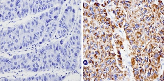
- Immunohistochemistry analysis of Bax showing positive staining in the cytoplasm of paraffin-treated Human hepatocarcinoma (right) compared with a negative control in the absence of primary antibody (left). To expose target proteins, antigen retrieval method was performed using 10mM sodium citrate (pH 6.0) microwaved for 8-15 min. Following antigen retrieval, tissues were blocked in 3% H2O2-methanol for 15 min at room temperature, washed with ddH2O and PBS, and then probed with a Bax monoclonal antibody (Product # MA5-14003) diluted by 3% BSA-PBS at a dilution of 1:100 overnight at 4°C in a humidified chamber. Tissues were washed extensively PBST and detection was performed using an HRP-conjugated secondary antibody followed by colorimetric detection using a DAB kit. Tissues were counterstained with hematoxylin and dehydrated with ethanol and xylene to prep for mounting.
Supportive validation
- Submitted by
- Invitrogen Antibodies (provider)
- Main image
- Experimental details
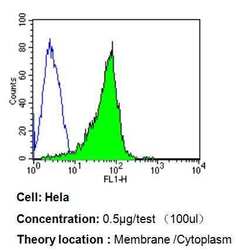
- Flow cytometry analysis of Bax in Hela cells compared to an isotype control (blue). Cells were harvested, adjusted to a concentration of 1-5x10^6 cells/mL, fixed with 2% paraformaldehyde, washed with PBS, and incubated with Bax monoclonal antibody (Product # MA5-14003) at a dilution of 0.5 µg/test for 60 min at room temperature. Cells were then blocked in a solution of 2% BSA-PBS for 30 min at room temperature, incubated for 40 min at room temperature in the dark using a Dylight 488-conjugated goat anti-mouse IgG (H+L) secondary antibody, and re-suspended in PBS for FACS analysis.

- Submitted by
- Invitrogen Antibodies (provider)
- Main image
- Experimental details
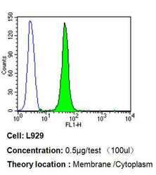
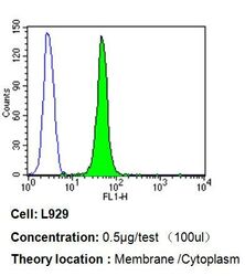
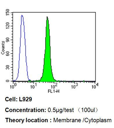
- Flow cytometry analysis of Bax in L929 cells compared to an isotype control (blue). Cells were harvested, adjusted to a concentration of 1-5x10^6 cells/mL, fixed with 2% paraformaldehyde, washed with PBS, and incubated with Bax monoclonal antibody (Product # MA5-14003) at a dilution of 0.5 µg/test for 60 min at room temperature. Cells were then blocked in a solution of 2% BSA-PBS for 30 min at room temperature, incubated for 40 min at room temperature in the dark using a Dylight 488-conjugated goat anti-mouse IgG (H+L) secondary antibody, and re-suspended in PBS for FACS analysis.

- Submitted by
- Invitrogen Antibodies (provider)
- Main image
- Experimental details
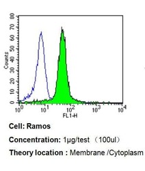
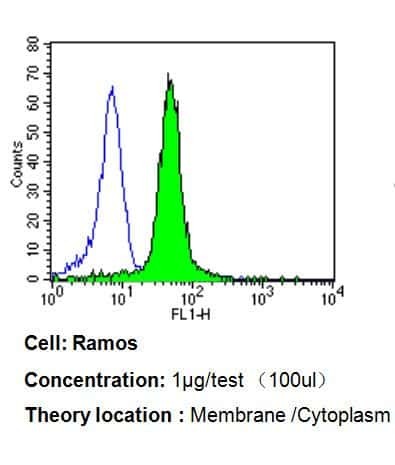
- Flow cytometry analysis of Bax in Ramos cells compared to an isotype control (blue). Cells were harvested, adjusted to a concentration of 1-5x10^6 cells/mL, fixed with 2% paraformaldehyde, washed with PBS, and incubated with Bax monoclonal antibody (Product # MA5-14003) at a dilution of 1 µg/test for 60 min at room temperature. Cells were then blocked in a solution of 2% BSA-PBS for 30 min at room temperature, incubated for 40 min at room temperature in the dark using a Dylight 488-conjugated goat anti-mouse IgG (H+L) secondary antibody, and re-suspended in PBS for FACS analysis.
- Submitted by
- Invitrogen Antibodies (provider)
- Main image

- Experimental details
- Flow cytometry analysis of Bax in Hela cells compared to an isotype control (blue). Cells were harvested, adjusted to a concentration of 1-5x10^6 cells/mL, fixed with 2% paraformaldehyde, washed with PBS, and incubated with Bax monoclonal antibody (Product # MA5-14003) at a dilution of 0.5 µg/test for 60 min at room temperature. Cells were then blocked in a solution of 2% BSA-PBS for 30 min at room temperature, incubated for 40 min at room temperature in the dark using a Dylight 488-conjugated goat anti-mouse IgG (H+L) secondary antibody, and re-suspended in PBS for FACS analysis.

- Submitted by
- Invitrogen Antibodies (provider)
- Main image
- Experimental details
- Flow cytometry analysis of Bax in L929 cells compared to an isotype control (blue). Cells were harvested, adjusted to a concentration of 1-5x10^6 cells/mL, fixed with 2% paraformaldehyde, washed with PBS, and incubated with Bax monoclonal antibody (Product # MA5-14003) at a dilution of 0.5 µg/test for 60 min at room temperature. Cells were then blocked in a solution of 2% BSA-PBS for 30 min at room temperature, incubated for 40 min at room temperature in the dark using a Dylight 488-conjugated goat anti-mouse IgG (H+L) secondary antibody, and re-suspended in PBS for FACS analysis.
- Submitted by
- Invitrogen Antibodies (provider)
- Main image

- Experimental details
- Flow cytometry analysis of Bax in Ramos cells compared to an isotype control (blue). Cells were harvested, adjusted to a concentration of 1-5x10^6 cells/mL, fixed with 2% paraformaldehyde, washed with PBS, and incubated with Bax monoclonal antibody (Product # MA5-14003) at a dilution of 1 µg/test for 60 min at room temperature. Cells were then blocked in a solution of 2% BSA-PBS for 30 min at room temperature, incubated for 40 min at room temperature in the dark using a Dylight 488-conjugated goat anti-mouse IgG (H+L) secondary antibody, and re-suspended in PBS for FACS analysis.

Supportive validation
- Submitted by
- Invitrogen Antibodies (provider)
- Main image
- Experimental details
- NULL
- Submitted by
- Invitrogen Antibodies (provider)
- Main image
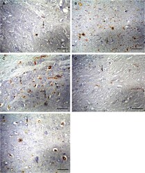
- Experimental details
- Figure 7 Photomicrographs of sections in the cerebral cortex of adult male albino rats. (A) control group showing weak positive immunoreaction for Bax (arrow). (B) diabetic group showing strong positive immunoreaction for Bax (arrows). (C) diabetic cerebral ischemic-reperfused group showing strong positive immunoreaction for Bax (arrows). (D) diabetic group treated with resveratrol showing moderate immunoreaction for Bax (arrows). (E) diabetic cerebral ischemic-reperfused group treated with resveratrol showing moderate immunoreaction for Bax (arrows). [A, B, C, D, E using Avidin-biotin peroxidase stain with Hx counter stain x400 (scale bars represent 10 mum)].

- Submitted by
- Invitrogen Antibodies (provider)
- Main image
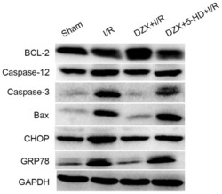
- Experimental details
- Figure 5. Pretreatment with DZX modulates the protein expression of Bcl-2, caspase-12, caspase-3, Bax, CHOP and GRP78 in rats subjected to I/R. Western blot analysis was used to detect the protein expression levels of Bcl-2, caspase-12, caspase-3, Bax, CHOP and GRP78 in hippocampal cells isolated from I/R-treated rats. Rats were treated with DZX with or without 5-HD and subjected to cerebral ischemia for 2 h followed by 12 h of reperfusion. Sham rats were not subjected to cerebral ischemia. DZX, diazoxide; Bcl-2, B-cell lymphoma-2; Bax, Bcl-2-associated X protein; CHOP, C/EBP homologous protein; GRP78, 78 kDa glucose-regulated protein; I/R, ischemia/reperfusion; 5-HD, 5-hydroxydecanoate.

- Submitted by
- Invitrogen Antibodies (provider)
- Main image
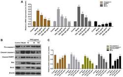
- Experimental details
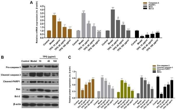
- Figure 3. TPG regulates apoptosis-associated factors in H9C2 cells. H9C2 cells were treated with TPG (10, 40 and 160 ug/ml) for 24 h and subjected to I/R injury. (A) Relative mRNA expression levels of caspase-3, PARP1, Bax and Bcl-2 were investigated by reverse transcription-quantitative polymerase chain reaction. (B) Relative protein expression levels of caspase-3, PARP1, Bax and Bcl-2 were evaluated by western blotting and normalized to beta-actin expression. (C) Blot results were semi-quantified. *P
- Submitted by
- Invitrogen Antibodies (provider)
- Main image
- Experimental details
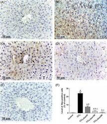
- 4 Effect of DAS (50, 100, and 200 mg/kg) on Bax immunohistochemical expression in CCL 4 -intoxicated rats. Hepatic sections (scale bar = 50) obtained from (A) control group, (B) CCl 4 group, (C) CCl 4 + DAS50 group, (D) CCl 4 + DAS100 group and (E) CCl 4 +DAS200 group. (F) Quantification of Bax immune-positive cells to the total area of microscopic field across six fields. Values are expressed as mean +- SD of six rats. a P < 0.05 compared with control group; b P < 0.05 compared with CCl 4 group; c P < 0.05 compared with corresponding CCl 4 +DAS50; d P < 0.05 compared with corresponding CCl 4 +DAS100 group. CCl4, carbon tetrachloride; DAS, diallyl sulfide; SD, standard deviation

- Submitted by
- Invitrogen Antibodies (provider)
- Main image
- Experimental details
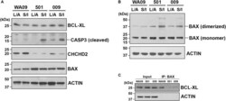
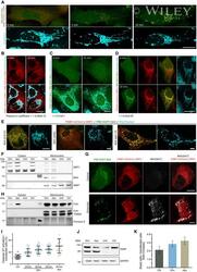
- FIGURE 6 The expression of CHCHD2, the dimerization of BAX, and the interaction between BCL-XL and BAX in the aggregates of UEC-derived hiPSCs and WA09 hESCs that were cultured in LDN193189/A83-01-containing and SB431542/IWR-1-containing media. (A) The expression and activation of CHCHD2, BAX, BCL-XL, and CASP3 were revealed by western blotting in the indicated cell aggregate samples that were cultured in the indicated media. (B) Dimerized BAX (~40 kDa) with electromobility shift from BAX monomer (~20 kDa) was detected by western blotting in the cell aggregate samples. (C) BCL-XL interacted and co-immunoprecipitated with BAX in the cell aggregate samples cultured in the SB431542/IWR-1-containing medium was detected by western blotting. 501: UEC715i-501 hiPSCs. 009: UEC001i-009 hiPSCs. L/A: LDN193189/A83-01-containing medium. S/I: SB431542/IWR-1-containing medium. All the cell aggregates were cultured in the indicated media for 6 days prior to sample collection and analysis.

- Submitted by
- Invitrogen Antibodies (provider)
- Main image

- Experimental details
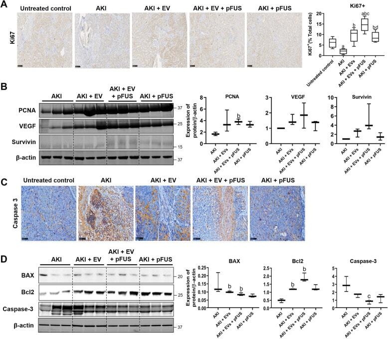
- Fig. 5 Proliferation and apoptosis. a Immunohistochemical staining for proliferation marker Ki67 in kidney tissue (left), alongside quantification of the percentage of Ki67-positive cells (right). b Western blot on kidney tissue measuring proliferation markers PCNA, VEGF, survivin, and beta-actin (left) alongside quantification (right). c Immunohistochemical staining for apoptosis marker Caspase-3 in kidney tissue. d Western blot on kidney tissue measuring apoptosis markers BAX, Bcl-2, and Caspase-3 (left) alongside quantification (right). Each group has n = 3 mice. Significant difference a p < 0.05: relative to untreated control; b p < 0.05: relative to AKI; c p < 0.05: relative to AKI-EV; d p < 0.05: relative to AKI-EV-pFUS
- Submitted by
- Invitrogen Antibodies (provider)
- Main image
- Experimental details
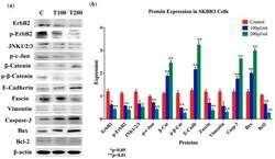
- Figure 7 ( a , b ) Protein expression and molecular mechanisms of EA inhibitory actions in SKBR3 cell line. This plant extract induces an overexpression of E-cadherin, beta-catenin, and downregulation of vimentin and fascin, while upregulating pro-apoptotic markers (Bax and Caspase-3), in comparison with their control and inhibiting anti-apoptotic markers (Bcl-2). Furthermore, EA plant extract inhibits the phosphorylation of ErbB2 and beta-catenin, as well as the expression of JNK1/2/3. beta-actin was used as a control for the proteins amount in this assay. Cells were treated with 100 and 200 muL/mL of EA extract for 48 h, as explained in the materials and methods and the results sections. ( a ) Blot image and ( b ) quantification of bands.

- Submitted by
- Invitrogen Antibodies (provider)
- Main image
- Experimental details
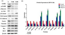
- Figure 8 ( a , b ) Protein expression and molecular mechanisms of EA inhibitory actions in ZR75 cell line. This plant extract induces an overexpression of E-cadherin, beta-catenin, and downregulation of vimentin and fascin; in addition, pro-apoptotic markers Bax and Caspase-3 are upregulated in comparison with their control, while anti-apoptotic marker Bcl-2 is inhibited. Furthermore, EA plant extract inhibits the phosphorylation of ErbB2 and beta-catenin, as well as JNK1/2/3 expression. beta-actin served as a control in this assay. Cells were treated with 100 and 200 muL/mL of EA extract for 48 h, as explained in the materials and Methods section. ( a ) Blot image and ( b ) quantification of bands.

- Submitted by
- Invitrogen Antibodies (provider)
- Main image

- Experimental details
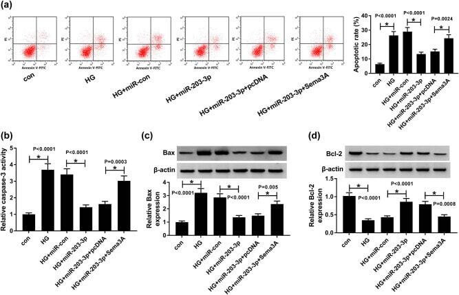
- Figure 7 MiR-203-3p and Sema3A regulated the apoptosis of HG-induced podocytes. HG-induced podocytes were transfected with miR-con, miR-203-3p, miR-203-3p + pcDNA or miR-203-3p + Sema3A. (a) Podocyte apoptosis was evaluated using flow cytometry. (b) The activity of caspase-3 was measured by the Caspase-3 Activity Detection Kit. (c and d) The WB analysis determined the protein levels of apoptosis-associated markers Bax and Bcl-2. * P < 0.05.
- Submitted by
- Invitrogen Antibodies (provider)
- Main image
- Experimental details
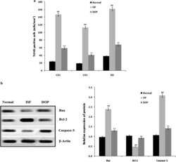
- Fig. 3 Desoxyrhapontigenin ameliorates neuronal apoptosis in the brain tissues of rats with anaesthesia-induced neuronal injury. a TUNEL-positive cells; b Protein expression of caspase-2, Bcl-2, and Bax by Western blotting. Mean +- SEM (n = 15); ## p < 0.01 vs. controls; ** p < 0.01 vs. ISF group

- Submitted by
- Invitrogen Antibodies (provider)
- Main image
- Experimental details
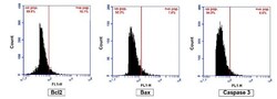
- Figure 6 Flow cytometry analysis of negative control cells acquired without stain for Bcl2, Bax and Caspase 3.
- Submitted by
- Invitrogen Antibodies (provider)
- Main image
- Experimental details
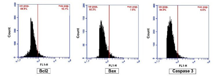
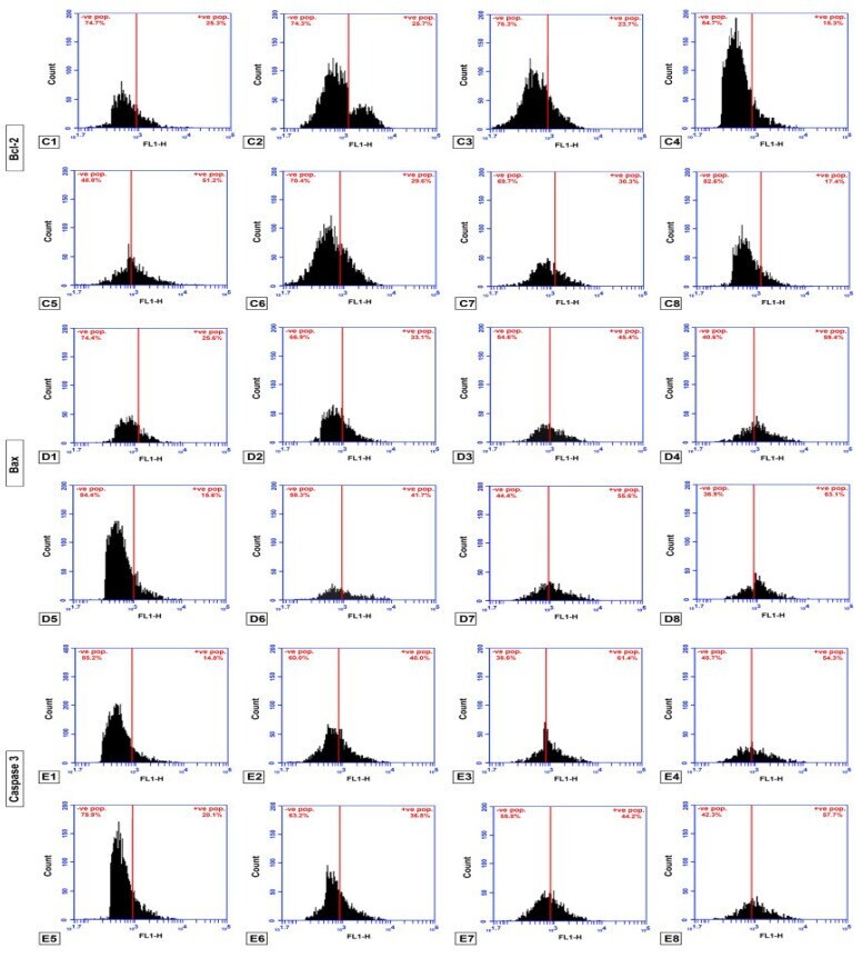
- Figure 7 ( C ) Flow cytometry analysis of cell counts, detecting the number of Bcl-2 positive cells. ( D ) Flow cytometry analysis of cell counts, detecting the number of Bax positive cells. ( E ) Flow cytometry analysis of cell counts, detecting the number of Caspase 3 positive cells. 1: GMSCs control; 2: GMSCs treated with Schiff base complex; 3: GMSCs treated with cisplatin; 4: GMSCs treated with Schiff base complex and cisplatin together; 5: SCC cells; 6: SCC treated with Schiff base complex; 7: SCC treated with cisplatin; 8: SCC treated with Schiff base complex and cisplatin together.
- Submitted by
- Invitrogen Antibodies (provider)
- Main image

- Experimental details
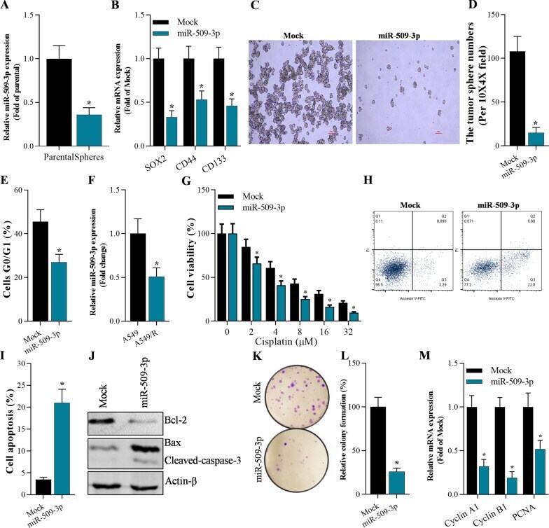
- Fig. 4 Overexpression of miR-509-3p inhibited stemness, cisplatin resistance and cell growth, and promoted apoptosis of A549-dereived stem cells. A qRT-PCR analysis on miR-509-3pin spheres of A549-derived stem cells, * p < 0.01 compared with parental group. B qRT-PCR on the expression of stemness-associated gens (SOX2, CD44, and CD133) in A549cell spheres 48 h after transfection with mimic miRNAs, * p < 0.01 compared with Mock. C , D Number of tumor spheres in miR-509-3p overexpressing A459 stem cells, * p < 0.01 compared with Mock. E Flow cytometry analysis of cell cycle in A549-dereived stem cells 48 h after overexpression of miR-509-3p, * p < 0.01 compared with Mock. F qRT-PCR analysis of miR-509-3pin A549 and cisplatin-resistant A549/R cells. * p < 0.01 compared with A549. G CCK-8 assay on the cell viability of miR-509-3p-overexpressing A549-dereived stem cells treated with cisplatin at various concentration (0 muM, 4 muM, 8 muM, 16 muM, 32 muM), * p < 0.01 compared with Mock. H , I Annexin V-FITC/PI staining of A549-dereived stem cells 48 h after miR-509-3p overexpression, * p < 0.01 compared with Mock. J Western blotting analysis on expression of apoptosis-associated genes (Bax, Bcl-2 and cleaved-caspase-3) 48 h after transfection with miR-509-3p or Mock, * p < 0.01 compared with Mock. K , L Colony formation assay on cell growth of A549-dereived stem cells after 15 days culture, * p < 0.01 compared with Mock. M qRT-PCR analysis on proliferation-associated genes (Cyclin
- Submitted by
- Invitrogen Antibodies (provider)
- Main image
- Experimental details
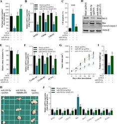
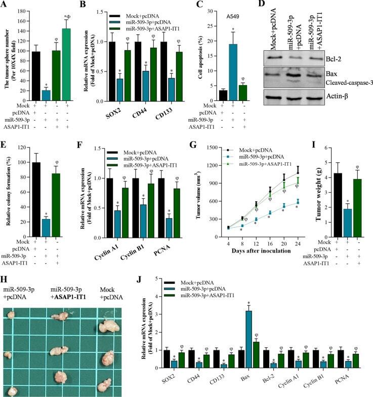
- Fig. 5 Overexpression of ASAP1-IT1 blocked miR-509-3p-mediated cell functions in cancer stem cells both in vitro and in vivo . A Number of A549 cell tumor spheres after expression of Mock, pcDNA, miR-509-3p, and ASAP1-IT1, * p < 0.01 compared with Mock + pcDNA, phi p < 0.01 compared with miR-509-3p + pcDNA, F p < 0.01 compared with miR-509-3p + ASAP1-IT1. B qRT-PCR analysis on stemness-related genes (SOX2, CD44, and CD133) in A549 stem cells, * p < 0.01 compared with Mock + pcDNA, phi p < 0.01compared with miR-509-3p + pcDNA. C Annexin V-FITC/PI staining of A549 stem cells with Mock, pcDNA, miR-509-3p, and ASAP1-IT1, * p < 0.01 compared with Mock + pcDNA, phi p < 0.01 compared with miR-509-3p + pcDNA. D Western blotting analysis on apoptosis-associated genes (Bax, Bcl-2, and cleaved-caspase-3) in A549 stem cells, * p < 0.01 compared with Mock + pcDNA, phi p < 0.01 compared with miR-509-3p + pcDNA. E Colony formation assay on cell growth after 15 days culture, * p < 0.01 compared with Mock + pcDNA, phi p < 0.01 compared with miR-509-3p + pcDNA. F qRT-PCR analysis of growth-associated genes (Cyclin A1, Cyclin B1, and PCNA) in A549 stem cells, * p < 0.01 compared with Mock + pcDNA, phi p < 0.01 compared with miR-509-3p + pcDNA. G Tumor volume in nude mice at the indicated time after inoculation, * p < 0.01 compared with Mock + pcDNA, phi p < 0.01 compared with miR-509-3p + pcDNA. H Photo of harvested tumors at the 24th day after inoculation. I Tumor weight at the 24th day after
- Submitted by
- Invitrogen Antibodies (provider)
- Main image
- Experimental details
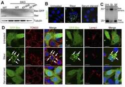
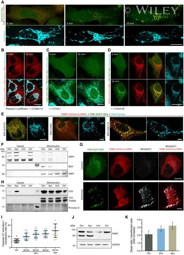
- EV5 Figure Induced dimerization of BAX and DRP1 induces apoptosis A Confocal microscopy of U2OS BAX/BAK DKO cells transfected with FKBP-mCherry-DRP1 (red) and FRB-EGFP-BAX (green) and stained with MitoTracker Deep Red FM (cyan). Images were acquired before (0 min) and after induction of BAX/DRP1 dimerization (5, 10 min). Scale bar 20 um. B-D Induced dimerization of DRP1 to itself (B), BAX to itself (C) or BAX to TOM20 (D) in U2OS BAX/BAK DKO cells transected with FKBP- and FRB-mCherry DRP1 (B, red), FKBP- and FRB-EGFP-BAX (C, green) or TOM20-mCherry-FKBP (red) and FRB-EGFP-BAX (green, D) before (0 min) and after induced dimerization (10 min). Mitochondria were stained using MitoTracker Deep Red FM or MitoSpy NIR (cyan) as indicated. Scale bar 20 um. Pearson's correlation coefficient was calculated between the induced dimerization signal of BAX/BAX, DRP1/DRP1 and BAX/TOM20, respectively, and mitochondria based on MitoTracker Deep Red TM signal as depicted below the images. E Induced dimerization of DRP1 and BAX mutants L63E, Delta19-37 or DeltaL1-2. Confocal microscopy of U2OS BAX/BAK DKO cells transfected with FKBP-mCherry-DRP1 (red) and mutant variants FRB-EGFP-BAX (green) as indicated and stained with MitoTracker Deep Red FM (cyan). Images were acquired 10 min after induction of BAX/DRP1 dimerization. Scale bar 20 um. All images are representative of n = 3 independent experiments. F-H Dimerization of BAX and DRP1 induces their translocation to mitochondria, exposure of the
- Submitted by
- Invitrogen Antibodies (provider)
- Main image
- Experimental details
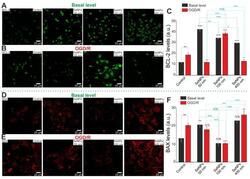
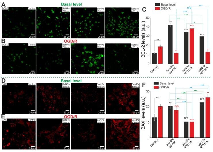
- Immunocytochemical staining of cortical cell culture with antibodies against BCL-2 ( A , B ) and BAX ( D , E ), regulators of apoptosis, after 24-h incubation with 3 mug/mL of different-sized SeNPs without OGD/R ( A , D ) and 24 h after OGD/R ( B , E ). ( C , F )--Intensity levels of BCL-2 ( C ) and BAX ( F ) were determined by confocal imaging. We analyzed individual cells that had fluorescence of secondary antibodies. The quantative data reflecting the level of BCL-2 or BAX expression are presented as fluorescence intensity values in summary bar charts (mean +- SEM). The values were averaged by 150 cells for each column. The results obtained after immunostaining agree with the data of fluorescence presented in panels ( A , B , D , E ). Each value is the mean +- SE ( n >= 3, p < 0.05). Statistical significance was assessed using paired t -test. Black asterisks represent comparison of baseline expression levels after incubation with different sized nanoparticles versus control. Red asterisks represent comparison of expression level after incubation with different-sized SeNPs versus OGD/R-induced levels; n/s--data not significant ( p > 0.05), * p < 0.05, ** p < 0.01, *** p < 0.001.
- Submitted by
- Invitrogen Antibodies (provider)
- Main image
- Experimental details
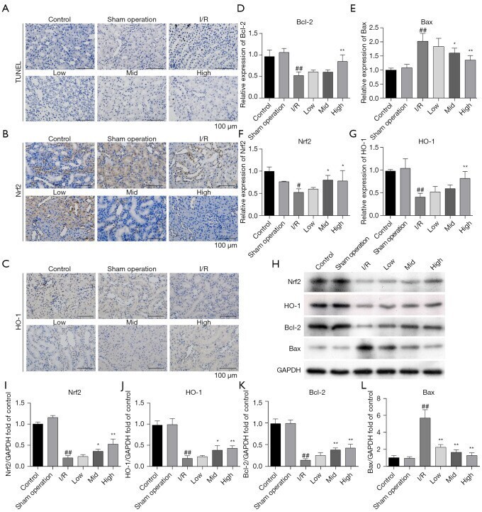
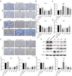
- Figure 2 AS-IV treatment relieved I/R-induced apoptosis and regulated the Nrf2/HO-1 pathway in rat kidney tissues. (A) Apoptosis in the renal tissues of AKI rats as detected by TUNEL staining. Magnification x400. (B,C) Nrf2 and HO-1 were detected by IHC. Magnification x400. Bcl-2, Bax, Nrf2, and HO-1 expression were evaluated by qRT-PCR (D-G) and western blotting (H-L). # P