Explore
 Validate
 Learn
Learn Western blot
Antibody data
- Antibody Data
- Antigen structure
- References [3]
- Comments [0]
- Validations
- Western blot [2]
- Immunohistochemistry [1]
- Flow cytometry [1]
Submit
Validation data
Reference
Comment
Report error
- Product number
- AP1302a - Provider product page

- Provider
- Abcepta
- Proper citation
- Abgent Cat#AP1302a, RRID:AB_2227716
- Product name
- Bax Antibody (BH3 Domain Specific)
- Antibody type
- Polyclonal
- Antigen
- Synthetic peptide
- Description
- Purified Rabbit Polyclonal Antibody (Pab)
- Reactivity
- Human
- Host
- Rabbit
- Isotype
- IgG
- Vial size
- 400 µl
- Concentration
- 1.6 mg/ml
- Storage
- Maintain refrigerated at 2-8°C for up to 6 months. For long term storage store at -20°C in small aliquots to prevent freeze-thaw cycles.
Submitted references Oridonin phosphate-induced autophagy effectively enhances cell apoptosis of human breast cancer cells.
BH3-triggered structural reorganization drives the activation of proapoptotic BAX.
Ceramide 1-phosphate inhibits serine palmitoyltransferase and blocks apoptosis in alveolar macrophages.
Li Y, Wang Y, Wang S, Gao Y, Zhang X, Lu C
Medical oncology (Northwood, London, England) 2015 Jan;32(1):365
Medical oncology (Northwood, London, England) 2015 Jan;32(1):365
BH3-triggered structural reorganization drives the activation of proapoptotic BAX.
Gavathiotis E, Reyna DE, Davis ML, Bird GH, Walensky LD
Molecular cell 2010 Nov 12;40(3):481-92
Molecular cell 2010 Nov 12;40(3):481-92
Ceramide 1-phosphate inhibits serine palmitoyltransferase and blocks apoptosis in alveolar macrophages.
Granado MH, Gangoiti P, Ouro A, Arana L, Gómez-Muñoz A
Biochimica et biophysica acta 2009 Apr;1791(4):263-72
Biochimica et biophysica acta 2009 Apr;1791(4):263-72
No comments: Submit comment
Supportive validation
- Submitted by
- Abcepta (provider)
- Main image
- Experimental details
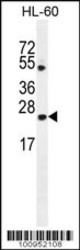
- Bax Antibody(BH3)(Cat. #AP1302a) western blot analysis in HL-60 cell line lysates (35ug/lane).This demonstrates the Bax antibody detected the Bax protein (arrow).
- Primary Ab dilution
- 1:1000

- Submitted by
- Abcepta (provider)
- Main image
- Experimental details
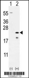
- Western blot analysis of Bax (arrow) using rabbit polyclonal Bax Antibody (BH3) (Cat. #AP1302a). 293 cell lysates (2 ug/lane) either nontransfected (Lane 1) or transiently transfected (Lane 2) with the Bax gene.
- Primary Ab dilution
- 1:1000

Supportive validation
- Submitted by
- Abcepta (provider)
- Main image
- Experimental details
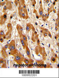
- "Formalin-fixed and paraffin-embedded human hepatocarcinoma reacted with Bax Antibody (BH3 Domain Specific), which was peroxidase-conjugated to the secondary antibody, followed by DAB staining. This data demonstrates the use of this antibody for immunohistochemistry; clinical relevance has not been evaluated."
- Primary Ab dilution
- 1:50~100

Supportive validation
- Submitted by
- Abcepta (provider)
- Main image

- Experimental details
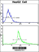
- Flow cytometric analysis of HepG2 cells using Bax Antibody (BH3 Domain Specific)(bottom histogram) compared to a negative control cell (top histogram). FITC-conjugated goat-anti-rabbit secondary antibodies were used for the analysis.
- Primary Ab dilution
- 1:10~50