Explore
 Validate
 Learn
Learn Western blot
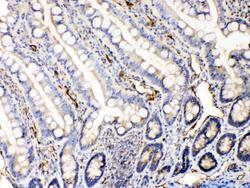
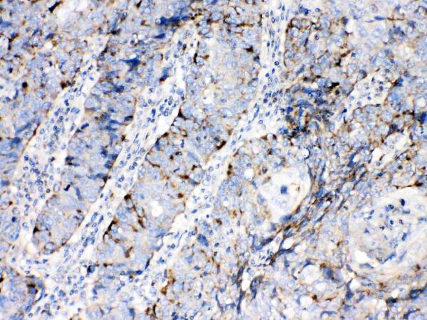
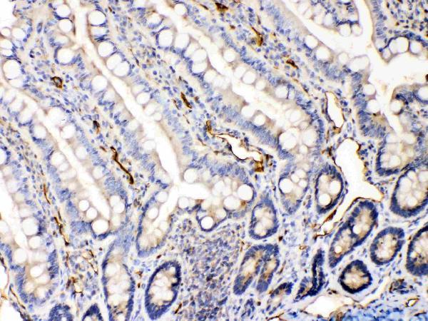
 Immunohistochemistry
Antibody data
- Antibody Data
- Antigen structure
- References [262]
- Comments [0]
- Validations
- Immunohistochemistry [1]
Submit
Validation data
Reference
Comment
Report error
- Product number
- A00183 - Provider product page

- Provider
- Boster Biological Technology
- Product name
- Anti-Bax Antibody Picoband™
- Antibody type
- Polyclonal
- Description
- Polyclonal antibody for BAX detection. Host: Rabbit.Size: 100μg/vial. Tested applications: IHC-P. Reactive species: Human. BAX information: Molecular Weight: 21184 MW; Subcellular Localization: Isoform Alpha: Mitochondrion membrane; Single-pass membrane protein. Cytoplasm. Colocalizes with 14- 3-3 proteins in the cytoplasm. Under stress conditions, undergoes a conformation change that causes release from JNK-phosphorylated 14-3-3 proteins and translocation to the mitochondrion membrane; Tissue Specificity: Expressed in a wide variety of tissues. Isoform Psi is found in glial tumors. Isoform Alpha is expressed in spleen, breast, ovary, testis, colon and brain, and at low levels in skin and lung. Isoform Sigma is expressed in spleen, breast, ovary, testis, lung, colon, brain and at low levels in skin. Isoform Alpha and isoform Sigma are expressed in pro- myelocytic leukemia, histiocytic lymphoma, Burkitt's lymphoma, T- cell lymphoma, lymphoblastic leukemia, breast adenocarcinoma, ovary adenocarcinoma, prostate carcinoma, prostate adenocarcinoma, lung carcinoma, epidermoid carcinoma, small cell lung carcinoma and colon adenocarcinoma cell lines.
- Reactivity
- Human, Mouse, Rat
- Host
- Rabbit
- Vial size
- 100μg/vial
- Concentration
- Add 0.2ml of distilled water will yield a concentration of 500ug/ml.
- Storage
- At -20°C for one year. After reconstitution, at 4°C for one month. It can also be aliquoted and stored frozen at -20°C for a longer time. Avoid repeated freezing and thawing.
- Handling
- Add 0.2ml of distilled water will yield a concentration of 500ug/ml.
Submitted references The protective effect of Macrostemonoside T from Allium macrostemon Bunge against Isoproterenol-Induced myocardial injury via the PI3K/Akt/mTOR signaling pathway.
Circ_0002331 Interacts with ELAVL1 to Improve ox-LDL-Induced Vascular Endothelial Cell Dysfunction via Regulating CCND2 mRNA Stability.
Cytisine-N-methylene-(5,7,4(')-trihydroxy)- isoflavone ameliorates ischemic stroke-induced brain injury in mouse by regulating the oxidative stress and BDNF-Trkb/Akt pathway.
Regulatory role of oxidative stress in retrorsine - Induced apoptosis and autophagy in primary rat hepatocytes.
FAXDC2 inhibits the proliferation and invasion of human liver cancer HepG2 cells.
PANoptosis-like death in acute-on-chronic liver failure injury.
Borneol-Modified Schisandrin B Micelles Cross the Blood-Brain Barrier To Treat Alzheimer's Disease in Aged Mice.
Rosavin Alleviates LPS-Induced Acute Lung Injure by Modulating the TLR-4/NF-κB/MAPK Singnaling Pathways.
Declined circular RNA mitofusin 2 constrains the deterioration of Wilms tumor via modulating microRNA-372-3p/transforming growth factor-β receptor type 2 axis.
Adiponectin attenuates H2O2-induced apoptosis in chicken skeletal myoblasts through the lysosomal-mitochondrial axis.
Hepatitis E virus causes apoptosis of ovarian cells in hens and resulting in a decrease in egg production.
Mitochondrial Fission in Nickel Nanoparticle-Induced Reproductive Toxicity: An In Vitro GC-1 Cell Study.
Comparison of acute kidney injury following brain death between male and female rats.
Effects of Cooling Interventions with Different Target Temperatures on Heat Stroke Rats.
Mechanisms underlying citrinin-induced toxicity via oxidative stress and apoptosis-mediated by mitochondrial-dependent pathway in SH-SY5Y cells.
Co-exposure to polystyrene microplastics and microcystin-LR aggravated male reproductive toxicity in mice.
Deciphering anti-benign prostatic hyperplasia potential of liangwanoside II based on metabolite profile characterization combined with targeted network pharmacology.
A Testlet Diagnostic Classification Model with Attribute Hierarchies.
Synthesis, biological activity and mechanism of action of novel allosecurinine derivatives as potential antitumor agents.
The combined analgesic, sedative, and anti-gastric cancer mechanisms of Tinospora sagittata var. yunnanensis (S. Y. Hu) H. S. Lo based on integrated ethnopharmacological data.
An experimental rat model of electric shock injury with isolated electric shock and water conduction: the histopathological changes on the skin and internal organs and the effect on biochemical parameters.
Connexin 43-modified bone marrow stromal cells reverse the imatinib resistance of K562 cells via Ca 2+ -dependent gap junction intercellular communication.
Hsp27, Hsp60, Hsp70, or Hsp90 depletion enhances the antitumor effects of resveratrol via oxidative and ER stress response in human glioblastoma cells.
Exosomal circCNOT6L Regulates Astrocyte Apoptotic Signals Induced by Hypoxia Exposure Through miR99a-5p/SERPINE1 and Alleviates Ischemic Stroke Injury.
Structural identification of an polysaccharide isolated from Epimedium brevicornum and its beneficial effect on promoting osteogenesis in osteoblasts induced by high glucose.
miR-466 Contributes to the Enhanced Antitumor Effect of Bortezomib on Non-Small-Cell Lung Cancer by Inhibiting CCND1.
Protective role of exosomes derived from regulatory T cells against inflammation and apoptosis of BV-2 microglia under oxygen-glucose deprivation/reperfusion challenge.
lncRNA HOTTIP Recruits EZH2 to Inhibit PTEN Expression and Participates in IM Resistance in Chronic Myeloid Leukemia.
Soluble Epoxide Hydrolase Inhibition Protected against Diabetic Cardiomyopathy through Inducing Autophagy and Reducing Apoptosis Relying on Nrf2 Upregulation and Transcription Activation.
Aerobic Exercise Regulates Apoptosis through the PI3K/Akt/GSK-3β Signaling Pathway to Improve Cognitive Impairment in Alzheimer's Disease Mice.
Intravenous Immunoglobulin Inhibits Liver Cancer Progression by Promoting p38MAPK-Associated Apoptosis.
TGF-β1 Promotes Autophagy and Inhibits Apoptosis in Breast Cancer by Targeting TP63.
MiR-325-3p Alleviates Acute Pancreatitis via Targeting RIPK3.
Glabridin from Glycyrrhiza glabra Possesses a Therapeutic Role against Keloid via Attenuating PI3K/Akt and Transforming Growth Factor-β1/SMAD Signaling Pathways.
Trilobatin, an Active Dihydrochalcone from Lithocarpus polystachyus, Prevents Cisplatin-Induced Nephrotoxicity via Mitogen-Activated Protein Kinase Pathway-Mediated Apoptosis in Mice.
Low-dose 5-aza-2'-deoxycytidine protects against early renal injury by increasing klotho expression.
Nicorandil, an ATP-sensitive potassium channel activation, attenuates myocardial injury in rats with ischemic cardiomyopathy.
Aconitine induces cell apoptosis via mitochondria and death receptor signaling pathways in hippocampus cell line.
Cryptotanshinone ameliorates MPP(+)-induced oxidative stress and apoptosis of SH-SY5Y neuroblastoma cells: the role of STAT3 in Parkinson's disease.
MiR-153 downregulation alleviates PTSD-like behaviors and reduces cell apoptosis by upregulating the Sigma-1 receptor in the hippocampus of rats exposed to single-prolonged stress.
SEMA4D/PlexinB1 promotes AML progression via activation of PI3K/Akt signaling.
Reprogramming of Rat Fibroblasts into Induced Neurons by Small-Molecule Compounds In Vitro and In Vivo.
Propofol induces the apoptosis of neural stem cells via microRNA-9-5p / chemokine CXC receptor 4 signaling pathway.
Prenatal exposure to environmentally relevant levels of PBDE-99 leads to testicular dysgenesis with steroidogenesis disorders.
Thiazolidinedione-Based Structure Modification of Celastrol Provides Thiazolidinedione-Conjugated Derivatives as Potent Agents against Non-Small-Cell Lung Cancer Cells through a Mitochondria-Mediated Apoptotic Pathway.
The Inhibition of Gastric Cancer Cells' Progression by 23,24-Dihydrocucurbitacin E through Disruption of the Ras/Raf/ERK/MMP9 Signaling Pathway.
Agomelatine Softens Depressive-Like Behavior through the Regulation of Autophagy and Apoptosis.
Ferritinophagy-Mediated ROS Production Contributed to Proliferation Inhibition, Apoptosis, and Ferroptosis Induction in Action of Mechanism of 2-Pyridylhydrazone Dithiocarbamate Acetate.
Mechanism of Action of Xiaoyao San in Treatment of Ischemic Stroke is Related to Anti-Apoptosis and Activation of PI3K/Akt Pathway.
Protective Effect of Dictyophora Polysaccharides on Sodium Arsenite-Induced Hepatotoxicity: A Proteomics Study.
Anti-Myocardial Ischemia Reperfusion Injury Mechanism of Dried Ginger-Aconite Decoction Based on Network Pharmacology.
Continuous artificial light at night exacerbates diisononyl phthalate-induced learning and memory impairment in mice: Toxicological evidence.
Activation of NADPH oxidase mediates mitochondrial oxidative stress and atrial remodeling in diabetic rabbits.
Insulin injections inhibits PTZ-induced mitochondrial dysfunction, oxidative stress and neurological deficits via the SIRT1/PGC-1α/SIRT3 pathway.
Metformin decreased myocardial fibrosis and apoptosis in hyperhomocysteinemia -induced cardiac hypertrophy.
CircHIPK3 regulates the autophagy and apoptosis of hypoxia/reoxygenation-stimulated cardiomyocytes via the miR-20b-5p/ATG7 axis.
Mechanisms underlying reproductive toxicity induced by nickel nanoparticles identified by comprehensive gene expression analysis in GC-1 spg cells.
Gastroprotective Activity of the Total Flavones from Abelmoschus manihot (L.) Medic Flowers.
System Prediction and Validation of TCM for Chronic Myeloid Leukemia Treatment from the Perspective of Low-Toxicity Chemotherapy: A Stilbene α-Viniferin Has a Proapoptotic Effect on K562 Cells via the Mitochondrial Pathway.
CircPOSTN/miR-361-5p/TPX2 axis regulates cell growth, apoptosis and aerobic glycolysis in glioma cells.
Neuroprotective effect of intermittent hypobaric hypoxia preconditioning on cerebral ischemia/reperfusion in rats.
Schizandrol A protects against Aβ(1-42)-induced autophagy via activation of PI3K/AKT/mTOR pathway in SH-SY5Y cells and primary hippocampal neurons.
Effect and mechanism of PI3K/AKT/mTOR signaling pathway in the apoptosis of GC-1 cells induced by nickel nanoparticles.
COX-2-PGE(2) signaling pathway contributes to hippocampal neuronal injury and cognitive impairment in PTZ-kindled epilepsy mice.
Allicin suppresses growth and metastasis of gastric carcinoma: the key role of microRNA-383-5p-mediated inhibition of ERBB4 signaling.
Knockdown of SERPINE1 reverses resistance of triple‑negative breast cancer to paclitaxel via suppression of VEGFA.
Programmable prodrug micelle with size-shrinkage and charge-reversal for chemotherapy-improved IDO immunotherapy.
Antihypertensive and renal protective effect of Shunaoxin pill combined with captopril on spontaneous hypertension rats.
Inhibition Effect of Dictyophora Polysaccharides on Human Hepatocellular Carcinoma Cell Line HCC-LM3.
Clioquinol improves motor and non-motor deficits in MPTP-induced monkey model of Parkinson's disease through AKT/mTOR pathway.
The PI3K/Akt and NF-κB signaling pathways are involved in the protective effects of Lithocarpus polystachyus (sweet tea) on APAP-induced oxidative stress injury in mice.
Triptolide improves spinal cord injury by promoting autophagy and inhibiting apoptosis.
Icariin improves cognitive deficits by reducing the deposition of β-amyloid peptide and inhibition of neurons apoptosis in SAMP8 mice.
Antioxidant preconditioning improves therapeutic outcomes of adipose tissue-derived mesenchymal stem cells through enhancing intrahepatic engraftment efficiency in a mouse liver fibrosis model.
The study of effect and mechanism of 630-nm laser on human lung adenocarcinoma cell xenograft model in nude mice mediated by hematoporphyrin derivatives.
LncRNA NEAT1 promotes the progression of sepsis-induced myocardial cell injury by sponging miR-144-3p.
P53 Plays an Important Role in the Early Stage of Autologous Heterotopic Transplantation of Ovaries into the Backs of Mice.
Piperine Inhibits Cell Proliferation and Induces Apoptosis of Human Gastric Cancer Cells by Downregulating Phosphatidylinositol 3-Kinase (PI3K)/Akt Pathway.
MicroRNA-375 exacerbates knee osteoarthritis through repressing chondrocyte autophagy by targeting ATG2B.
Polysaccharide from Echinacea purpurea reduce the oxidant stress in vitro and in vivo.
Qingxin kaiqiao fang ameliorates memory impairment and inhibits apoptosis in APP/PS1 double transgenic mice through the MAPK pathway.
Alteronol Enhances the Anti-tumor Activity and Reduces the Toxicity of High-Dose Adriamycin in Breast Cancer.
Extraction, Characterization, Antitumor and Immunological Activities of Hemicellulose Polysaccharide from Astragalus radix Herb Residue.
Medical findings of nasopharyngeal carcinoma patients and anti-tumor benefits of formononetin.
Effects of rosmarinic acid on immunoregulatory activity and hepatocellular carcinoma cell apoptosis in H22 tumor-bearing mice.
H(2)S attenuates sepsis-induced cardiac dysfunction via a PI3K/Akt-dependent mechanism.
Noncoding transcribed ultraconserved region (T-UCR) UC.48+ is a novel regulator of high-fat diet induced myocardial ischemia/reperfusion injury.
Inotodiol inhibits cells migration and invasion and induces apoptosis via p53-dependent pathway in HeLa cells.
LaSota Strain Expressing The Rabies Virus Glycoprotein (rL-RVG) Suppresses Gastric Cancer by Inhibiting the Alpha 7 Nicotinic Acetylcholine Receptor (α7 nAChR)/Phosphoinositide 3-Kinase (PI3K)/AKT Pathway.
c‑Jun N‑terminal kinase inhibition attenuates early brain injury induced neuronal apoptosis via decreasing p53 phosphorylation and mitochondrial apoptotic pathway activation in subarachnoid hemorrhage rats.
Downregulation of long non-coding RNA Opa interacting protein 5-antisense RNA 1 inhibits breast cancer progression by targeting sex-determining region Y-box 2 by microRNA-129-5p upregulation.
Polyamine analogue QMA attenuated ischemic injury in MCAO rats via ERK and Akt activated Nrf2/HO-1 signaling pathway.
Metabolomic evidence for the therapeutic effect of gentiopicroside in a corticosterone-induced model of depression.
Protective effects of pentadecapeptide derived from Cyclaina sinensis against cyclophosphamide-induced hepatotoxicity.
Total flavonoids, extracted from Polygonum knotweed L, exert beneficial hepatoprotection against liver injury.
The Increased Endogenous Sulfur Dioxide Acts as a Compensatory Mechanism for the Downregulated Endogenous Hydrogen Sulfide Pathway in the Endothelial Cell Inflammation.
Newcastle Disease Virus V Protein Inhibits Cell Apoptosis and Promotes Viral Replication by Targeting CacyBP/SIP.
Artemisinin Ameliorates Osteoarthritis by Inhibiting the Wnt/β-Catenin Signaling Pathway.
Perfluorooctanoic acid exposure during pregnancy alters the apoptosis of uterine cells in pregnant mice.
Ginsenoside Rb1 inhibit apoptosis in rat model of Alzheimer's disease induced by Aβ(1-40).
RRM2 promotes the progression of human glioblastoma.
Newcastle disease virus V protein inhibits apoptosis in DF-1 cells by downregulating TXNL1.
The protective effects of maltol on cisplatin-induced nephrotoxicity through the AMPK-mediated PI3K/Akt and p53 signaling pathways.
Platycodin D protects acetaminophen-induced hepatotoxicity by inhibiting hepatocyte MAPK pathway and apoptosis in C57BL/6J mice.
Adolescent cocaine exposure induces prolonged synaptic modifications in medial prefrontal cortex of adult rats.
Improvement of Cisplatin-induced renal dysfunction by Schisandra chinensis stems via anti-inflammation and anti-apoptosis effects.
Combined Phycocyanin and Hematoporphyrin Monomethyl Ether for Breast Cancer Treatment via Photosensitizers Modified Fe(3)O(4) Nanoparticles Inhibiting the Proliferation and Migration of MCF-7 Cells.
Down-regulation of long non-coding RNA ESCCAL_1 inhibits tumor growth of esophageal squamous cell carcinoma in a xenograft mouse model.
Replication of hepatitis E virus in the ovary and promotion of oocyte apoptosis in rabbits infected with HEV-4.
Neuroprotective effects of a triple GLP-1/GIP/glucagon receptor agonist in the APP/PS1 transgenic mouse model of Alzheimer's disease.
Rho Kinase Inhibitor, Fasudil, Attenuates Contrast-induced Acute Kidney Injury.
Protective effects of a Ganoderma atrum polysaccharide against acrylamide induced oxidative damage via a mitochondria mediated intrinsic apoptotic pathway in IEC-6 cells.
The impact of acute thermal stress on green mussel Perna viridis: Oxidative damage and responses.
Lithium chloride (LiCl) induced autophagy and downregulated expression of transforming growth factor β-induced protein (TGFBI) in granular corneal dystrophy.
microRNA-129-5p suppresses Adriamycin resistance in breast cancer by targeting SOX2.
12-Lipoxygenase and 12-hydroxyeicosatetraenoic acid regulate hypoxic angiogenesis and survival of pulmonary artery endothelial cells via PI3K/Akt pathway.
Inhibition of glioblastoma growth and invasion by (125)I brachytherapy in rat glioma model.
Lipoxin A4 pretreatment mitigates skeletal muscle ischemia-reperfusion injury in rats.
Celastrol Attenuates Multiple Sclerosis and Optic Neuritis in an Experimental Autoimmune Encephalomyelitis Model.
Inhibition by Multifunctional Magnetic Nanoparticles Loaded with Alpha-Synuclein RNAi Plasmid in a Parkinson's Disease Model.
Glaucocalyxin B Alleviates Lipopolysaccharide-Induced Parkinson's Disease by Inhibiting TLR/NF-κB and Activating Nrf2/HO-1 Pathway.
The Protective Effects of Sika Deer Antler Protein on Cisplatin-Induced Nephrotoxicity.
Dexmedetomidine Alleviates Hyperoxia-Induced Acute Lung Injury via Inhibiting NLRP3 Inflammasome Activation.
MicroRNA-210 alleviates oxidative stress-associated cardiomyocyte apoptosis by regulating BNIP3.
Active targeting co-delivery system based on hollow mesoporous silica nanoparticles for antitumor therapy in ovarian cancer stem-like cells.
Synergistic anticancer effect of curcumin and chemotherapy regimen FP in human gastric cancer MGC-803 cells.
Targeting protein for Xenopus kinesin-like protein 2 knockdown enhances radiation sensitivity of human lung squamous carcinoma cell.
Down-regulation of long non-coding RNA MALAT1 by RNA interference inhibits proliferation and induces apoptosis in multiple myeloma.
miR-124 downregulates BACE 1 and alters autophagy in APP/PS1 transgenic mice.
Maspin suppresses growth, proliferation and invasion in cutaneous squamous cell carcinoma cells.
Transforming growth factor-beta1 suppresses hepatocellular carcinoma proliferation via activation of Hippo signaling.
Calcium release induced by 2-pyridinecarboxaldehyde thiosemicarbazone and its copper complex contributes to tumor cell death.
Protection mechanism of Se-containing protein hydrolysates from Se-enriched rice on Pb(2+)-induced apoptosis in PC12 and RAW264.7 cells.
Cisatracurium-induced proliferation impairment and death of colorectal cancer cells, HCT116 is mediated by p53 dependent intrinsic apoptotic pathway in vitro.
Silencing of astrocyte elevated gene-1 inhibits proliferation and migration of melanoma cells and induces apoptosis.
MiR-429 suppresses glioblastoma multiforme by targeting SOX2.
In vivo inhibitory activity of andrographolide derivative ADN-9 against liver cancer and its mechanisms involved in inhibition of tumor angiogenesis.
Study on antitumor, antioxidant and immunoregulatory activities of the purified polyphenols from pinecone of Pinus koraiensis on tumor-bearing S180 mice in vivo.
Inhibition of cerebral ischemia/reperfusion injury-induced apoptosis: nicotiflorin and JAK2/STAT3 pathway.
4-Methylcatechol inhibits cell growth and testosterone production in TM3 Leydig cells by reducing mitochondrial activity.
Osthole prevents cerebral ischemia-reperfusion injury via the Notch signaling pathway.
Augmentation of the Nipples Reconstructed with Modified Top-Hat Flap Using Dermal Grafts in Implant-Based Breast Reconstruction: A Comparative Study.
Experimental study on the therapeutic effect and underlining mechanisms of positron in pancreatic cancer cells.
KLK4 silencing inhibits the growth of oral squamous cell carcinoma through Wnt/β-catenin signaling pathway.
Baicalin Attenuates Alcoholic Liver Injury through Modulation of Hepatic Oxidative Stress, Inflammation and Sonic Hedgehog Pathway in Rats.
Inhibition of CPU0213, a Dual Endothelin Receptor Antagonist, on Apoptosis via Nox4-Dependent ROS in HK-2 Cells.
Glycyrrhizic Acid Attenuates Sepsis-Induced Acute Kidney Injury by Inhibiting NF-κB Signaling Pathway.
miR-21 Reduces Hydrogen Peroxide-Induced Apoptosis in c-kit(+) Cardiac Stem Cells In Vitro through PTEN/PI3K/Akt Signaling.
Ginsenoside-Rh2 Inhibits Proliferation and Induces Apoptosis of Human Gastric Cancer SGC-7901 Side Population Cells.
Methacryloxylethyl Cetyl Ammonium Chloride Induces DNA Damage and Apoptosis in Human Dental Pulp Cells via Generation of Oxidative Stress.
Cryptolepine derivative-6h inhibits liver fibrosis in TGF-β1-induced HSC-T6 cells by targeting the Shh pathway.
Valproate Attenuates Nitroglycerin-Induced Trigeminovascular Activation by Preserving Mitochondrial Function in a Rat Model of Migraine.
Photosensitizer enhanced disassembly of amphiphilic micelle for ROS-response targeted tumor therapy in vivo.
Role of Cyt-C/caspases-9,3, Bax/Bcl-2 and the FAS death receptor pathway in apoptosis induced by zinc oxide nanoparticles in human aortic endothelial cells and the protective effect by alpha-lipoic acid.
Knockdown of High Mobility Group-Box 3 (HMGB3) Expression Inhibits Proliferation, Reduces Migration, and Affects Chemosensitivity in Gastric Cancer Cells.
Vitamin E TPGS modified liposomes enhance cellular uptake and targeted delivery of luteolin: An in vivo/in vitro evaluation.
Dioscin suppresses hepatocellular carcinoma tumor growth by inducing apoptosis and regulation of TP53, BAX, BCL2 and cleaved CASP3.
The role of cofilin-l in vulvar squamous cell carcinoma: A marker of carcinogenesis, progression and targeted therapy.
A novel polysaccharide from Sargassum integerrimum induces apoptosis in A549 cells and prevents angiogensis in vitro and in vivo.
Redox cycling of a copper complex with benzaldehyde nitrogen mustard-2-pyridine carboxylic acid hydrazone contributes to its enhanced antitumor activity, but no change in the mechanism of action occurs after chelation.
SPOCK1 promotes the proliferation, migration and invasion of glioma cells through PI3K/AKT and Wnt/β-catenin signaling pathways.
NOB1 silencing inhibits the growth and metastasis of laryngeal cancer cells through the regulation of JNK signaling pathway.
Ultrasonic diagnosis of patients with clonorchiasis and preliminary study of pathogenic mechanism.
ECRG4 as a novel tumor suppressor gene inhibits colorectal cancer cell growth in vitro and in vivo.
The natural secolignan peperomin E induces apoptosis of human gastric carcinoma cells via the mitochondrial and PI3K/Akt signaling pathways in vitro and in vivo.
Glycyrrhizic Acid Prevents Sepsis-Induced Acute Lung Injury and Mortality in Rats.
Curcumin alleviates glucocorticoid-induced osteoporosis by protecting osteoblasts from apoptosis in vivo and in vitro.
Effect of quercetin on the expression of Bcl-2/Bax apoptotic proteins in endometrial cells of lipopolysaccharide-induced-abortion.
PUMA and survivin are involved in the apoptosis of HepG2 cells induced by microcystin-LR via mitochondria-mediated pathway.
Copper Ion Attenuated the Antiproliferative Activity of Di-2-pyridylhydrazone Dithiocarbamate Derivative; However, There Was a Lack of Correlation between ROS Generation and Antiproliferative Activity.
Resveratrol fails to affect cocaine conditioned place preference behavior, but alleviates anxiety-like behaviors in cocaine withdrawn rats.
Fenofibrate, a PPARα agonist, protect proximal tubular cells from albumin-bound fatty acids induced apoptosis via the activation of NF-kB.
Effect of curcumin on Bcl-2 and Bax expression in nude mice prostate cancer.
Alpha-lipoic acid attenuates trinitrobenzene sulfonic acid-induced ulcerative colitis in mice.
Afatinib inhibits proliferation and invasion and promotes apoptosis of the T24 bladder cancer cell line.
Design of magnetic nanoparticles for hepatocellular carcinoma treatment using the control mechanisms of the cell internal nucleus and external membrane.
shRNA-mediated EMMPRIN silencing inhibits human leukemic monocyte lymphoma U937 cell proliferation and increases chemosensitivity to adriamycin.
Hispidulin potentiates the antitumor effect of sunitinib against human renal cell carcinoma in laboratory models.
Cycloartan-24-ene-1α,2α,3β-triol, a cycloartane-type triterpenoid from the resinous exudates of Commiphora myrrha, induces apoptosis in human prostatic cancer PC-3 cells.
Cooperative effect of Bifidobacteria lipoteichoic acid combined with 5-fluorouracil on hepatoma-22 cells growth and apoptosis.
Hispidulin inhibits proliferation and enhances chemosensitivity of gallbladder cancer cells by targeting HIF-1α.
Doxorubicin and curcumin co-delivery by lipid nanoparticles for enhanced treatment of diethylnitrosamine-induced hepatocellular carcinoma in mice.
The role of NF-κB in PARP-inhibitor-mediated sensitization and detoxification of arsenic trioxide in hepatocellular carcinoma cells.
Radioprotective activity of neutral polysaccharides isolated from the fruiting bodies of Hohenbuehelia serotina.
Protective Effects of Baicalin on Aβ₁₋₄₂-Induced Learning and Memory Deficit, Oxidative Stress, and Apoptosis in Rat.
Critical role of prohibitin in endothelial cell apoptosis caused by glycated low-density lipoproteins and protective effects of grape seed procyanidin B2.
Transplantation of olfactory ensheathing cells attenuates acute carbon monoxide poisoning-induced brain damages in rats.
Forsythiaside protects against hydrogen peroxide-induced oxidative stress and apoptosis in PC12 cell.
Apoptotic induction of lung adenocarcinoma A549 cells infected by recombinant RVG Newcastle disease virus (rL-RVG) in vitro.
The neuroprotective effects of β-hydroxybutyrate on Aβ-injected rat hippocampus in vivo and in Aβ-treated PC-12 cells in vitro.
Interleukin-6 enhances acid-induced apoptosis via upregulating acid-sensing ion channel 1a expression and function in rat articular chondrocytes.
Silent information regulator 1 (SIRT1) ameliorates liver fibrosis via promoting activated stellate cell apoptosis and reversion.
Autophagy is involved in recombinant Newcastle disease virus (rL-RVG)-induced cell death of stomach adenocarcinoma cells in vitro.
Transient receptor potential vanilloid 4 inhibits rat HSC-T6 apoptosis through induction of autophagy.
Mono-(2-ethylhexyl) phthalate induces injury in human umbilical vein endothelial cells.
Pathway of programmed cell death and oxidative stress induced by β-hydroxybutyrate in dairy cow abomasum smooth muscle cells and in mouse gastric smooth muscle.
Involvement of substance p/neurokinin-1 receptor in the analgesic and anticancer activities of minimally toxic fraction from the traditional Chinese medicine Liu-Shen-Wan in vitro.
Enhanced antitumor efficacy with combined administration of astragalus and pterostilbene for melanoma.
The viral oncoprotein HBx of Hepatitis B virus promotes the growth of hepatocellular carcinoma through cooperating with the cellular oncoprotein RMP.
Protective effects of phillyrin on H2O 2-induced oxidative stress and apoptosis in PC12 cells.
Inhibitory effects of long noncoding RNA MEG3 on hepatic stellate cells activation and liver fibrogenesis.
Laminarin-induced apoptosis in human colon cancer LoVo cells.
Colistin-induced apoptosis in PC12 cells: involvement of the mitochondrial apoptotic and death receptor pathways.
Protective effects of exogenous β-hydroxybutyrate on paraquat toxicity in rat kidney.
Protective effects of total flavonoids from Epimedium on the male mouse reproductive system against cyclophosphamide-induced oxidative injury by up-regulating the expressions of SOD3 and GPX1.
Glutamate microinjection into the hypothalamic paraventricular nucleus attenuates ulcerative colitis in rats.
The effects of escitalopram on myocardial apoptosis and the expression of Bax and Bcl-2 during myocardial ischemia/reperfusion in a model of rats with depression.
Effects of varying tissue sizes on the efficiency of baboon ovarian tissue vitrification.
Knockdown of RLIP76 expression by RNA interference inhibits proliferation, enhances apoptosis, and increases chemosensitivity to daunorubicin in U937 leukemia cells.
The toxicity mechanisms of action of Aβ25-35 in isolated rat cardiac myocytes.
Intracranial injection of recombinant stromal-derived factor-1 alpha (SDF-1α) attenuates traumatic brain injury in rats.
Antitumor effects of artesunate on human breast carcinoma MCF-7 cells and IGF-IR expression in nude mice xenografts.
Does dynamic immobilization reduce chondrocyte apoptosis and disturbance to the femoral head perfusion?
Exhaustive training increases uncoupling protein 2 expression and decreases Bcl-2/Bax ratio in rat skeletal muscle.
Toxicological effects of cigarette smoke on Ana-1 macrophages in vitro.
Effects of fluoride on liver apoptosis and Bcl-2, Bax protein expression in freshwater teleost, Cyprinus carpio.
Hepatosteatosis and hepatic insulin resistance are blunted by argirein, an anti-inflammatory agent, through normalizing endoplasmic reticulum stress and apoptosis in diabetic liver.
Induction of apoptosis by costunolide in bladder cancer cells is mediated through ROS generation and mitochondrial dysfunction.
8-hydroxy-2-(di-n-propylamino)tetralin intervenes with neural cell apoptosis following diffuse axonal injury.
The effects of apoptosis vulnerability markers on the myocardium in depression after myocardial infarction.
Olive leaf extract inhibits lead poisoning-induced brain injury.
Reactive oxygen species mediate isoalantolactone-induced apoptosis in human prostate cancer cells.
Xiao-Chai-Hu Tang in treating model mice with D-galactosamine-induced liver injury.
Effect of the tumor suppressor gene ING4 on the proliferation of MCF-7 human breast cancer cells.
Overexpression of the hydatidiform mole-related gene F10 inhibits apoptosis in A549 cells through downregulation of BCL2-associated X protein and caspase-3.
Protective effect of Danhong injection on cerebral ischemia-reperfusion injury in rats.
Matrine inhibits proliferation and induces apoptosis of the androgen‑independent prostate cancer cell line PC-3.
Preventative effects of 4,4'-diphenylmethane-bis(methyl) carbamate isolated from cortex mori on human umbilical vein endothelial cell dysfunction induced by advanced glycation end products.
PCSK9 siRNA inhibits HUVEC apoptosis induced by ox-LDL via Bcl/Bax-caspase9-caspase3 pathway.
Wogonin induces apoptosis and down-regulates survivin in human breast cancer MCF-7 cells by modulating PI3K-AKT pathway.
Neuroprotective effects of tanshinone IIA and/or tetramethylpyrazine in cerebral ischemic injury in vivo and in vitro.
Picroside II protects cardiomyocytes from hypoxia/reoxygenation-induced apoptosis by activating the PI3K/Akt and CREB pathways.
Erythropoietin protects pancreatic β-cell line NIT-1 cells against cytokine-induced apoptosis via phosphatidylinositol 3-kinase/Akt signaling.
Oleanolic acid from Prunella Vulgaris L. induces SPC-A-1 cell line apoptosis via regulation of Bax, Bad and Bcl-2 expression.
Roux-en-Y gastric bypass-induced improvement of glucose tolerance and insulin resistance in type 2 diabetic rats are mediated by glucagon-like peptide-1.
Effect of dietary high molybdenum on the cell cycle and apoptosis of kidney in broilers.
Modulating Bcl-2 family proteins and caspase-3 in induction of apoptosis by paeoniflorin in human cervical cancer cells.
Phytoestrogen calycosin-7-O-β-D-glucopyranoside ameliorates advanced glycation end products-induced HUVEC damage.
Calycosin stimulates proliferation of estrogen receptor-positive human breast cancer cells through downregulation of Bax gene expression and upregulation of Bcl-2 gene expression at low concentrations.
RPB5-mediating protein is required for the proliferation of hepatocellular carcinoma cells.
Effects of Ad-p27mt gene transfer on the expression of Bax, Bcl-2, VEGF and MMP-9 in the transplanted liver tumors in nude mice.
Elevated expression of Dickkopf-1 increases the sensitivity of human glioma cell line SHG44 to BCNU.
Effects of matrine on HepG2 cell proliferation and expression of tumor relevant proteins in vitro.
Maternal diabetes increases apoptosis in mice oocytes, not 2-cell embryos.
SCLM, total saponins extracted from Chaihu-jia-longgu-muli-tang, reduces chronic mild stress-induced apoptosis in the hippocampus in mice.
Mechanism of intracellular signal transduction during injury of renal tubular cells induced by postasphyxial serum in neonates with asphyxia.
Epidermal growth factor-induced proliferation of chicken primordial germ cells: involvement of calcium/protein kinase C and NFKB1.
Antiproliferation and apoptosis induced by evodiamine in human colorectal carcinoma cells (COLO-205).
Is caspase inhibition a valid therapeutic strategy in cryopreservation of ovarian tissue?
In vitro and in vivo antitumor effects of acetylshikonin isolated from Arnebia euchroma (Royle) Johnst (Ruanzicao) cell suspension cultures.
Downregulation of survivin and activation of caspase-3 through the PI3K/Akt pathway in ursolic acid-induced HepG2 cell apoptosis.
Immunoregulatory and anti-tumor effects of polysaccharopeptide and Astragalus polysaccharides on tumor-bearing mice.
Effects of strontium fructose 1,6-diphosphate on expression of apoptosis-related genes and oxidative stress in testes of diabetic rats.
The effect of targeted magnetic nanopaticles on hepatoma and the expression of bcl-2/bax protein.
Neuroprotective effects of tetramethylpyrazine on hydrogen peroxide-induced apoptosis in PC12 cells.
ACTX-8, a cytotoxic L-amino acid oxidase isolated from Agkistrodon acutus snake venom, induces apoptosis in Hela cervical cancer cells.
Effect of targeted magnetic nanoparticles containing 5-FU on expression of bcl-2, bax and caspase 3 in nude mice with transplanted human liver cancer.
Regulation of multidrug resistance by MGr1-antigen in gastric cancer cells.
Effects of garlicin on apoptosis in rat model of colitis.
Effects of antidigoxin antiserum on endoxin levels, apoptosis and the expression of Bax and Bcl-2 protein in ischaemia-reperfusion myocardium.
Anti-proliferative effects of oridonin on SPC-A-1 cells and its mechanism of action.
Ribosomal proteins S13 and L23 promote multidrug resistance in gastric cancer cells by suppressing drug-induced apoptosis.
Wu J, Cui Y, Ding W, Zhang J, Wang L
International immunopharmacology 2024 May 30;133:112086
International immunopharmacology 2024 May 30;133:112086
Circ_0002331 Interacts with ELAVL1 to Improve ox-LDL-Induced Vascular Endothelial Cell Dysfunction via Regulating CCND2 mRNA Stability.
Chen F, Yu X
Cardiovascular toxicology 2024 Jul;24(7):625-636
Cardiovascular toxicology 2024 Jul;24(7):625-636
Cytisine-N-methylene-(5,7,4(')-trihydroxy)- isoflavone ameliorates ischemic stroke-induced brain injury in mouse by regulating the oxidative stress and BDNF-Trkb/Akt pathway.
Li Y, Fan F, Liu Q
European journal of pharmacology 2024 Jul 5;974:176512
European journal of pharmacology 2024 Jul 5;974:176512
Regulatory role of oxidative stress in retrorsine - Induced apoptosis and autophagy in primary rat hepatocytes.
Zhu Y, Zhang S, Shao Y, Tang L, Zhang C, Tang S, Lu H
Ecotoxicology and environmental safety 2024 Jul 1;279:116515
Ecotoxicology and environmental safety 2024 Jul 1;279:116515
FAXDC2 inhibits the proliferation and invasion of human liver cancer HepG2 cells.
Peng Z, Xu S, Zhang Q, Yang X, Yuan W, Wang Y, Li Y, Zhu P, Wu X, Jiang Z, Li F, Fan X
Experimental and therapeutic medicine 2024 Jan;27(1):27
Experimental and therapeutic medicine 2024 Jan;27(1):27
PANoptosis-like death in acute-on-chronic liver failure injury.
Ye Q, Wang H, Chen Y, Zheng Y, Du Y, Ma C, Zhang Q
Scientific reports 2024 Jan 3;14(1):392
Scientific reports 2024 Jan 3;14(1):392
Borneol-Modified Schisandrin B Micelles Cross the Blood-Brain Barrier To Treat Alzheimer's Disease in Aged Mice.
Li FR, Yu Y, Du YM, Kong L, Liu Y, Wang JH, Chen MH, Liu M, Zhang ZX, Li XT, Ju RJ
ACS chemical neuroscience 2024 Feb 7;15(3):593-607
ACS chemical neuroscience 2024 Feb 7;15(3):593-607
Rosavin Alleviates LPS-Induced Acute Lung Injure by Modulating the TLR-4/NF-κB/MAPK Singnaling Pathways.
Liu QH, Zhang K, Feng SS, Zhang LJ, Li SY, Wang HY, Wang JH
International journal of molecular sciences 2024 Feb 3;25(3)
International journal of molecular sciences 2024 Feb 3;25(3)
Declined circular RNA mitofusin 2 constrains the deterioration of Wilms tumor via modulating microRNA-372-3p/transforming growth factor-β receptor type 2 axis.
Xiao M, Pu X, Tong W, Xiao X
Cellular and molecular biology (Noisy-le-Grand, France) 2024 Feb 29;70(2):143-149
Cellular and molecular biology (Noisy-le-Grand, France) 2024 Feb 29;70(2):143-149
Adiponectin attenuates H2O2-induced apoptosis in chicken skeletal myoblasts through the lysosomal-mitochondrial axis.
Wang H, Li C, Zhu L, Liu Z, Li N, Zheng Z, Liang S, Yan J
In vitro cellular & developmental biology. Animal 2024 Aug;60(7):805-814
In vitro cellular & developmental biology. Animal 2024 Aug;60(7):805-814
Hepatitis E virus causes apoptosis of ovarian cells in hens and resulting in a decrease in egg production.
Zhang Y, Gao X, Cao M, Xu H, Liu H, Zhao Q, Zhou EM, Chen Y, Liu B
Poultry science 2024 Apr;103(4):103501
Poultry science 2024 Apr;103(4):103501
Mitochondrial Fission in Nickel Nanoparticle-Induced Reproductive Toxicity: An In Vitro GC-1 Cell Study.
Zheng H, Liang G, Guan C, Liu L, Dong J, Zhao J, Tang M, Kong L
Nanomaterials (Basel, Switzerland) 2024 Apr 17;14(8)
Nanomaterials (Basel, Switzerland) 2024 Apr 17;14(8)
Comparison of acute kidney injury following brain death between male and female rats.
Armstrong-Jr R, Ricardo-da-Silva FY, Vidal-Dos-Santos M, da Anunciação LF, Ottens PJ, Correia CJ, Moreira LFP, Leuvenink HGD, Breithaupt-Faloppa AC
Clinics (Sao Paulo, Brazil) 2023;78:100222
Clinics (Sao Paulo, Brazil) 2023;78:100222
Effects of Cooling Interventions with Different Target Temperatures on Heat Stroke Rats.
Wu C, Wang P, Wang B, Nijiati M, Hou M
Journal of inflammation research 2023;16:2345-2355
Journal of inflammation research 2023;16:2345-2355
Mechanisms underlying citrinin-induced toxicity via oxidative stress and apoptosis-mediated by mitochondrial-dependent pathway in SH-SY5Y cells.
Abudayyak M, Karaman EF, Ozden S
Drug and chemical toxicology 2023 Nov;46(5):944-954
Drug and chemical toxicology 2023 Nov;46(5):944-954
Co-exposure to polystyrene microplastics and microcystin-LR aggravated male reproductive toxicity in mice.
Liu H, Jin H, Pan C, Chen Y, Li D, Ding J, Han X
Food and chemical toxicology : an international journal published for the British Industrial Biological Research Association 2023 Nov;181:114104
Food and chemical toxicology : an international journal published for the British Industrial Biological Research Association 2023 Nov;181:114104
Deciphering anti-benign prostatic hyperplasia potential of liangwanoside II based on metabolite profile characterization combined with targeted network pharmacology.
Fan L, Peng Y, Sun C, Ma P, Peng C, Sun A, Li X
Journal of ethnopharmacology 2023 Nov 15;316:116725
Journal of ethnopharmacology 2023 Nov 15;316:116725
A Testlet Diagnostic Classification Model with Attribute Hierarchies.
Ma W, Wang C, Xiao J
Applied psychological measurement 2023 May;47(3):183-199
Applied psychological measurement 2023 May;47(3):183-199
Synthesis, biological activity and mechanism of action of novel allosecurinine derivatives as potential antitumor agents.
Xu XL, Lan JX, Huang H, Dai W, Peng XP, Liu SL, Chen WM, Huang LJ, Liu J, Li XJ, Zeng JL, Huang XH, Zhao GN, Hou W
Bioorganic & medicinal chemistry 2023 Mar 15;82:117234
Bioorganic & medicinal chemistry 2023 Mar 15;82:117234
The combined analgesic, sedative, and anti-gastric cancer mechanisms of Tinospora sagittata var. yunnanensis (S. Y. Hu) H. S. Lo based on integrated ethnopharmacological data.
Wang QQ, Sun QR, Ji XY, Tang Y, Zhang K, Wang XQ, Li HR, Huang XZ, Zhang B
Journal of ethnopharmacology 2023 Mar 1;303:115990
Journal of ethnopharmacology 2023 Mar 1;303:115990
An experimental rat model of electric shock injury with isolated electric shock and water conduction: the histopathological changes on the skin and internal organs and the effect on biochemical parameters.
Dündar AS, Oruç M, Celbiş O, Şamdancı ET, Akatlı AN, Okumuş H, Taşkapan Ç, Özhan O, Parlakpınar H
International journal of legal medicine 2023 Jan;137(1):215-226
International journal of legal medicine 2023 Jan;137(1):215-226
Connexin 43-modified bone marrow stromal cells reverse the imatinib resistance of K562 cells via Ca 2+ -dependent gap junction intercellular communication.
Li X, Xiao Y, Wang X, Huang R, Wang R, Deng Y, Rao J, Gao Q, Yang S, Zhang X
Chinese medical journal 2023 Jan 20;136(2):194-206
Chinese medical journal 2023 Jan 20;136(2):194-206
Hsp27, Hsp60, Hsp70, or Hsp90 depletion enhances the antitumor effects of resveratrol via oxidative and ER stress response in human glioblastoma cells.
Önay Uçar E, Şengelen A, Mertoğlu Kamalı E
Biochemical pharmacology 2023 Feb;208:115409
Biochemical pharmacology 2023 Feb;208:115409
Exosomal circCNOT6L Regulates Astrocyte Apoptotic Signals Induced by Hypoxia Exposure Through miR99a-5p/SERPINE1 and Alleviates Ischemic Stroke Injury.
He W, Gu L, Yang J, Zhang R, Long J, Peng W, Liang B, Zhu L, Lv M, Nan A, Su L
Molecular neurobiology 2023 Dec;60(12):7118-7135
Molecular neurobiology 2023 Dec;60(12):7118-7135
Structural identification of an polysaccharide isolated from Epimedium brevicornum and its beneficial effect on promoting osteogenesis in osteoblasts induced by high glucose.
Lei SS, Li B, Huang XW, Wang XP, Xiong S, Duan R, Li LZ
Biomedicine & pharmacotherapy = Biomedecine & pharmacotherapie 2023 Dec 31;169:115893
Biomedicine & pharmacotherapy = Biomedecine & pharmacotherapie 2023 Dec 31;169:115893
miR-466 Contributes to the Enhanced Antitumor Effect of Bortezomib on Non-Small-Cell Lung Cancer by Inhibiting CCND1.
Wang WH, Zhan JM, Tang YL, Zhou N, Liu WY, Jiang DW
Chemotherapy 2022;67(2):110-122
Chemotherapy 2022;67(2):110-122
Protective role of exosomes derived from regulatory T cells against inflammation and apoptosis of BV-2 microglia under oxygen-glucose deprivation/reperfusion challenge.
Yang C, Yuan F, Shao W, Yao L, Jin S, Han F
Genetics and molecular biology 2022;45(4):e20220119
Genetics and molecular biology 2022;45(4):e20220119
lncRNA HOTTIP Recruits EZH2 to Inhibit PTEN Expression and Participates in IM Resistance in Chronic Myeloid Leukemia.
Liu J, Yang L, Liu X, Liu L, Liu M, Feng X, Luo J
Stem cells international 2022;2022:9993393
Stem cells international 2022;2022:9993393
Soluble Epoxide Hydrolase Inhibition Protected against Diabetic Cardiomyopathy through Inducing Autophagy and Reducing Apoptosis Relying on Nrf2 Upregulation and Transcription Activation.
Fang Q, Liu X, Ding J, Zhang Z, Chen G, Du T, Wang Y, Xu R
Oxidative medicine and cellular longevity 2022;2022:3773415
Oxidative medicine and cellular longevity 2022;2022:3773415
Aerobic Exercise Regulates Apoptosis through the PI3K/Akt/GSK-3β Signaling Pathway to Improve Cognitive Impairment in Alzheimer's Disease Mice.
Peng Y, Chi R, Liu G, Tian W, Zhang J, Zhang R
Neural plasticity 2022;2022:1500710
Neural plasticity 2022;2022:1500710
Intravenous Immunoglobulin Inhibits Liver Cancer Progression by Promoting p38MAPK-Associated Apoptosis.
Xu F, Lin R, Liu J, Chen Z, Zhuo H, Liu X
Journal of oncology 2022;2022:1300989
Journal of oncology 2022;2022:1300989
TGF-β1 Promotes Autophagy and Inhibits Apoptosis in Breast Cancer by Targeting TP63.
Wang Y, Lu H, Wang Z, Li Y, Chen X
Frontiers in oncology 2022;12:865067
Frontiers in oncology 2022;12:865067
MiR-325-3p Alleviates Acute Pancreatitis via Targeting RIPK3.
Jia A, Yang ZW, Shi JY, Liu JM, Zhang K, Cui YF
Digestive diseases and sciences 2022 Sep;67(9):4471-4483
Digestive diseases and sciences 2022 Sep;67(9):4471-4483
Glabridin from Glycyrrhiza glabra Possesses a Therapeutic Role against Keloid via Attenuating PI3K/Akt and Transforming Growth Factor-β1/SMAD Signaling Pathways.
Zhang Q, Qian D, Tang DD, Liu J, Wang LY, Chen W, Wu CJ, Peng W
Journal of agricultural and food chemistry 2022 Sep 7;70(35):10782-10793
Journal of agricultural and food chemistry 2022 Sep 7;70(35):10782-10793
Trilobatin, an Active Dihydrochalcone from Lithocarpus polystachyus, Prevents Cisplatin-Induced Nephrotoxicity via Mitogen-Activated Protein Kinase Pathway-Mediated Apoptosis in Mice.
Duan YY, Mi XJ, Su WY, Tang S, Jiang S, Wang Z, Zhao LC, Li W
ACS omega 2022 Oct 25;7(42):37401-37409
ACS omega 2022 Oct 25;7(42):37401-37409
Low-dose 5-aza-2'-deoxycytidine protects against early renal injury by increasing klotho expression.
Zhao Y, Zeng X, Xu X, Wang W, Xu L, Wu Y, Li H
Epigenomics 2022 Nov;14(22):1411-1425
Epigenomics 2022 Nov;14(22):1411-1425
Nicorandil, an ATP-sensitive potassium channel activation, attenuates myocardial injury in rats with ischemic cardiomyopathy.
Shaoqing L, Ting Z, Hao L, He Z, Wang Y, Ming Z
Medical molecular morphology 2022 Mar;55(1):41-46
Medical molecular morphology 2022 Mar;55(1):41-46
Aconitine induces cell apoptosis via mitochondria and death receptor signaling pathways in hippocampus cell line.
Wang H, Liu Y, Guo Z, Wu K, Zhang Y, Tian Y, Zhao B, Lu H
Research in veterinary science 2022 Mar;143:124-133
Research in veterinary science 2022 Mar;143:124-133
Cryptotanshinone ameliorates MPP(+)-induced oxidative stress and apoptosis of SH-SY5Y neuroblastoma cells: the role of STAT3 in Parkinson's disease.
Wang Q, Liu Y
Metabolic brain disease 2022 Jun;37(5):1477-1485
Metabolic brain disease 2022 Jun;37(5):1477-1485
MiR-153 downregulation alleviates PTSD-like behaviors and reduces cell apoptosis by upregulating the Sigma-1 receptor in the hippocampus of rats exposed to single-prolonged stress.
Chen YL, Tong L, Chen Y, Fu CH, Peng JB, Ji LL
Experimental neurology 2022 Jun;352:114034
Experimental neurology 2022 Jun;352:114034
SEMA4D/PlexinB1 promotes AML progression via activation of PI3K/Akt signaling.
Liu L, Yang L, Liu X, Liu M, Liu J, Feng X, Nie Z, Luo J
Journal of translational medicine 2022 Jul 6;20(1):304
Journal of translational medicine 2022 Jul 6;20(1):304
Reprogramming of Rat Fibroblasts into Induced Neurons by Small-Molecule Compounds In Vitro and In Vivo.
Wang X, Wu J, Wang W, Zhang Y, He D, Xiao B, Zhang H, Song A, Xing Y, Li B
ACS chemical neuroscience 2022 Jul 20;13(14):2099-2109
ACS chemical neuroscience 2022 Jul 20;13(14):2099-2109
Propofol induces the apoptosis of neural stem cells via microRNA-9-5p / chemokine CXC receptor 4 signaling pathway.
Zhang W, Liu Q, Zhu H, Ma C, Luo Q, Ji M, Liu L
Bioengineered 2022 Jan;13(1):1062-1072
Bioengineered 2022 Jan;13(1):1062-1072
Prenatal exposure to environmentally relevant levels of PBDE-99 leads to testicular dysgenesis with steroidogenesis disorders.
Zhao T, Tang X, Li D, Zhao J, Zhou R, Shu F, Jia W, Fu W, Xia H, Liu G
Journal of hazardous materials 2022 Feb 15;424(Pt B):127547
Journal of hazardous materials 2022 Feb 15;424(Pt B):127547
Thiazolidinedione-Based Structure Modification of Celastrol Provides Thiazolidinedione-Conjugated Derivatives as Potent Agents against Non-Small-Cell Lung Cancer Cells through a Mitochondria-Mediated Apoptotic Pathway.
Fu X, Mao Q, Zhang B, Lv J, Ping K, Zhang P, Lin F, Zhao J, Feng Y, Yang J, Wang H, Zhang L, Mou Y, Wang S
Journal of natural products 2022 Apr 22;85(4):1147-1156
Journal of natural products 2022 Apr 22;85(4):1147-1156
The Inhibition of Gastric Cancer Cells' Progression by 23,24-Dihydrocucurbitacin E through Disruption of the Ras/Raf/ERK/MMP9 Signaling Pathway.
Liu H, Wang H, Dong A, Huo X, Wang H, Wang J, Si J
Molecules (Basel, Switzerland) 2022 Apr 22;27(9)
Molecules (Basel, Switzerland) 2022 Apr 22;27(9)
Agomelatine Softens Depressive-Like Behavior through the Regulation of Autophagy and Apoptosis.
Chen F, Chen S, Liu J, Amin N, Jin W, Fang M
BioMed research international 2021;2021:6664591
BioMed research international 2021;2021:6664591
Ferritinophagy-Mediated ROS Production Contributed to Proliferation Inhibition, Apoptosis, and Ferroptosis Induction in Action of Mechanism of 2-Pyridylhydrazone Dithiocarbamate Acetate.
Li L, Li H, Li Y, Feng J, Guan D, Zhang Y, Fu Y, Li S, Li C
Oxidative medicine and cellular longevity 2021;2021:5594059
Oxidative medicine and cellular longevity 2021;2021:5594059
Mechanism of Action of Xiaoyao San in Treatment of Ischemic Stroke is Related to Anti-Apoptosis and Activation of PI3K/Akt Pathway.
Xu Y, Chen W, Chen Z, Huang M, Yang F, Zhang Y
Drug design, development and therapy 2021;15:753-767
Drug design, development and therapy 2021;15:753-767
Protective Effect of Dictyophora Polysaccharides on Sodium Arsenite-Induced Hepatotoxicity: A Proteomics Study.
Hu T, Shen L, Huang Q, Wu C, Zhang H, Zeng Q, Wang G, Wei S, Zhang S, Zhang J, Khan NU, Shen X, Luo P
Frontiers in pharmacology 2021;12:749035
Frontiers in pharmacology 2021;12:749035
Anti-Myocardial Ischemia Reperfusion Injury Mechanism of Dried Ginger-Aconite Decoction Based on Network Pharmacology.
Xie F, Wu YY, Duan GJ, Wang B, Gao F, Wei PF, Chen L, Liu AP, Li M
Frontiers in pharmacology 2021;12:609702
Frontiers in pharmacology 2021;12:609702
Continuous artificial light at night exacerbates diisononyl phthalate-induced learning and memory impairment in mice: Toxicological evidence.
Song P, Yan B, Lei F, Qiu Z, Zhang C, Wu Y, Chen S, Yang X, Shen D, Ma P
Food and chemical toxicology : an international journal published for the British Industrial Biological Research Association 2021 May;151:112102
Food and chemical toxicology : an international journal published for the British Industrial Biological Research Association 2021 May;151:112102
Activation of NADPH oxidase mediates mitochondrial oxidative stress and atrial remodeling in diabetic rabbits.
Zhou L, Liu Y, Wang Z, Liu D, Xie B, Zhang Y, Yuan M, Tse G, Li G, Xu G, Liu T
Life sciences 2021 May 1;272:119240
Life sciences 2021 May 1;272:119240
Insulin injections inhibits PTZ-induced mitochondrial dysfunction, oxidative stress and neurological deficits via the SIRT1/PGC-1α/SIRT3 pathway.
Cheng Y, Zeng X, Mai Q, Bai X, Jiang Y, Li J, Fan S, Ding H
Biochimica et biophysica acta. Molecular basis of disease 2021 Jun 1;1867(6):166124
Biochimica et biophysica acta. Molecular basis of disease 2021 Jun 1;1867(6):166124
Metformin decreased myocardial fibrosis and apoptosis in hyperhomocysteinemia -induced cardiac hypertrophy.
Zhao Q, Song W, Huang J, Wang D, Xu C
Current research in translational medicine 2021 Jan;69(1):103270
Current research in translational medicine 2021 Jan;69(1):103270
CircHIPK3 regulates the autophagy and apoptosis of hypoxia/reoxygenation-stimulated cardiomyocytes via the miR-20b-5p/ATG7 axis.
Qiu Z, Wang Y, Liu W, Li C, Zhao R, Long X, Rong J, Deng W, Shen C, Yuan J, Chen W, Shi B
Cell death discovery 2021 Apr 6;7(1):64
Cell death discovery 2021 Apr 6;7(1):64
Mechanisms underlying reproductive toxicity induced by nickel nanoparticles identified by comprehensive gene expression analysis in GC-1 spg cells.
Kong L, Wu Y, Hu W, Liu L, Xue Y, Liang G
Environmental pollution (Barking, Essex : 1987) 2021 Apr 15;275:116556
Environmental pollution (Barking, Essex : 1987) 2021 Apr 15;275:116556
Gastroprotective Activity of the Total Flavones from Abelmoschus manihot (L.) Medic Flowers.
Zhang J, Fu ZL, Chu ZX, Song BW
Evidence-based complementary and alternative medicine : eCAM 2020;2020:6584945
Evidence-based complementary and alternative medicine : eCAM 2020;2020:6584945
System Prediction and Validation of TCM for Chronic Myeloid Leukemia Treatment from the Perspective of Low-Toxicity Chemotherapy: A Stilbene α-Viniferin Has a Proapoptotic Effect on K562 Cells via the Mitochondrial Pathway.
Chai BY, Gong FK, Chen ZH, Li ZX, Zhang B
Evidence-based complementary and alternative medicine : eCAM 2020;2020:1986962
Evidence-based complementary and alternative medicine : eCAM 2020;2020:1986962
CircPOSTN/miR-361-5p/TPX2 axis regulates cell growth, apoptosis and aerobic glycolysis in glioma cells.
Long N, Chu L, Jia J, Peng S, Gao Y, Yang H, Yang Y, Zhao Y, Liu J
Cancer cell international 2020;20:374
Cancer cell international 2020;20:374
Neuroprotective effect of intermittent hypobaric hypoxia preconditioning on cerebral ischemia/reperfusion in rats.
Yue W, Cunlin G, Lu H, Yuanqing Z, Yanjun T, Qiong W
International journal of clinical and experimental pathology 2020;13(11):2860-2869
International journal of clinical and experimental pathology 2020;13(11):2860-2869
Schizandrol A protects against Aβ(1-42)-induced autophagy via activation of PI3K/AKT/mTOR pathway in SH-SY5Y cells and primary hippocampal neurons.
Song L, Yao L, Zhang L, Piao Z, Lu Y
Naunyn-Schmiedeberg's archives of pharmacology 2020 Sep;393(9):1739-1752
Naunyn-Schmiedeberg's archives of pharmacology 2020 Sep;393(9):1739-1752
Effect and mechanism of PI3K/AKT/mTOR signaling pathway in the apoptosis of GC-1 cells induced by nickel nanoparticles.
Wu Y, Ma J, Sun Y, Tang M, Kong L
Chemosphere 2020 Sep;255:126913
Chemosphere 2020 Sep;255:126913
COX-2-PGE(2) signaling pathway contributes to hippocampal neuronal injury and cognitive impairment in PTZ-kindled epilepsy mice.
Zhu X, Yao Y, Yang J, Zhengxie J, Li X, Hu S, Zhang A, Dong J, Zhang C, Gan G
International immunopharmacology 2020 Oct;87:106801
International immunopharmacology 2020 Oct;87:106801
Allicin suppresses growth and metastasis of gastric carcinoma: the key role of microRNA-383-5p-mediated inhibition of ERBB4 signaling.
Lv Q, Xia Q, Li J, Wang Z
Bioscience, biotechnology, and biochemistry 2020 Oct;84(10):1997-2004
Bioscience, biotechnology, and biochemistry 2020 Oct;84(10):1997-2004
Knockdown of SERPINE1 reverses resistance of triple‑negative breast cancer to paclitaxel via suppression of VEGFA.
Zhang Q, Lei L, Jing D
Oncology reports 2020 Nov;44(5):1875-1884
Oncology reports 2020 Nov;44(5):1875-1884
Programmable prodrug micelle with size-shrinkage and charge-reversal for chemotherapy-improved IDO immunotherapy.
Dai L, Li X, Yao M, Niu P, Yuan X, Li K, Chen M, Fu Z, Duan X, Liu H, Cai K, Yang H
Biomaterials 2020 May;241:119901
Biomaterials 2020 May;241:119901
Antihypertensive and renal protective effect of Shunaoxin pill combined with captopril on spontaneous hypertension rats.
Man S, Yang L, Xiang H, Lu G, Wang Y, Liu C, Gao W
Biomedicine & pharmacotherapy = Biomedecine & pharmacotherapie 2020 May;125:109977
Biomedicine & pharmacotherapy = Biomedecine & pharmacotherapie 2020 May;125:109977
Inhibition Effect of Dictyophora Polysaccharides on Human Hepatocellular Carcinoma Cell Line HCC-LM3.
Hu T, Zhang K, Pan D, Pan X, Yang H, Xiao J, Shen X, Luo P
Medical science monitor : international medical journal of experimental and clinical research 2020 May 6;26:e918870
Medical science monitor : international medical journal of experimental and clinical research 2020 May 6;26:e918870
Clioquinol improves motor and non-motor deficits in MPTP-induced monkey model of Parkinson's disease through AKT/mTOR pathway.
Shi L, Huang C, Luo Q, Xia Y, Liu W, Zeng W, Cheng A, Shi R, Zhengli C
Aging 2020 May 18;12(10):9515-9533
Aging 2020 May 18;12(10):9515-9533
The PI3K/Akt and NF-κB signaling pathways are involved in the protective effects of Lithocarpus polystachyus (sweet tea) on APAP-induced oxidative stress injury in mice.
Yang JY, Zhong YT, Hao WN, Liu XX, Shen Q, Li YF, Ren S, Wang Z, Li W, Zhao LC
RSC advances 2020 May 10;10(31):18044-18053
RSC advances 2020 May 10;10(31):18044-18053
Triptolide improves spinal cord injury by promoting autophagy and inhibiting apoptosis.
Zhu N, Ruan J, Yang X, Huang Y, Jiang Y, Wang Y, Cai D, Geng Y, Fang M
Cell biology international 2020 Mar;44(3):785-794
Cell biology international 2020 Mar;44(3):785-794
Icariin improves cognitive deficits by reducing the deposition of β-amyloid peptide and inhibition of neurons apoptosis in SAMP8 mice.
Wu J, Qu JQ, Zhou YJ, Zhou YJ, Li YY, Huang NQ, Deng CM, Luo Y
Neuroreport 2020 Jun 7;31(9):663-671
Neuroreport 2020 Jun 7;31(9):663-671
Antioxidant preconditioning improves therapeutic outcomes of adipose tissue-derived mesenchymal stem cells through enhancing intrahepatic engraftment efficiency in a mouse liver fibrosis model.
Liao N, Shi Y, Wang Y, Liao F, Zhao B, Zheng Y, Zeng Y, Liu X, Liu J
Stem cell research & therapy 2020 Jun 16;11(1):237
Stem cell research & therapy 2020 Jun 16;11(1):237
The study of effect and mechanism of 630-nm laser on human lung adenocarcinoma cell xenograft model in nude mice mediated by hematoporphyrin derivatives.
Lin C, Zhang Y, Wang J, Sui A, Xiu L, Zhu X
Lasers in medical science 2020 Jul;35(5):1085-1094
Lasers in medical science 2020 Jul;35(5):1085-1094
LncRNA NEAT1 promotes the progression of sepsis-induced myocardial cell injury by sponging miR-144-3p.
Wei JL, Wu CJ, Chen JJ, Shang FT, Guo SG, Zhang XC, Liu H
European review for medical and pharmacological sciences 2020 Jan;24(2):851-861
European review for medical and pharmacological sciences 2020 Jan;24(2):851-861
P53 Plays an Important Role in the Early Stage of Autologous Heterotopic Transplantation of Ovaries into the Backs of Mice.
Cao H, Lin W, Xie C, Yao L
Transplantation proceedings 2020 Jan-Feb;52(1):406-413
Transplantation proceedings 2020 Jan-Feb;52(1):406-413
Piperine Inhibits Cell Proliferation and Induces Apoptosis of Human Gastric Cancer Cells by Downregulating Phosphatidylinositol 3-Kinase (PI3K)/Akt Pathway.
Chen H, Sheng H, Zhao Y, Zhu G
Medical science monitor : international medical journal of experimental and clinical research 2020 Dec 31;26:e928403
Medical science monitor : international medical journal of experimental and clinical research 2020 Dec 31;26:e928403
MicroRNA-375 exacerbates knee osteoarthritis through repressing chondrocyte autophagy by targeting ATG2B.
Li H, Li Z, Pi Y, Chen Y, Mei L, Luo Y, Xie J, Mao X
Aging 2020 Apr 26;12(8):7248-7261
Aging 2020 Apr 26;12(8):7248-7261
Polysaccharide from Echinacea purpurea reduce the oxidant stress in vitro and in vivo.
Hou R, Xu T, Li Q, Yang F, Wang C, Huang T, Hao Z
International journal of biological macromolecules 2020 Apr 15;149:41-50
International journal of biological macromolecules 2020 Apr 15;149:41-50
Qingxin kaiqiao fang ameliorates memory impairment and inhibits apoptosis in APP/PS1 double transgenic mice through the MAPK pathway.
Gao S, Lin J, Wang T, Shen Y, Li Y, Yang W, Zhou K, Hu H
Drug design, development and therapy 2019;13:459-475
Drug design, development and therapy 2019;13:459-475
Alteronol Enhances the Anti-tumor Activity and Reduces the Toxicity of High-Dose Adriamycin in Breast Cancer.
Ren B, Ye L, Gong J, Ren H, Ding Y, Chen X, Liu X, Lu P, Wei F, Xu W, Zheng Q, Li D
Frontiers in pharmacology 2019;10:285
Frontiers in pharmacology 2019;10:285
Extraction, Characterization, Antitumor and Immunological Activities of Hemicellulose Polysaccharide from Astragalus radix Herb Residue.
Li K, Li S, Wang D, Li X, Wu X, Liu X, Du G, Li X, Qin X, Du Y
Molecules (Basel, Switzerland) 2019 Oct 9;24(20)
Molecules (Basel, Switzerland) 2019 Oct 9;24(20)
Medical findings of nasopharyngeal carcinoma patients and anti-tumor benefits of formononetin.
Ying K, Liu Y, Zhang C, Shangguan M
European journal of pharmacology 2019 Oct 15;861:172619
European journal of pharmacology 2019 Oct 15;861:172619
Effects of rosmarinic acid on immunoregulatory activity and hepatocellular carcinoma cell apoptosis in H22 tumor-bearing mice.
Cao W, Mo K, Wei S, Lan X, Zhang W, Jiang W
The Korean journal of physiology & pharmacology : official journal of the Korean Physiological Society and the Korean Society of Pharmacology 2019 Nov;23(6):501-508
The Korean journal of physiology & pharmacology : official journal of the Korean Physiological Society and the Korean Society of Pharmacology 2019 Nov;23(6):501-508
H(2)S attenuates sepsis-induced cardiac dysfunction via a PI3K/Akt-dependent mechanism.
Liu J, Li J, Tian P, Guli B, Weng G, Li L, Cheng Q
Experimental and therapeutic medicine 2019 May;17(5):4064-4072
Experimental and therapeutic medicine 2019 May;17(5):4064-4072
Noncoding transcribed ultraconserved region (T-UCR) UC.48+ is a novel regulator of high-fat diet induced myocardial ischemia/reperfusion injury.
Ding L, Gong C, Zhao J, Liu X, Li T, Rao S, Wang S, Liu Y, Peng S, Xiao W, Xiong C, Wang R, Liang S, Xu H
Journal of cellular physiology 2019 Jun;234(6):9849-9861
Journal of cellular physiology 2019 Jun;234(6):9849-9861
Inotodiol inhibits cells migration and invasion and induces apoptosis via p53-dependent pathway in HeLa cells.
Zhang SD, Yu L, Wang P, Kou P, Li J, Wang LT, Wang W, Yao LP, Zhao XH, Fu YJ
Phytomedicine : international journal of phytotherapy and phytopharmacology 2019 Jul;60:152957
Phytomedicine : international journal of phytotherapy and phytopharmacology 2019 Jul;60:152957
LaSota Strain Expressing The Rabies Virus Glycoprotein (rL-RVG) Suppresses Gastric Cancer by Inhibiting the Alpha 7 Nicotinic Acetylcholine Receptor (α7 nAChR)/Phosphoinositide 3-Kinase (PI3K)/AKT Pathway.
Bu X, Yin C, Zhang X, Zhang A, Shao X, Zhang Y, Yan Y
Medical science monitor : international medical journal of experimental and clinical research 2019 Jul 24;25:5482-5492
Medical science monitor : international medical journal of experimental and clinical research 2019 Jul 24;25:5482-5492
c‑Jun N‑terminal kinase inhibition attenuates early brain injury induced neuronal apoptosis via decreasing p53 phosphorylation and mitochondrial apoptotic pathway activation in subarachnoid hemorrhage rats.
Ling GQ, Li XF, Lei XH, Wang ZY, Ma DY, Wang YN, Ye W
Molecular medicine reports 2019 Jan;19(1):327-337
Molecular medicine reports 2019 Jan;19(1):327-337
Downregulation of long non-coding RNA Opa interacting protein 5-antisense RNA 1 inhibits breast cancer progression by targeting sex-determining region Y-box 2 by microRNA-129-5p upregulation.
Zeng H, Wang J, Chen T, Zhang K, Chen J, Wang L, Li H, Tuluhong D, Li J, Wang S
Cancer science 2019 Jan;110(1):289-302
Cancer science 2019 Jan;110(1):289-302
Polyamine analogue QMA attenuated ischemic injury in MCAO rats via ERK and Akt activated Nrf2/HO-1 signaling pathway.
Cen J, Zhao N, Huang WW, Liu L, Xie YY, Gan Y, Wang CJ, Ji BS
European journal of pharmacology 2019 Feb 5;844:165-174
European journal of pharmacology 2019 Feb 5;844:165-174
Metabolomic evidence for the therapeutic effect of gentiopicroside in a corticosterone-induced model of depression.
Yao T, Cui Q, Liu Z, Wang C, Zhang Q, Wang G
Biomedicine & pharmacotherapy = Biomedecine & pharmacotherapie 2019 Dec;120:109549
Biomedicine & pharmacotherapy = Biomedecine & pharmacotherapie 2019 Dec;120:109549
Protective effects of pentadecapeptide derived from Cyclaina sinensis against cyclophosphamide-induced hepatotoxicity.
Jiang X, Yang F, Zhao Q, Tian D, Tang Y
Biochemical and biophysical research communications 2019 Dec 3;520(2):392-398
Biochemical and biophysical research communications 2019 Dec 3;520(2):392-398
Total flavonoids, extracted from Polygonum knotweed L, exert beneficial hepatoprotection against liver injury.
Xu L, Huang G, Guo X, Zhou Q, He S
Journal of cellular biochemistry 2019 Aug;120(8):12677-12683
Journal of cellular biochemistry 2019 Aug;120(8):12677-12683
The Increased Endogenous Sulfur Dioxide Acts as a Compensatory Mechanism for the Downregulated Endogenous Hydrogen Sulfide Pathway in the Endothelial Cell Inflammation.
Zhang D, Wang X, Tian X, Zhang L, Yang G, Tao Y, Liang C, Li K, Yu X, Tang X, Tang C, Zhou J, Kong W, Du J, Huang Y, Jin H
Frontiers in immunology 2018;9:882
Frontiers in immunology 2018;9:882
Newcastle Disease Virus V Protein Inhibits Cell Apoptosis and Promotes Viral Replication by Targeting CacyBP/SIP.
Chu Z, Wang C, Tang Q, Shi X, Gao X, Ma J, Lu K, Han Q, Jia Y, Wang X, Adam FEA, Liu H, Xiao S, Wang X, Yang Z
Frontiers in cellular and infection microbiology 2018;8:304
Frontiers in cellular and infection microbiology 2018;8:304
Artemisinin Ameliorates Osteoarthritis by Inhibiting the Wnt/β-Catenin Signaling Pathway.
Zhong G, Liang R, Yao J, Li J, Jiang T, Liu J, Le Y, Shen C, Long H, Lu H, Ma S, Luo J, Miao Z, Su W, Zheng L, Zhao J
Cellular physiology and biochemistry : international journal of experimental cellular physiology, biochemistry, and pharmacology 2018;51(6):2575-2590
Cellular physiology and biochemistry : international journal of experimental cellular physiology, biochemistry, and pharmacology 2018;51(6):2575-2590
Perfluorooctanoic acid exposure during pregnancy alters the apoptosis of uterine cells in pregnant mice.
Li D, Song P, Liu L, Wang X
International journal of clinical and experimental pathology 2018;11(12):5602-5611
International journal of clinical and experimental pathology 2018;11(12):5602-5611
Ginsenoside Rb1 inhibit apoptosis in rat model of Alzheimer's disease induced by Aβ(1-40).
Wang Y, Li Y, Yang W, Gao S, Lin J, Wang T, Zhou K, Hu H
American journal of translational research 2018;10(3):796-805
American journal of translational research 2018;10(3):796-805
RRM2 promotes the progression of human glioblastoma.
Li C, Zheng J, Chen S, Huang B, Li G, Feng Z, Wang J, Xu S
Journal of cellular physiology 2018 Oct;233(10):6759-6767
Journal of cellular physiology 2018 Oct;233(10):6759-6767
Newcastle disease virus V protein inhibits apoptosis in DF-1 cells by downregulating TXNL1.
Wang C, Chu Z, Liu W, Pang Y, Gao X, Tang Q, Ma J, Lu K, Adam FEA, Dang R, Xiao S, Wang X, Yang Z
Veterinary research 2018 Oct 5;49(1):102
Veterinary research 2018 Oct 5;49(1):102
The protective effects of maltol on cisplatin-induced nephrotoxicity through the AMPK-mediated PI3K/Akt and p53 signaling pathways.
Mi XJ, Hou JG, Wang Z, Han Y, Ren S, Hu JN, Chen C, Li W
Scientific reports 2018 Oct 29;8(1):15922
Scientific reports 2018 Oct 29;8(1):15922
Platycodin D protects acetaminophen-induced hepatotoxicity by inhibiting hepatocyte MAPK pathway and apoptosis in C57BL/6J mice.
Fu CL, Liu Y, Leng J, Zhang J, He YF, Chen C, Wang Z, Li W
Biomedicine & pharmacotherapy = Biomedecine & pharmacotherapie 2018 Nov;107:867-877
Biomedicine & pharmacotherapy = Biomedecine & pharmacotherapie 2018 Nov;107:867-877
Adolescent cocaine exposure induces prolonged synaptic modifications in medial prefrontal cortex of adult rats.
Zhu W, Ge X, Gao P, Li M, Guan Y, Guan X
Brain structure & function 2018 May;223(4):1829-1838
Brain structure & function 2018 May;223(4):1829-1838
Improvement of Cisplatin-induced renal dysfunction by Schisandra chinensis stems via anti-inflammation and anti-apoptosis effects.
Li YZ, Ren S, Yan XT, Li HP, Li W, Zheng B, Wang Z, Liu YY
Journal of ethnopharmacology 2018 May 10;217:228-237
Journal of ethnopharmacology 2018 May 10;217:228-237
Combined Phycocyanin and Hematoporphyrin Monomethyl Ether for Breast Cancer Treatment via Photosensitizers Modified Fe(3)O(4) Nanoparticles Inhibiting the Proliferation and Migration of MCF-7 Cells.
Du SW, Zhang LK, Han K, Chen S, Hu Z, Chen W, Hu K, Yin L, Wu B, Guan YQ
Biomacromolecules 2018 Jan 8;19(1):31-41
Biomacromolecules 2018 Jan 8;19(1):31-41
Down-regulation of long non-coding RNA ESCCAL_1 inhibits tumor growth of esophageal squamous cell carcinoma in a xenograft mouse model.
Cui Y, Wu W, Lv P, Zhang J, Bai B, Cao W
Oncotarget 2018 Jan 2;9(1):783-790
Oncotarget 2018 Jan 2;9(1):783-790
Replication of hepatitis E virus in the ovary and promotion of oocyte apoptosis in rabbits infected with HEV-4.
An J, Liu T, She R, Wu Q, Tian J, Shi R, Hao W, Ren X, Yang Y, Lu Y, Yang Y, Wu Y
Oncotarget 2018 Jan 12;9(4):4475-4484
Oncotarget 2018 Jan 12;9(4):4475-4484
Neuroprotective effects of a triple GLP-1/GIP/glucagon receptor agonist in the APP/PS1 transgenic mouse model of Alzheimer's disease.
Tai J, Liu W, Li Y, Li L, Hölscher C
Brain research 2018 Jan 1;1678:64-74
Brain research 2018 Jan 1;1678:64-74
Rho Kinase Inhibitor, Fasudil, Attenuates Contrast-induced Acute Kidney Injury.
Wang Y, Zhang H, Yang Z, Miao D, Zhang D
Basic & clinical pharmacology & toxicology 2018 Feb;122(2):278-287
Basic & clinical pharmacology & toxicology 2018 Feb;122(2):278-287
Protective effects of a Ganoderma atrum polysaccharide against acrylamide induced oxidative damage via a mitochondria mediated intrinsic apoptotic pathway in IEC-6 cells.
Jiang G, Zhang L, Wang H, Chen Q, Wu X, Yan X, Chen Y, Xie M
Food & function 2018 Feb 21;9(2):1133-1143
Food & function 2018 Feb 21;9(2):1133-1143
The impact of acute thermal stress on green mussel Perna viridis: Oxidative damage and responses.
Wang J, Dong B, Yu ZX, Yao CL
Comparative biochemistry and physiology. Part A, Molecular & integrative physiology 2018 Aug;222:7-15
Comparative biochemistry and physiology. Part A, Molecular & integrative physiology 2018 Aug;222:7-15
Lithium chloride (LiCl) induced autophagy and downregulated expression of transforming growth factor β-induced protein (TGFBI) in granular corneal dystrophy.
Nie D, Peng Y, Li M, Liu X, Zhu M, Ye L
Experimental eye research 2018 Aug;173:44-50
Experimental eye research 2018 Aug;173:44-50
microRNA-129-5p suppresses Adriamycin resistance in breast cancer by targeting SOX2.
Zeng H, Wang L, Wang J, Chen T, Li H, Zhang K, Chen J, Zhen S, Tuluhong D, Li J, Wang S
Archives of biochemistry and biophysics 2018 Aug 1;651:52-60
Archives of biochemistry and biophysics 2018 Aug 1;651:52-60
12-Lipoxygenase and 12-hydroxyeicosatetraenoic acid regulate hypoxic angiogenesis and survival of pulmonary artery endothelial cells via PI3K/Akt pathway.
Zhang C, Ma C, Yao H, Zhang L, Yu X, Liu Y, Shen T, Zhang L, Zhang F, Chen X, Zhu D
American journal of physiology. Lung cellular and molecular physiology 2018 Apr 1;314(4):L606-L616
American journal of physiology. Lung cellular and molecular physiology 2018 Apr 1;314(4):L606-L616
Inhibition of glioblastoma growth and invasion by (125)I brachytherapy in rat glioma model.
Chen F, Wang D
American journal of translational research 2017;9(5):2243-2254
American journal of translational research 2017;9(5):2243-2254
Lipoxin A4 pretreatment mitigates skeletal muscle ischemia-reperfusion injury in rats.
Zong H, Li X, Lin H, Hou C, Ma F
American journal of translational research 2017;9(3):1139-1150
American journal of translational research 2017;9(3):1139-1150
Celastrol Attenuates Multiple Sclerosis and Optic Neuritis in an Experimental Autoimmune Encephalomyelitis Model.
Yang H, Liu C, Jiang J, Wang Y, Zhang X
Frontiers in pharmacology 2017;8:44
Frontiers in pharmacology 2017;8:44
Inhibition by Multifunctional Magnetic Nanoparticles Loaded with Alpha-Synuclein RNAi Plasmid in a Parkinson's Disease Model.
Niu S, Zhang LK, Zhang L, Zhuang S, Zhan X, Chen WY, Du S, Yin L, You R, Li CH, Guan YQ
Theranostics 2017;7(2):344-356
Theranostics 2017;7(2):344-356
Glaucocalyxin B Alleviates Lipopolysaccharide-Induced Parkinson's Disease by Inhibiting TLR/NF-κB and Activating Nrf2/HO-1 Pathway.
Xu W, Zheng D, Liu Y, Li J, Yang L, Shang X
Cellular physiology and biochemistry : international journal of experimental cellular physiology, biochemistry, and pharmacology 2017;44(6):2091-2104
Cellular physiology and biochemistry : international journal of experimental cellular physiology, biochemistry, and pharmacology 2017;44(6):2091-2104
The Protective Effects of Sika Deer Antler Protein on Cisplatin-Induced Nephrotoxicity.
Yang H, Li W, Wang L, Li W, Sun H, He X, Zhang J
Cellular physiology and biochemistry : international journal of experimental cellular physiology, biochemistry, and pharmacology 2017;43(1):395-404
Cellular physiology and biochemistry : international journal of experimental cellular physiology, biochemistry, and pharmacology 2017;43(1):395-404
Dexmedetomidine Alleviates Hyperoxia-Induced Acute Lung Injury via Inhibiting NLRP3 Inflammasome Activation.
Zhang Q, Wu D, Yang Y, Liu T, Liu H
Cellular physiology and biochemistry : international journal of experimental cellular physiology, biochemistry, and pharmacology 2017;42(5):1907-1919
Cellular physiology and biochemistry : international journal of experimental cellular physiology, biochemistry, and pharmacology 2017;42(5):1907-1919
MicroRNA-210 alleviates oxidative stress-associated cardiomyocyte apoptosis by regulating BNIP3.
Diao H, Liu B, Shi Y, Song C, Guo Z, Liu N, Song X, Lu Y, Lin X, Li Z
Bioscience, biotechnology, and biochemistry 2017 Sep;81(9):1712-1720
Bioscience, biotechnology, and biochemistry 2017 Sep;81(9):1712-1720
Active targeting co-delivery system based on hollow mesoporous silica nanoparticles for antitumor therapy in ovarian cancer stem-like cells.
Guo X, Guo N, Zhao J, Cai Y
Oncology reports 2017 Sep;38(3):1442-1450
Oncology reports 2017 Sep;38(3):1442-1450
Synergistic anticancer effect of curcumin and chemotherapy regimen FP in human gastric cancer MGC-803 cells.
He B, Wei W, Liu J, Xu Y, Zhao G
Oncology letters 2017 Sep;14(3):3387-3394
Oncology letters 2017 Sep;14(3):3387-3394
Targeting protein for Xenopus kinesin-like protein 2 knockdown enhances radiation sensitivity of human lung squamous carcinoma cell.
Yang J, Gao F, Xu X, Wang Y, Zhu S
Clinical and experimental pharmacology & physiology 2017 Oct;44(10):1060-1068
Clinical and experimental pharmacology & physiology 2017 Oct;44(10):1060-1068
Down-regulation of long non-coding RNA MALAT1 by RNA interference inhibits proliferation and induces apoptosis in multiple myeloma.
Liu H, Wang H, Wu B, Yao K, Liao A, Miao M, Li Y, Yang W
Clinical and experimental pharmacology & physiology 2017 Oct;44(10):1032-1041
Clinical and experimental pharmacology & physiology 2017 Oct;44(10):1032-1041
miR-124 downregulates BACE 1 and alters autophagy in APP/PS1 transgenic mice.
Du X, Huo X, Yang Y, Hu Z, Botchway BOA, Jiang Y, Fang M
Toxicology letters 2017 Oct 5;280:195-205
Toxicology letters 2017 Oct 5;280:195-205
Maspin suppresses growth, proliferation and invasion in cutaneous squamous cell carcinoma cells.
Zhu H, Mao Q, Liu W, Yang Z, Jian X, Qu L, He C
Oncology reports 2017 May;37(5):2875-2882
Oncology reports 2017 May;37(5):2875-2882
Transforming growth factor-beta1 suppresses hepatocellular carcinoma proliferation via activation of Hippo signaling.
Zhang X, Fan Q, Li Y, Yang Z, Yang L, Zong Z, Wang B, Meng X, Li Q, Liu J, Li H
Oncotarget 2017 May 2;8(18):29785-29794
Oncotarget 2017 May 2;8(18):29785-29794
Calcium release induced by 2-pyridinecarboxaldehyde thiosemicarbazone and its copper complex contributes to tumor cell death.
Fu Y, Liu Y, Wang J, Li C, Zhou S, Yang Y, Zhou P, Lu C, Li C
Oncology reports 2017 Mar;37(3):1662-1670
Oncology reports 2017 Mar;37(3):1662-1670
Protection mechanism of Se-containing protein hydrolysates from Se-enriched rice on Pb(2+)-induced apoptosis in PC12 and RAW264.7 cells.
Fang Y, Xu Z, Shi Y, Pei F, Yang W, Ma N, Kimatu BM, Liu K, Qiu W, Hu Q
Food chemistry 2017 Mar 15;219:391-398
Food chemistry 2017 Mar 15;219:391-398
Cisatracurium-induced proliferation impairment and death of colorectal cancer cells, HCT116 is mediated by p53 dependent intrinsic apoptotic pathway in vitro.
Yabasin IB, Lu Z, Yu JC, Wen Q
Biomedicine & pharmacotherapy = Biomedecine & pharmacotherapie 2017 Jul;91:320-329
Biomedicine & pharmacotherapy = Biomedecine & pharmacotherapie 2017 Jul;91:320-329
Silencing of astrocyte elevated gene-1 inhibits proliferation and migration of melanoma cells and induces apoptosis.
Zhang Y, Peng G, Wang Y, Cui L, Wu W, Wang L, Liu C, Han X
Clinical and experimental pharmacology & physiology 2017 Jul;44(7):815-826
Clinical and experimental pharmacology & physiology 2017 Jul;44(7):815-826
MiR-429 suppresses glioblastoma multiforme by targeting SOX2.
Dong H, Hao X, Cui B, Guo M
Cell biochemistry and function 2017 Jul;35(5):260-268
Cell biochemistry and function 2017 Jul;35(5):260-268
In vivo inhibitory activity of andrographolide derivative ADN-9 against liver cancer and its mechanisms involved in inhibition of tumor angiogenesis.
Yang W, Zhao J, Wang Y, Xu H, Wu Z, Hu Y, Jiang K, Shen P, Ma C, Guan Z, Zhang Y, Ma J, Shang N, Yan G, Wang Z, Dai G
Toxicology and applied pharmacology 2017 Jul 15;327:1-12
Toxicology and applied pharmacology 2017 Jul 15;327:1-12
Study on antitumor, antioxidant and immunoregulatory activities of the purified polyphenols from pinecone of Pinus koraiensis on tumor-bearing S180 mice in vivo.
Yi J, Qu H, Wu Y, Wang Z, Wang L
International journal of biological macromolecules 2017 Jan;94(Pt A):735-744
International journal of biological macromolecules 2017 Jan;94(Pt A):735-744
Inhibition of cerebral ischemia/reperfusion injury-induced apoptosis: nicotiflorin and JAK2/STAT3 pathway.
Hu GQ, Du X, Li YJ, Gao XQ, Chen BQ, Yu L
Neural regeneration research 2017 Jan;12(1):96-102
Neural regeneration research 2017 Jan;12(1):96-102
4-Methylcatechol inhibits cell growth and testosterone production in TM3 Leydig cells by reducing mitochondrial activity.
Li CJ, Jiang YW, Chen SX, Li HJ, Chen L, Liu YT, Gao S, Zhao Y, Zhu XL, Wang HT, Wang FG, Zheng L, Zhou X
Andrologia 2017 Feb;49(1)
Andrologia 2017 Feb;49(1)
Osthole prevents cerebral ischemia-reperfusion injury via the Notch signaling pathway.
Guan J, Wei X, Qu S, Lv T, Fu Q, Yuan Y
Biochemistry and cell biology = Biochimie et biologie cellulaire 2017 Aug;95(4):459-467
Biochemistry and cell biology = Biochimie et biologie cellulaire 2017 Aug;95(4):459-467
Augmentation of the Nipples Reconstructed with Modified Top-Hat Flap Using Dermal Grafts in Implant-Based Breast Reconstruction: A Comparative Study.
Kim H, Kim SI, Ha W, Yoon CS, Lee TJ, Kim KN
Aesthetic plastic surgery 2017 Aug;41(4):800-805
Aesthetic plastic surgery 2017 Aug;41(4):800-805
Experimental study on the therapeutic effect and underlining mechanisms of positron in pancreatic cancer cells.
Wang Y, Li M, Diao R, Tung B, Zhang D, Li Y
Oncotarget 2017 Aug 1;8(31):51652-51662
Oncotarget 2017 Aug 1;8(31):51652-51662
KLK4 silencing inhibits the growth of oral squamous cell carcinoma through Wnt/β-catenin signaling pathway.
Cui Z, Cui Y, Yang S, Luo G, Wang Y, Lou Y, Sun X
Cell biology international 2017 Apr;41(4):392-404
Cell biology international 2017 Apr;41(4):392-404
Baicalin Attenuates Alcoholic Liver Injury through Modulation of Hepatic Oxidative Stress, Inflammation and Sonic Hedgehog Pathway in Rats.
Wang H, Zhang Y, Bai R, Wang M, Du S
Cellular physiology and biochemistry : international journal of experimental cellular physiology, biochemistry, and pharmacology 2016;39(3):1129-40
Cellular physiology and biochemistry : international journal of experimental cellular physiology, biochemistry, and pharmacology 2016;39(3):1129-40
Inhibition of CPU0213, a Dual Endothelin Receptor Antagonist, on Apoptosis via Nox4-Dependent ROS in HK-2 Cells.
Li Q, Li J, Shao H, Li XX, Yu F, Xu M
Cellular physiology and biochemistry : international journal of experimental cellular physiology, biochemistry, and pharmacology 2016;39(1):183-92
Cellular physiology and biochemistry : international journal of experimental cellular physiology, biochemistry, and pharmacology 2016;39(1):183-92
Glycyrrhizic Acid Attenuates Sepsis-Induced Acute Kidney Injury by Inhibiting NF-κB Signaling Pathway.
Zhao H, Zhao M, Wang Y, Li F, Zhang Z
Evidence-based complementary and alternative medicine : eCAM 2016;2016:8219287
Evidence-based complementary and alternative medicine : eCAM 2016;2016:8219287
miR-21 Reduces Hydrogen Peroxide-Induced Apoptosis in c-kit(+) Cardiac Stem Cells In Vitro through PTEN/PI3K/Akt Signaling.
Deng W, Wang Y, Long X, Zhao R, Wang Z, Liu Z, Cao S, Shi B
Oxidative medicine and cellular longevity 2016;2016:5389181
Oxidative medicine and cellular longevity 2016;2016:5389181
Ginsenoside-Rh2 Inhibits Proliferation and Induces Apoptosis of Human Gastric Cancer SGC-7901 Side Population Cells.
Qian J, Li J, Jia JG, Jin X, Yu DJ, Guo CX, Xie B, Qian LY
Asian Pacific journal of cancer prevention : APJCP 2016;17(4):1817-21
Asian Pacific journal of cancer prevention : APJCP 2016;17(4):1817-21
Methacryloxylethyl Cetyl Ammonium Chloride Induces DNA Damage and Apoptosis in Human Dental Pulp Cells via Generation of Oxidative Stress.
Jiao Y, Ma S, Wang Y, Li J, Shan L, Sun J, Chen J
International journal of biological sciences 2016;12(5):580-93
International journal of biological sciences 2016;12(5):580-93
Cryptolepine derivative-6h inhibits liver fibrosis in TGF-β1-induced HSC-T6 cells by targeting the Shh pathway.
He YH, Li Z, Ni MM, Zhang XY, Li MF, Meng XM, Huang C, Li J
Canadian journal of physiology and pharmacology 2016 Sep;94(9):987-95
Canadian journal of physiology and pharmacology 2016 Sep;94(9):987-95
Valproate Attenuates Nitroglycerin-Induced Trigeminovascular Activation by Preserving Mitochondrial Function in a Rat Model of Migraine.
Li R, Liu Y, Chen N, Zhang Y, Song G, Zhang Z
Medical science monitor : international medical journal of experimental and clinical research 2016 Sep 12;22:3229-37
Medical science monitor : international medical journal of experimental and clinical research 2016 Sep 12;22:3229-37
Photosensitizer enhanced disassembly of amphiphilic micelle for ROS-response targeted tumor therapy in vivo.
Dai L, Yu Y, Luo Z, Li M, Chen W, Shen X, Chen F, Sun Q, Zhang Q, Gu H, Cai K
Biomaterials 2016 Oct;104:1-17
Biomaterials 2016 Oct;104:1-17
Role of Cyt-C/caspases-9,3, Bax/Bcl-2 and the FAS death receptor pathway in apoptosis induced by zinc oxide nanoparticles in human aortic endothelial cells and the protective effect by alpha-lipoic acid.
Liang S, Sun K, Wang Y, Dong S, Wang C, Liu L, Wu Y
Chemico-biological interactions 2016 Oct 25;258:40-51
Chemico-biological interactions 2016 Oct 25;258:40-51
Knockdown of High Mobility Group-Box 3 (HMGB3) Expression Inhibits Proliferation, Reduces Migration, and Affects Chemosensitivity in Gastric Cancer Cells.
Guo S, Wang Y, Gao Y, Zhang Y, Chen M, Xu M, Hu L, Jing Y, Jing F, Li C, Wang Q, Zhu Z
Medical science monitor : international medical journal of experimental and clinical research 2016 Oct 24;22:3951-3960
Medical science monitor : international medical journal of experimental and clinical research 2016 Oct 24;22:3951-3960
Vitamin E TPGS modified liposomes enhance cellular uptake and targeted delivery of luteolin: An in vivo/in vitro evaluation.
Li J, Cheng X, Chen Y, He W, Ni L, Xiong P, Wei M
International journal of pharmaceutics 2016 Oct 15;512(1):262-272
International journal of pharmaceutics 2016 Oct 15;512(1):262-272
Dioscin suppresses hepatocellular carcinoma tumor growth by inducing apoptosis and regulation of TP53, BAX, BCL2 and cleaved CASP3.
Zhang G, Zeng X, Zhang R, Liu J, Zhang W, Zhao Y, Zhang X, Wu Z, Tan Y, Wu Y, Du B
Phytomedicine : international journal of phytotherapy and phytopharmacology 2016 Nov 15;23(12):1329-1336
Phytomedicine : international journal of phytotherapy and phytopharmacology 2016 Nov 15;23(12):1329-1336
The role of cofilin-l in vulvar squamous cell carcinoma: A marker of carcinogenesis, progression and targeted therapy.
Wu Q, Jiang Y, Cui S, Wang Y, Wu X
Oncology reports 2016 May;35(5):2743-54
Oncology reports 2016 May;35(5):2743-54
A novel polysaccharide from Sargassum integerrimum induces apoptosis in A549 cells and prevents angiogensis in vitro and in vivo.
Liu G, Kuang S, Wu S, Jin W, Sun C
Scientific reports 2016 May 24;6:26722
Scientific reports 2016 May 24;6:26722
Redox cycling of a copper complex with benzaldehyde nitrogen mustard-2-pyridine carboxylic acid hydrazone contributes to its enhanced antitumor activity, but no change in the mechanism of action occurs after chelation.
Yang Y, Li C, Fu Y, Liu Y, Zhang Y, Zhang Y, Zhou P, Yuan Y, Zhou S, Li S, Li C
Oncology reports 2016 Mar;35(3):1636-44
Oncology reports 2016 Mar;35(3):1636-44
SPOCK1 promotes the proliferation, migration and invasion of glioma cells through PI3K/AKT and Wnt/β-catenin signaling pathways.
Yang J, Yang Q, Yu J, Li X, Yu S, Zhang X
Oncology reports 2016 Jun;35(6):3566-76
Oncology reports 2016 Jun;35(6):3566-76
NOB1 silencing inhibits the growth and metastasis of laryngeal cancer cells through the regulation of JNK signaling pathway.
Gao X, Wang J, Bai W, Ji W, Wang L
Oncology reports 2016 Jun;35(6):3313-20
Oncology reports 2016 Jun;35(6):3313-20
Ultrasonic diagnosis of patients with clonorchiasis and preliminary study of pathogenic mechanism.
Fan M, Lu L, Su C, Xue M, Dou JM, Li P, Feng HQ, Fan YB
Asian Pacific journal of tropical medicine 2016 Jul;9(7):694-7
Asian Pacific journal of tropical medicine 2016 Jul;9(7):694-7
ECRG4 as a novel tumor suppressor gene inhibits colorectal cancer cell growth in vitro and in vivo.
Cai Z, Liang P, Xuan J, Wan J, Guo H
Tumour biology : the journal of the International Society for Oncodevelopmental Biology and Medicine 2016 Jul;37(7):9111-20
Tumour biology : the journal of the International Society for Oncodevelopmental Biology and Medicine 2016 Jul;37(7):9111-20
The natural secolignan peperomin E induces apoptosis of human gastric carcinoma cells via the mitochondrial and PI3K/Akt signaling pathways in vitro and in vivo.
Wang XZ, Cheng Y, Wu H, Li N, Liu R, Yang XL, Qiu YY, Wen HM, Liang JY
Phytomedicine : international journal of phytotherapy and phytopharmacology 2016 Jul 15;23(8):818-27
Phytomedicine : international journal of phytotherapy and phytopharmacology 2016 Jul 15;23(8):818-27
Glycyrrhizic Acid Prevents Sepsis-Induced Acute Lung Injury and Mortality in Rats.
Zhao H, Zhao M, Wang Y, Li F, Zhang Z
The journal of histochemistry and cytochemistry : official journal of the Histochemistry Society 2016 Feb;64(2):125-37
The journal of histochemistry and cytochemistry : official journal of the Histochemistry Society 2016 Feb;64(2):125-37
Curcumin alleviates glucocorticoid-induced osteoporosis by protecting osteoblasts from apoptosis in vivo and in vitro.
Chen Z, Xue J, Shen T, Ba G, Yu D, Fu Q
Clinical and experimental pharmacology & physiology 2016 Feb;43(2):268-76
Clinical and experimental pharmacology & physiology 2016 Feb;43(2):268-76
Effect of quercetin on the expression of Bcl-2/Bax apoptotic proteins in endometrial cells of lipopolysaccharide-induced-abortion.
Wang X, Yan Y, Yang L, Li M, Zhong X
Journal of traditional Chinese medicine = Chung i tsa chih ying wen pan 2016 Dec;36(6):737-42
Journal of traditional Chinese medicine = Chung i tsa chih ying wen pan 2016 Dec;36(6):737-42
PUMA and survivin are involved in the apoptosis of HepG2 cells induced by microcystin-LR via mitochondria-mediated pathway.
Ma J, Feng Y, Liu Y, Li X
Chemosphere 2016 Aug;157:241-9
Chemosphere 2016 Aug;157:241-9
Copper Ion Attenuated the Antiproliferative Activity of Di-2-pyridylhydrazone Dithiocarbamate Derivative; However, There Was a Lack of Correlation between ROS Generation and Antiproliferative Activity.
Wang T, Fu Y, Huang T, Liu Y, Wu M, Yuan Y, Li S, Li C
Molecules (Basel, Switzerland) 2016 Aug 20;21(8)
Molecules (Basel, Switzerland) 2016 Aug 20;21(8)
Resveratrol fails to affect cocaine conditioned place preference behavior, but alleviates anxiety-like behaviors in cocaine withdrawn rats.
Hu P, Zhu W, Zhu C, Jin L, Guan Y, Guan X
Psychopharmacology 2016 Apr;233(7):1279-87
Psychopharmacology 2016 Apr;233(7):1279-87
Fenofibrate, a PPARα agonist, protect proximal tubular cells from albumin-bound fatty acids induced apoptosis via the activation of NF-kB.
Zuo N, Zheng X, Liu H, Ma X
International journal of clinical and experimental pathology 2015;8(9):10653-61
International journal of clinical and experimental pathology 2015;8(9):10653-61
Effect of curcumin on Bcl-2 and Bax expression in nude mice prostate cancer.
Yang J, Ning J, Peng L, He D
International journal of clinical and experimental pathology 2015;8(8):9272-8
International journal of clinical and experimental pathology 2015;8(8):9272-8
Alpha-lipoic acid attenuates trinitrobenzene sulfonic acid-induced ulcerative colitis in mice.
Sun J, Zhang H, Guan L, Zhou H, Sun M
International journal of clinical and experimental medicine 2015;8(1):358-67
International journal of clinical and experimental medicine 2015;8(1):358-67
Afatinib inhibits proliferation and invasion and promotes apoptosis of the T24 bladder cancer cell line.
Tang Y, Zhang X, Qi F, Chen M, Li Y, Liu L, He W, Li Z, Zu X
Experimental and therapeutic medicine 2015 May;9(5):1851-1856
Experimental and therapeutic medicine 2015 May;9(5):1851-1856
Design of magnetic nanoparticles for hepatocellular carcinoma treatment using the control mechanisms of the cell internal nucleus and external membrane.
Zhan X, Guan YQ
Journal of materials chemistry. B 2015 May 28;3(20):4191-4204
Journal of materials chemistry. B 2015 May 28;3(20):4191-4204
shRNA-mediated EMMPRIN silencing inhibits human leukemic monocyte lymphoma U937 cell proliferation and increases chemosensitivity to adriamycin.
Gao H, Jiang Q, Han Y, Peng J, Wang C
Cell biochemistry and biophysics 2015 Mar;71(2):827-35
Cell biochemistry and biophysics 2015 Mar;71(2):827-35
Hispidulin potentiates the antitumor effect of sunitinib against human renal cell carcinoma in laboratory models.
Gao H, Jiang Q, Han Y, Peng J, Wang C
Cell biochemistry and biophysics 2015 Mar;71(2):757-64
Cell biochemistry and biophysics 2015 Mar;71(2):757-64
Cycloartan-24-ene-1α,2α,3β-triol, a cycloartane-type triterpenoid from the resinous exudates of Commiphora myrrha, induces apoptosis in human prostatic cancer PC-3 cells.
Gao W, Su X, Dong X, Chen Y, Zhou C, Xin P, Yu C, Wei T
Oncology reports 2015 Mar;33(3):1107-14
Oncology reports 2015 Mar;33(3):1107-14
Cooperative effect of Bifidobacteria lipoteichoic acid combined with 5-fluorouracil on hepatoma-22 cells growth and apoptosis.
Guo B, Xie N, Wang Y
Bulletin du cancer 2015 Mar;102(3):204-12
Bulletin du cancer 2015 Mar;102(3):204-12
Hispidulin inhibits proliferation and enhances chemosensitivity of gallbladder cancer cells by targeting HIF-1α.
Gao H, Xie J, Peng J, Han Y, Jiang Q, Han M, Wang C
Experimental cell research 2015 Mar 15;332(2):236-46
Experimental cell research 2015 Mar 15;332(2):236-46
Doxorubicin and curcumin co-delivery by lipid nanoparticles for enhanced treatment of diethylnitrosamine-induced hepatocellular carcinoma in mice.
Zhao X, Chen Q, Li Y, Tang H, Liu W, Yang X
European journal of pharmaceutics and biopharmaceutics : official journal of Arbeitsgemeinschaft fur Pharmazeutische Verfahrenstechnik e.V 2015 Jun;93:27-36
European journal of pharmaceutics and biopharmaceutics : official journal of Arbeitsgemeinschaft fur Pharmazeutische Verfahrenstechnik e.V 2015 Jun;93:27-36
The role of NF-κB in PARP-inhibitor-mediated sensitization and detoxification of arsenic trioxide in hepatocellular carcinoma cells.
Luo Q, Li Y, Lai Y, Zhang Z
The Journal of toxicological sciences 2015 Jun;40(3):349-63
The Journal of toxicological sciences 2015 Jun;40(3):349-63
Radioprotective activity of neutral polysaccharides isolated from the fruiting bodies of Hohenbuehelia serotina.
Li X, Wang L, Wang Z
Physica medica : PM : an international journal devoted to the applications of physics to medicine and biology : official journal of the Italian Association of Biomedical Physics (AIFB) 2015 Jun;31(4):352-9
Physica medica : PM : an international journal devoted to the applications of physics to medicine and biology : official journal of the Italian Association of Biomedical Physics (AIFB) 2015 Jun;31(4):352-9
Protective Effects of Baicalin on Aβ₁₋₄₂-Induced Learning and Memory Deficit, Oxidative Stress, and Apoptosis in Rat.
Ding H, Wang H, Zhao Y, Sun D, Zhai X
Cellular and molecular neurobiology 2015 Jul;35(5):623-32
Cellular and molecular neurobiology 2015 Jul;35(5):623-32
Critical role of prohibitin in endothelial cell apoptosis caused by glycated low-density lipoproteins and protective effects of grape seed procyanidin B2.
Yin W, Li B, Li X, Yu F, Cai Q, Zhang Z, Wang J, Zhang J, Zhou R, Cheng M, Gao H
Journal of cardiovascular pharmacology 2015 Jan;65(1):13-21
Journal of cardiovascular pharmacology 2015 Jan;65(1):13-21
Transplantation of olfactory ensheathing cells attenuates acute carbon monoxide poisoning-induced brain damages in rats.
Liu W, Zheng Q, Wang Y, Han X, Yuan L, Zhao M
Neurochemical research 2015 Jan;40(1):70-80
Neurochemical research 2015 Jan;40(1):70-80
Forsythiaside protects against hydrogen peroxide-induced oxidative stress and apoptosis in PC12 cell.
Huang C, Lin Y, Su H, Ye D
Neurochemical research 2015 Jan;40(1):27-35
Neurochemical research 2015 Jan;40(1):27-35
Apoptotic induction of lung adenocarcinoma A549 cells infected by recombinant RVG Newcastle disease virus (rL-RVG) in vitro.
Yan Y, Liang B, Zhang J, Liu Y, Bu X
Molecular medicine reports 2015 Jan;11(1):317-26
Molecular medicine reports 2015 Jan;11(1):317-26
The neuroprotective effects of β-hydroxybutyrate on Aβ-injected rat hippocampus in vivo and in Aβ-treated PC-12 cells in vitro.
Xie G, Tian W, Wei T, Liu F
Free radical research 2015 Feb;49(2):139-50
Free radical research 2015 Feb;49(2):139-50
Interleukin-6 enhances acid-induced apoptosis via upregulating acid-sensing ion channel 1a expression and function in rat articular chondrocytes.
Zhou R, Wu X, Wang Z, Ge J, Chen F
International immunopharmacology 2015 Dec;29(2):748-760
International immunopharmacology 2015 Dec;29(2):748-760
Silent information regulator 1 (SIRT1) ameliorates liver fibrosis via promoting activated stellate cell apoptosis and reversion.
Wu Y, Liu X, Zhou Q, Huang C, Meng X, Xu F, Li J
Toxicology and applied pharmacology 2015 Dec 1;289(2):163-76
Toxicology and applied pharmacology 2015 Dec 1;289(2):163-76
Autophagy is involved in recombinant Newcastle disease virus (rL-RVG)-induced cell death of stomach adenocarcinoma cells in vitro.
Bu XF, Wang MB, Zhang ZJ, Zhao YH, Li M, Yan YL
International journal of oncology 2015 Aug;47(2):679-89
International journal of oncology 2015 Aug;47(2):679-89
Transient receptor potential vanilloid 4 inhibits rat HSC-T6 apoptosis through induction of autophagy.
Zhan L, Yang Y, Ma TT, Huang C, Meng XM, Zhang L, Li J
Molecular and cellular biochemistry 2015 Apr;402(1-2):9-22
Molecular and cellular biochemistry 2015 Apr;402(1-2):9-22
Mono-(2-ethylhexyl) phthalate induces injury in human umbilical vein endothelial cells.
Ban JB, Fan XW, Huang Q, Li BF, Chen C, Zhang HC, Xu SQ
PloS one 2014;9(5):e97607
PloS one 2014;9(5):e97607
Pathway of programmed cell death and oxidative stress induced by β-hydroxybutyrate in dairy cow abomasum smooth muscle cells and in mouse gastric smooth muscle.
Tian W, Wei T, Li B, Wang Z, Zhang N, Xie G
PloS one 2014;9(5):e96775
PloS one 2014;9(5):e96775
Involvement of substance p/neurokinin-1 receptor in the analgesic and anticancer activities of minimally toxic fraction from the traditional Chinese medicine Liu-Shen-Wan in vitro.
Li XJ, Jia MM, Li YS, Yang YL, Mao XQ, Tang HB
Biological & pharmaceutical bulletin 2014;37(3):431-8
Biological & pharmaceutical bulletin 2014;37(3):431-8
Enhanced antitumor efficacy with combined administration of astragalus and pterostilbene for melanoma.
Huang XY, Zhang SZ, Wang WX
Asian Pacific journal of cancer prevention : APJCP 2014;15(3):1163-9
Asian Pacific journal of cancer prevention : APJCP 2014;15(3):1163-9
The viral oncoprotein HBx of Hepatitis B virus promotes the growth of hepatocellular carcinoma through cooperating with the cellular oncoprotein RMP.
Wang Q, Xu Y, Zhou W, Zhong L, Wen Z, Yu H, Chen S, Shen J, Chen H, She Q, Jiang J, Miao J, Wei W
International journal of biological sciences 2014;10(10):1181-92
International journal of biological sciences 2014;10(10):1181-92
Protective effects of phillyrin on H2O 2-induced oxidative stress and apoptosis in PC12 cells.
Wei T, Tian W, Yan H, Shao G, Xie G
Cellular and molecular neurobiology 2014 Nov;34(8):1165-73
Cellular and molecular neurobiology 2014 Nov;34(8):1165-73
Inhibitory effects of long noncoding RNA MEG3 on hepatic stellate cells activation and liver fibrogenesis.
He Y, Wu YT, Huang C, Meng XM, Ma TT, Wu BM, Xu FY, Zhang L, Lv XW, Li J
Biochimica et biophysica acta 2014 Nov;1842(11):2204-15
Biochimica et biophysica acta 2014 Nov;1842(11):2204-15
Laminarin-induced apoptosis in human colon cancer LoVo cells.
Ji CF, Ji YB
Oncology letters 2014 May;7(5):1728-1732
Oncology letters 2014 May;7(5):1728-1732
Colistin-induced apoptosis in PC12 cells: involvement of the mitochondrial apoptotic and death receptor pathways.
Jiang H, Li J, Zhou T, Wang C, Zhang H, Wang H
International journal of molecular medicine 2014 May;33(5):1298-304
International journal of molecular medicine 2014 May;33(5):1298-304
Protective effects of exogenous β-hydroxybutyrate on paraquat toxicity in rat kidney.
Wei T, Tian W, Liu F, Xie G
Biochemical and biophysical research communications 2014 May 16;447(4):666-71
Biochemical and biophysical research communications 2014 May 16;447(4):666-71
Protective effects of total flavonoids from Epimedium on the male mouse reproductive system against cyclophosphamide-induced oxidative injury by up-regulating the expressions of SOD3 and GPX1.
Yuan D, Wang H, He H, Jia L, He Y, Wang T, Zeng X, Li Y, Li S, Zhang C
Phytotherapy research : PTR 2014 Jan;28(1):88-97
Phytotherapy research : PTR 2014 Jan;28(1):88-97
Glutamate microinjection into the hypothalamic paraventricular nucleus attenuates ulcerative colitis in rats.
Li TT, Zhang JF, Fei SJ, Zhu SP, Zhu JZ, Qiao X, Liu ZB
Acta pharmacologica Sinica 2014 Feb;35(2):185-94
Acta pharmacologica Sinica 2014 Feb;35(2):185-94
The effects of escitalopram on myocardial apoptosis and the expression of Bax and Bcl-2 during myocardial ischemia/reperfusion in a model of rats with depression.
Wang Y, Zhang H, Chai F, Liu X, Berk M
BMC psychiatry 2014 Dec 4;14:349
BMC psychiatry 2014 Dec 4;14:349
Effects of varying tissue sizes on the efficiency of baboon ovarian tissue vitrification.
Lu XL, Yu J, Zhang G, Wei ZT, Li JT, Zhang JM
Cryobiology 2014 Aug;69(1):79-83
Cryobiology 2014 Aug;69(1):79-83
Knockdown of RLIP76 expression by RNA interference inhibits proliferation, enhances apoptosis, and increases chemosensitivity to daunorubicin in U937 leukemia cells.
Yao K, Xing H, Yang W, Liao A, Wu B, Li Y, Zhang R, Liu Z
Tumour biology : the journal of the International Society for Oncodevelopmental Biology and Medicine 2014 Aug;35(8):8023-31
Tumour biology : the journal of the International Society for Oncodevelopmental Biology and Medicine 2014 Aug;35(8):8023-31
The toxicity mechanisms of action of Aβ25-35 in isolated rat cardiac myocytes.
Zhang B, Bian X, He P, Fu X, Higuchi K, Yang X, Li D
Molecules (Basel, Switzerland) 2014 Aug 13;19(8):12242-57
Molecules (Basel, Switzerland) 2014 Aug 13;19(8):12242-57
Intracranial injection of recombinant stromal-derived factor-1 alpha (SDF-1α) attenuates traumatic brain injury in rats.
Sun W, Liu J, Huan Y, Zhang C
Inflammation research : official journal of the European Histamine Research Society ... [et al.] 2014 Apr;63(4):287-97
Inflammation research : official journal of the European Histamine Research Society ... [et al.] 2014 Apr;63(4):287-97
Antitumor effects of artesunate on human breast carcinoma MCF-7 cells and IGF-IR expression in nude mice xenografts.
Dong HY, Wang ZF
Chinese journal of cancer research = Chung-kuo yen cheng yen chiu 2014 Apr;26(2):200-7
Chinese journal of cancer research = Chung-kuo yen cheng yen chiu 2014 Apr;26(2):200-7
Does dynamic immobilization reduce chondrocyte apoptosis and disturbance to the femoral head perfusion?
Li LY, Zhang LJ, Jia JY, Zhao Q, Wang EB, Li QW
International journal of clinical and experimental pathology 2013;6(2):212-23
International journal of clinical and experimental pathology 2013;6(2):212-23
Exhaustive training increases uncoupling protein 2 expression and decreases Bcl-2/Bax ratio in rat skeletal muscle.
Liu WY, He W, Li H
Oxidative medicine and cellular longevity 2013;2013:780719
Oxidative medicine and cellular longevity 2013;2013:780719
Toxicological effects of cigarette smoke on Ana-1 macrophages in vitro.
Yuan F, Dong P, Wang X, Fu X, Dai M, Zhang W
Experimental and toxicologic pathology : official journal of the Gesellschaft fur Toxikologische Pathologie 2013 Nov;65(7-8):1011-8
Experimental and toxicologic pathology : official journal of the Gesellschaft fur Toxikologische Pathologie 2013 Nov;65(7-8):1011-8
Effects of fluoride on liver apoptosis and Bcl-2, Bax protein expression in freshwater teleost, Cyprinus carpio.
Cao J, Chen J, Wang J, Jia R, Xue W, Luo Y, Gan X
Chemosphere 2013 May;91(8):1203-12
Chemosphere 2013 May;91(8):1203-12
Hepatosteatosis and hepatic insulin resistance are blunted by argirein, an anti-inflammatory agent, through normalizing endoplasmic reticulum stress and apoptosis in diabetic liver.
Shi FH, Wu Y, Dai DZ, Cong XD, Zhang YM, Dai Y
The Journal of pharmacy and pharmacology 2013 Jun;65(6):916-27
The Journal of pharmacy and pharmacology 2013 Jun;65(6):916-27
Induction of apoptosis by costunolide in bladder cancer cells is mediated through ROS generation and mitochondrial dysfunction.
Rasul A, Bao R, Malhi M, Zhao B, Tsuji I, Li J, Li X
Molecules (Basel, Switzerland) 2013 Jan 24;18(2):1418-33
Molecules (Basel, Switzerland) 2013 Jan 24;18(2):1418-33
8-hydroxy-2-(di-n-propylamino)tetralin intervenes with neural cell apoptosis following diffuse axonal injury.
Mao Z, Song Z, Li G, Lv W, Zhao X, Li B, Feng X, Chen Y
Neural regeneration research 2013 Jan 15;8(2):133-42
Neural regeneration research 2013 Jan 15;8(2):133-42
The effects of apoptosis vulnerability markers on the myocardium in depression after myocardial infarction.
Wang Y, Liu X, Zhang D, Chen J, Liu S, Berk M
BMC medicine 2013 Feb 8;11:32
BMC medicine 2013 Feb 8;11:32
Olive leaf extract inhibits lead poisoning-induced brain injury.
Wang Y, Wang S, Cui W, He J, Wang Z, Yang X
Neural regeneration research 2013 Aug 5;8(22):2021-9
Neural regeneration research 2013 Aug 5;8(22):2021-9
Reactive oxygen species mediate isoalantolactone-induced apoptosis in human prostate cancer cells.
Rasul A, Di J, Millimouno FM, Malhi M, Tsuji I, Ali M, Li J, Li X
Molecules (Basel, Switzerland) 2013 Aug 5;18(8):9382-96
Molecules (Basel, Switzerland) 2013 Aug 5;18(8):9382-96
Xiao-Chai-Hu Tang in treating model mice with D-galactosamine-induced liver injury.
Zhou YX, Qiu YQ, Xu LQ, Guo J, Li LJ
African journal of traditional, complementary, and alternative medicines : AJTCAM 2012;9(3):405-11
African journal of traditional, complementary, and alternative medicines : AJTCAM 2012;9(3):405-11
Effect of the tumor suppressor gene ING4 on the proliferation of MCF-7 human breast cancer cells.
Wei Q, He W, Lu Y, Yao J, Cao X
Oncology letters 2012 Sep;4(3):438-442
Oncology letters 2012 Sep;4(3):438-442
Overexpression of the hydatidiform mole-related gene F10 inhibits apoptosis in A549 cells through downregulation of BCL2-associated X protein and caspase-3.
Song Y, Zhang G, Zhu X, Pang Z, Xing F, Quan S
Oncology letters 2012 Sep;4(3):419-422
Oncology letters 2012 Sep;4(3):419-422
Protective effect of Danhong injection on cerebral ischemia-reperfusion injury in rats.
He Y, Wan H, Du Y, Bie X, Zhao T, Fu W, Xing P
Journal of ethnopharmacology 2012 Nov 21;144(2):387-94
Journal of ethnopharmacology 2012 Nov 21;144(2):387-94
Matrine inhibits proliferation and induces apoptosis of the androgen‑independent prostate cancer cell line PC-3.
Zhang P, Wang Z, Chong T, Ji Z
Molecular medicine reports 2012 Mar;5(3):783-7
Molecular medicine reports 2012 Mar;5(3):783-7
Preventative effects of 4,4'-diphenylmethane-bis(methyl) carbamate isolated from cortex mori on human umbilical vein endothelial cell dysfunction induced by advanced glycation end products.
Feng L, Xu YH, Wang SS, Au-Yeung W, Zheng ZG, Wang RS, Zhu Q, Xiang P
Phytotherapy research : PTR 2012 Mar;26(3):412-9
Phytotherapy research : PTR 2012 Mar;26(3):412-9
PCSK9 siRNA inhibits HUVEC apoptosis induced by ox-LDL via Bcl/Bax-caspase9-caspase3 pathway.
Wu CY, Tang ZH, Jiang L, Li XF, Jiang ZS, Liu LS
Molecular and cellular biochemistry 2012 Jan;359(1-2):347-58
Molecular and cellular biochemistry 2012 Jan;359(1-2):347-58
Wogonin induces apoptosis and down-regulates survivin in human breast cancer MCF-7 cells by modulating PI3K-AKT pathway.
Huang KF, Zhang GD, Huang YQ, Diao Y
International immunopharmacology 2012 Feb;12(2):334-41
International immunopharmacology 2012 Feb;12(2):334-41
Neuroprotective effects of tanshinone IIA and/or tetramethylpyrazine in cerebral ischemic injury in vivo and in vitro.
Tang Q, Han R, Xiao H, Shen J, Luo Q, Li J
Brain research 2012 Dec 7;1488:81-91
Brain research 2012 Dec 7;1488:81-91
Picroside II protects cardiomyocytes from hypoxia/reoxygenation-induced apoptosis by activating the PI3K/Akt and CREB pathways.
Meng FJ, Jiao SM, Yu B
International journal of molecular medicine 2012 Aug;30(2):263-70
International journal of molecular medicine 2012 Aug;30(2):263-70
Erythropoietin protects pancreatic β-cell line NIT-1 cells against cytokine-induced apoptosis via phosphatidylinositol 3-kinase/Akt signaling.
Shuai H, Zhang J, Zhang J, Xie J, Zhang M, Yu Y, Zhang L
Endocrine research 2011;36(1):25-34
Endocrine research 2011;36(1):25-34
Oleanolic acid from Prunella Vulgaris L. induces SPC-A-1 cell line apoptosis via regulation of Bax, Bad and Bcl-2 expression.
Feng L, Au-Yeung W, Xu YH, Wang SS, Zhu Q, Xiang P
Asian Pacific journal of cancer prevention : APJCP 2011;12(2):403-8
Asian Pacific journal of cancer prevention : APJCP 2011;12(2):403-8
Roux-en-Y gastric bypass-induced improvement of glucose tolerance and insulin resistance in type 2 diabetic rats are mediated by glucagon-like peptide-1.
Liu Y, Zhou Y, Wang Y, Geng D, Liu J
Obesity surgery 2011 Sep;21(9):1424-31
Obesity surgery 2011 Sep;21(9):1424-31
Effect of dietary high molybdenum on the cell cycle and apoptosis of kidney in broilers.
Xiao J, Cui HM, Yang F, Peng X, Cui Y
Biological trace element research 2011 Sep;142(3):523-31
Biological trace element research 2011 Sep;142(3):523-31
Modulating Bcl-2 family proteins and caspase-3 in induction of apoptosis by paeoniflorin in human cervical cancer cells.
Zhang L, Zhang S
Phytotherapy research : PTR 2011 Oct;25(10):1551-7
Phytotherapy research : PTR 2011 Oct;25(10):1551-7
Phytoestrogen calycosin-7-O-β-D-glucopyranoside ameliorates advanced glycation end products-induced HUVEC damage.
Xu Y, Feng L, Wang S, Zhu Q, Lin J, Lou C, Xiang P, He B, Zheng Z, Tang D, Zuo G
Journal of cellular biochemistry 2011 Oct;112(10):2953-65
Journal of cellular biochemistry 2011 Oct;112(10):2953-65
Calycosin stimulates proliferation of estrogen receptor-positive human breast cancer cells through downregulation of Bax gene expression and upregulation of Bcl-2 gene expression at low concentrations.
Chen J, Xiong WB, Xiong Y, Wu YY, Chen XJ, Shao ZJ, Liu LT, Kuang WJ, Tan XS, Zhou LM
JPEN. Journal of parenteral and enteral nutrition 2011 Nov;35(6):763-9
JPEN. Journal of parenteral and enteral nutrition 2011 Nov;35(6):763-9
RPB5-mediating protein is required for the proliferation of hepatocellular carcinoma cells.
Yang H, Gu J, Zheng Q, Li M, Lian X, Miao J, Jiang J, Wei W
The Journal of biological chemistry 2011 Apr 1;286(13):11865-74
The Journal of biological chemistry 2011 Apr 1;286(13):11865-74
Effects of Ad-p27mt gene transfer on the expression of Bax, Bcl-2, VEGF and MMP-9 in the transplanted liver tumors in nude mice.
Chen X, Wang W, Zhang L, He Z, Cai Q, Liu X, Cheng C, Wu L
Journal of Huazhong University of Science and Technology. Medical sciences = Hua zhong ke ji da xue xue bao. Yi xue Ying De wen ban = Huazhong keji daxue xuebao. Yixue Yingdewen ban 2010 Oct;30(5):611-4
Journal of Huazhong University of Science and Technology. Medical sciences = Hua zhong ke ji da xue xue bao. Yi xue Ying De wen ban = Huazhong keji daxue xuebao. Yixue Yingdewen ban 2010 Oct;30(5):611-4
Elevated expression of Dickkopf-1 increases the sensitivity of human glioma cell line SHG44 to BCNU.
Zhou Y, Li W, Xu Q, Huang Y
Journal of experimental & clinical cancer research : CR 2010 Oct 4;29(1):131
Journal of experimental & clinical cancer research : CR 2010 Oct 4;29(1):131
Effects of matrine on HepG2 cell proliferation and expression of tumor relevant proteins in vitro.
Qin XG, Hua Z, Shuang W, Wang YH, Cui YD
Pharmaceutical biology 2010 Mar;48(3):275-81
Pharmaceutical biology 2010 Mar;48(3):275-81
Maternal diabetes increases apoptosis in mice oocytes, not 2-cell embryos.
Lin S, Lin K, Li W, Zhou X, Huang T
Endocrine 2010 Jun;37(3):460-6
Endocrine 2010 Jun;37(3):460-6
SCLM, total saponins extracted from Chaihu-jia-longgu-muli-tang, reduces chronic mild stress-induced apoptosis in the hippocampus in mice.
Liu Y, Ma S, Qu R
Pharmaceutical biology 2010 Aug;48(8):840-8
Pharmaceutical biology 2010 Aug;48(8):840-8
Mechanism of intracellular signal transduction during injury of renal tubular cells induced by postasphyxial serum in neonates with asphyxia.
Zhao J, Dong WB, Li PY, Deng CL
Neonatology 2009;96(1):33-42
Neonatology 2009;96(1):33-42
Epidermal growth factor-induced proliferation of chicken primordial germ cells: involvement of calcium/protein kinase C and NFKB1.
Ge C, Yu M, Petitte JN, Zhang C
Biology of reproduction 2009 Mar;80(3):528-36
Biology of reproduction 2009 Mar;80(3):528-36
Antiproliferation and apoptosis induced by evodiamine in human colorectal carcinoma cells (COLO-205).
Yang ZG, Chen AQ, Liu B
Chemistry & biodiversity 2009 Jun;6(6):924-33
Chemistry & biodiversity 2009 Jun;6(6):924-33
Is caspase inhibition a valid therapeutic strategy in cryopreservation of ovarian tissue?
Zhang JM, Li LX, Yang YX, Liu XL, Wan XP
Journal of assisted reproduction and genetics 2009 Jul;26(7):415-20
Journal of assisted reproduction and genetics 2009 Jul;26(7):415-20
In vitro and in vivo antitumor effects of acetylshikonin isolated from Arnebia euchroma (Royle) Johnst (Ruanzicao) cell suspension cultures.
Xiong W, Luo G, Zhou L, Zeng Y, Yang W
Chinese medicine 2009 Jul 11;4:14
Chinese medicine 2009 Jul 11;4:14
Downregulation of survivin and activation of caspase-3 through the PI3K/Akt pathway in ursolic acid-induced HepG2 cell apoptosis.
Tang C, Lu YH, Xie JH, Wang F, Zou JN, Yang JS, Xing YY, Xi T
Anti-cancer drugs 2009 Apr;20(4):249-58
Anti-cancer drugs 2009 Apr;20(4):249-58
Immunoregulatory and anti-tumor effects of polysaccharopeptide and Astragalus polysaccharides on tumor-bearing mice.
Li J, Bao Y, Lam W, Li W, Lu F, Zhu X, Liu J, Wang H
Immunopharmacology and immunotoxicology 2008;30(4):771-82
Immunopharmacology and immunotoxicology 2008;30(4):771-82
Effects of strontium fructose 1,6-diphosphate on expression of apoptosis-related genes and oxidative stress in testes of diabetic rats.
Tang XY, Zhang Q, Dai DZ, Ying HJ, Wang QJ, Dai Y
International journal of urology : official journal of the Japanese Urological Association 2008 Mar;15(3):251-6
International journal of urology : official journal of the Japanese Urological Association 2008 Mar;15(3):251-6
The effect of targeted magnetic nanopaticles on hepatoma and the expression of bcl-2/bax protein.
Wang J, Xiao B, Zheng J, Zou S
Journal of Huazhong University of Science and Technology. Medical sciences = Hua zhong ke ji da xue xue bao. Yi xue Ying De wen ban = Huazhong keji daxue xuebao. Yixue Yingdewen ban 2008 Aug;28(4):443-6
Journal of Huazhong University of Science and Technology. Medical sciences = Hua zhong ke ji da xue xue bao. Yi xue Ying De wen ban = Huazhong keji daxue xuebao. Yixue Yingdewen ban 2008 Aug;28(4):443-6
Neuroprotective effects of tetramethylpyrazine on hydrogen peroxide-induced apoptosis in PC12 cells.
Cheng XR, Zhang L, Hu JJ, Sun L, Du GH
Cell biology international 2007 May;31(5):438-43
Cell biology international 2007 May;31(5):438-43
ACTX-8, a cytotoxic L-amino acid oxidase isolated from Agkistrodon acutus snake venom, induces apoptosis in Hela cervical cancer cells.
Zhang L, Wei LJ
Life sciences 2007 Mar 6;80(13):1189-97
Life sciences 2007 Mar 6;80(13):1189-97
Effect of targeted magnetic nanoparticles containing 5-FU on expression of bcl-2, bax and caspase 3 in nude mice with transplanted human liver cancer.
Wang JM, Xiao BL, Zheng JW, Chen HB, Zou SQ
World journal of gastroenterology 2007 Jun 21;13(23):3171-5
World journal of gastroenterology 2007 Jun 21;13(23):3171-5
Regulation of multidrug resistance by MGr1-antigen in gastric cancer cells.
Sun L, Shi Y, Guo C, Yao L, Lin T, Du J, Han Q, Han Y, Fan D
Tumour biology : the journal of the International Society for Oncodevelopmental Biology and Medicine 2006;27(1):27-35
Tumour biology : the journal of the International Society for Oncodevelopmental Biology and Medicine 2006;27(1):27-35
Effects of garlicin on apoptosis in rat model of colitis.
Xu XM, Yu JP, He XF, Li JH, Yu LL, Yu HG
World journal of gastroenterology 2005 Aug 7;11(29):4579-82
World journal of gastroenterology 2005 Aug 7;11(29):4579-82
Effects of antidigoxin antiserum on endoxin levels, apoptosis and the expression of Bax and Bcl-2 protein in ischaemia-reperfusion myocardium.
Ke YS, Liu ZF, Wang DG, Wang HG
Clinical and experimental pharmacology & physiology 2004 Oct;31(10):691-5
Clinical and experimental pharmacology & physiology 2004 Oct;31(10):691-5
Anti-proliferative effects of oridonin on SPC-A-1 cells and its mechanism of action.
Liu JJ, Huang RW, Lin DJ, Peng J, Wu XY, Pan XL, Li MQ, Lin Q
The Journal of international medical research 2004 Nov-Dec;32(6):617-25
The Journal of international medical research 2004 Nov-Dec;32(6):617-25
Ribosomal proteins S13 and L23 promote multidrug resistance in gastric cancer cells by suppressing drug-induced apoptosis.
Shi Y, Zhai H, Wang X, Han Z, Liu C, Lan M, Du J, Guo C, Zhang Y, Wu K, Fan D
Experimental cell research 2004 Jun 10;296(2):337-46
Experimental cell research 2004 Jun 10;296(2):337-46
No comments: Submit comment
Supportive validation
- Submitted by
- Boster Biological Technology (provider)
- Main image
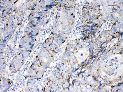
- Experimental details
- IHC analysis of Bax using anti-Bax antibody (A00183). Bax was detected in paraffin-embedded section of human lung cancer tissues. Heat mediated antigen retrieval was performed in citrate buffer (pH6, epitope retrieval solution) for 20 mins. The tissue section was blocked with 10% goat serum. The tissue section was then incubated with 1μg/ml rabbit anti-Bax Antibody (A00183) overnight at 4°C. Biotinylated goat anti-rabbit IgG was used as secondary antibody and incubated for 30 minutes at 37°C. The tissue section was developed using Strepavidin-Biotin-Complex (SABC)(Catalog # SA1022) with DAB as the chromogen.
- Additional image