Explore
 Validate
 Learn
Learn Western blot
 Immunocytochemistry
 Immunoprecipitation
Antibody data
- Antibody Data
- Antigen structure
- References [0]
- Comments [0]
- Validations
- Western blot [2]
- Immunoprecipitation [2]
- Immunohistochemistry [9]
Submit
Validation data
Reference
Comment
Report error
- Product number
- LS-C747226 - Provider product page

- Provider
- LSBio
- Product name
- BAX Antibody LS-C747226
- Antibody type
- Polyclonal
- Description
- Affinity purified
- Reactivity
- Human, Mouse, Rat
- Host
- Rabbit
- Isotype
- IgG
- Storage
- Store at -20°C. Avoid freeze-thaw cycles.
No comments: Submit comment
Enhanced validation
- Submitted by
- LSBio (provider)
- Enhanced method
- Genetic validation
- Main image

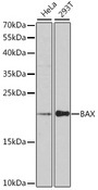
- Experimental details
- Western blot analysis of extracts of various cell lines, using BAX antibody at 1:1000 dilution. The secondary antibody used was an HRP Goat Anti-Rabbit IgG (H+L) at 1:10000 dilution. Lysates were loaded 25ug per lane and 3% nonfat dry milk in TBST was used for blocking. An ECL Kit was used for detection and the exposure time was 90s.
- Submitted by
- LSBio (provider)
- Enhanced method
- Genetic validation
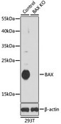
- Main image

- Experimental details
- Western blot analysis of extracts from normal (control) and BAX knockout (KO) 293T cells, using BAX antibody at 1:1000 dilution. The secondary antibody used was an HRP Goat Anti-Rabbit IgG (H+L) at 1:10000 dilution. Lysates were loaded 25ug per lane and 3% nonfat dry milk in TBST was used for blocking. An ECL Kit was used for detection and the exposure time was 90s.
Supportive validation
- Submitted by
- LSBio (provider)
- Enhanced method
- Genetic validation
- Main image
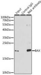
- Experimental details
- Immunoprecipitation analysis of 200ug extracts of 293T cells, using 3 ug BAX antibody. Western blot was performed from the immunoprecipitate using BAX antibody at a dilition of 1:1000.

- Submitted by
- LSBio (provider)
- Main image

- Experimental details
- Immunoprecipitation analysis of 200ug extracts of 293T cells, using 3 ug BAX antibody. Western blot was performed from the immunoprecipitate using BAX antibody at a dilition of 1:1000.

Supportive validation
- Submitted by
- LSBio (provider)
- Main image
- Experimental details
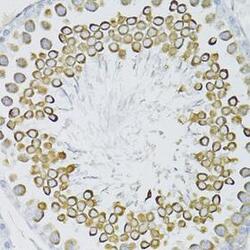
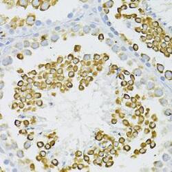
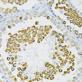
- Immunohistochemistry of paraffin-embedded rat testis using BAX antibody at dilution of 1:100 (40x lens).
- Submitted by
- LSBio (provider)
- Main image
- Experimental details
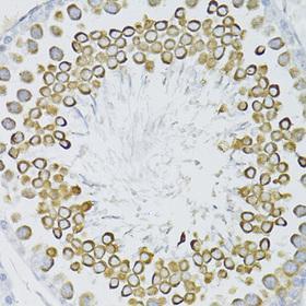
- Immunohistochemistry of paraffin-embedded mouse testis using BAX antibody at dilution of 1:100 (40x lens).
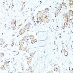
- Submitted by
- LSBio (provider)
- Main image
- Experimental details
- Immunohistochemistry of paraffin-embedded rat ovary using BAX antibody at dilution of 1:100 (40x lens).

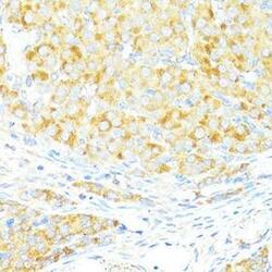
- Submitted by
- LSBio (provider)
- Main image
- Experimental details
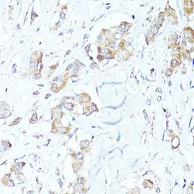
- Immunohistochemistry of paraffin-embedded human lung cancer using BAX antibody at dilution of 1:100 (40x lens).
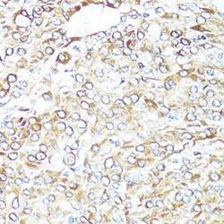
- Submitted by
- LSBio (provider)
- Main image
- Experimental details
- Immunohistochemistry of paraffin-embedded human mammary cancer using BAX antibody at dilution of 1:100 (40x lens).
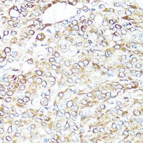
- Submitted by
- LSBio (provider)
- Main image

- Experimental details
- Immunohistochemistry of paraffin-embedded human stomach using BAX antibody at dilution of 1:100 (40x lens).
- Submitted by
- LSBio (provider)
- Main image
- Experimental details
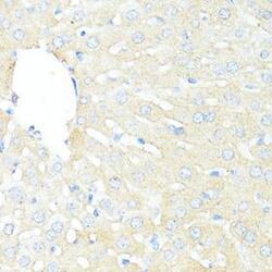
- Immunohistochemistry of paraffin-embedded mouse liver using BAX antibody at dilution of 1:100 (40x lens).
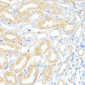
- Submitted by
- LSBio (provider)
- Main image
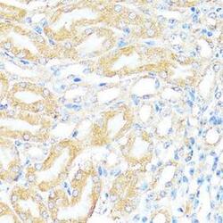
- Experimental details
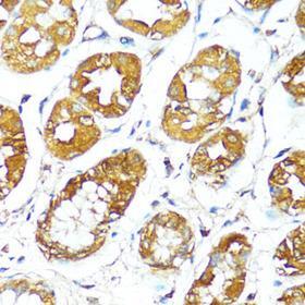
- Immunohistochemistry of paraffin-embedded mouse kidney using BAX antibody at dilution of 1:100 (40x lens).
- Submitted by
- LSBio (provider)
- Main image
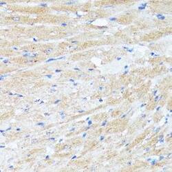
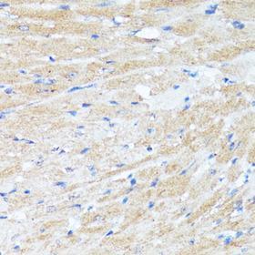
- Experimental details
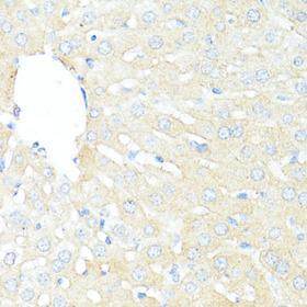
- Immunohistochemistry of paraffin-embedded mouse heart using BAX antibody at dilution of 1:100 (40x lens).