 Explore
 Validate
 Learn
Learn Western blot
Antibody data
- Antibody Data
- Antigen structure
- References [1]
- Comments [0]
- Validations
- Western blot [2]
- Immunocytochemistry [1]
Submit
Validation data
Reference
Comment
Report error
- Product number
- MAB846 - Provider product page

- Provider
- R&D Systems
- Product name
- Human Bax Minus C-Terminus Antibody
- Antibody type
- Monoclonal
- Description
- Protein A or G purified from hybridoma culture supernatant. Detects full-length human Bax in Western blots.
- Reactivity
- Human
- Host
- Mouse
- Conjugate
- Unconjugated
- Antigen sequence
Q07812- Isotype
- IgG
- Antibody clone number
- 127606
- Vial size
- 100 ug
- Concentration
- LYOPH
- Storage
- Use a manual defrost freezer and avoid repeated freeze-thaw cycles. 12 months from date of receipt, -20 to -70 °C as supplied. 1 month, 2 to 8 °C under sterile conditions after reconstitution. 6 months, -20 to -70 °C under sterile conditions after reconstitution.
Submitted references The role of nerve growth factor in hyperosmolar stress induced apoptosis.
Chang EJ, Im YS, Kay EP, Kim JY, Lee JE, Lee HK
Journal of cellular physiology 2008 Jul;216(1):69-77
Journal of cellular physiology 2008 Jul;216(1):69-77
No comments: Submit comment
Supportive validation
- Submitted by
- R&D Systems (provider)
- Main image

- Experimental details
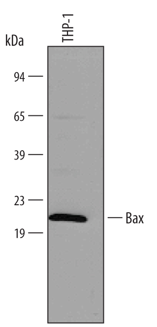
- Detection of Human Bax Minus C-Terminus by Western Blot. Western blot shows lysate of THP-1 human acute monocytic leukemia cell line. PVDF membrane was probed with 1 µg/mL of Human Bax Minus C-Terminus Monoclonal Antibody (Catalog # MAB846) followed by HRP-conjugated Anti-Mouse IgG Secondary Antibody (Catalog # HAF007). A specific band was detected for Bax Minus C-Terminus at approximately 21 kDa (as indicated). This experiment was conducted under reducing conditions and using Immunoblot Buffer Group 4.
- Submitted by
- R&D Systems (provider)
- Main image
- Experimental details
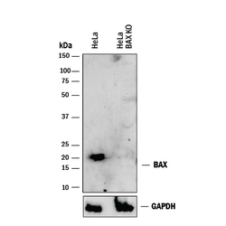
- Western Blot Shows Human Bax Specificity by Using Knockout Cell Line. Western blot shows lysates of HeLa human cervical epithelial carcinoma parental cell line and Bax knockout HeLa cell line (KO). PVDF membrane was probed with 1 µg/mL of Mouse Anti-Human Bax Minus C-Terminus Monoclonal Antibody (Catalog # MAB846) followed by HRP-conjugated Anti-Mouse IgG Secondary Antibody (Catalog # HAF018). A specific band was detected for Bax at approximately 20 kDa (as indicated) in the parental HeLa cell line, but is not detectable in knockout HeLa cell line. GAPDH (Catalog # MAB5718) is shown as a loading control. This experiment was conducted under reducing conditions and using Immunoblot Buffer Group 1.
Supportive validation
- Submitted by
- R&D Systems (provider)
- Main image
- Experimental details
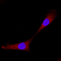
- Bax in A549 Human Cell Line. Bax was detected in immersion fixed A549 human lung carcinoma cell line using Mouse Anti-Human Bax Minus C-Terminus Monoclonal Antibody (Catalog # MAB846) at 8 µg/mL for 3 hours at room temperature. Cells were stained using the NorthernLights™ 557-conjugated Anti-Mouse IgG Secondary Antibody (red; Catalog # NL007) and counterstained with DAPI (blue). Specific staining was localized to cytoplasm. View our protocol for Fluorescent ICC Staining of Cells on Coverslips.
