Explore
 Validate
 Learn
Learn720129
antibody from Invitrogen Antibodies
Targeting: SMARCA4
BAF190, BRG1, FLJ39786, hSNF2b, SNF2, SNF2-BETA, SNF2L4, SNF2LB, SWI2
 Western blot
 Immunocytochemistry
Antibody data
- Antibody Data
- Antigen structure
- References [1]
- Comments [0]
- Validations
- Immunocytochemistry [2]
- Flow cytometry [1]
Submit
Validation data
Reference
Comment
Report error
- Product number
- 720129 - Provider product page

- Provider
- Invitrogen Antibodies
- Product name
- BRG1 Polyclonal Antibody
- Antibody type
- Polyclonal
- Antigen
- Recombinant full-length protein
- Description
- These Polyclonal antibodies are of rabbit origin developed by immunizing animals with proteins or peptides. The polyclonal antibody is purified by affinity purification from the rabbit sera generated after immunizing the rabbits with a specific type of protein or peptide. The purified antibody is tested for its functionality in various relevant research applications. The antibody is developed for Research Use Only and is non-hazardous or non-infectious in nature. This antibody is predicted to react with Monkey, Pig, Mouse and Rat.
- Reactivity
- Human, Mouse
- Host
- Rabbit
- Isotype
- IgG
- Vial size
- 100 μg
- Concentration
- 0.5 mg/mL
- Storage
- Store at 4°C short term. For long term storage, store at -20°C, avoiding freeze/thaw cycles.
Submitted references A cytoskeletal function for PBRM1 reading methylated microtubules.
Karki M, Jangid RK, Anish R, Seervai RNH, Bertocchio JP, Hotta T, Msaouel P, Jung SY, Grimm SL, Coarfa C, Weissman BE, Ohi R, Verhey KJ, Hodges HC, Burggren W, Dere R, Park IY, Prasad BVV, Rathmell WK, Walker CL, Tripathi DN
Science advances 2021 Apr;7(14)
Science advances 2021 Apr;7(14)
No comments: Submit comment
Supportive validation
- Submitted by
- Invitrogen Antibodies (provider)
- Main image

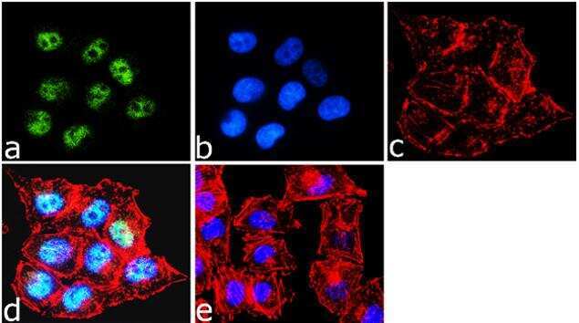
- Experimental details
- Immunofluorescence was performed on fixed and permeabilized A375 cells for detection of BRG1 using Anti-BRG1 Rabbit Polyclonal Antibody (Product # 720129, 1 µg/mL) and labeled with Goat anti-Rabbit IgG (H+L) Superclonal Secondary Antibody, Alexa Fluor® 488 conjugate (Product # A27034, 1:2000). Panel a) shows representative cells that were stained for detection and localization of BRG1 protein (green), Panel b) is stained for nuclei (blue) using SlowFade® Gold Antifade Mountant with DAPI (Product # S36938). Panel c) represents cytoskeletal F-actin staining using Alexa Fluor® 555 Rhodamine Phalloidin (Product # R415, 1:300). Panel d) is a composite image of Panels a, b and c clearly demonstrating nuclear localization of BRG1. Panel e) represents control cells with no primary Antibody to assess background.
- Submitted by
- Invitrogen Antibodies (provider)
- Main image
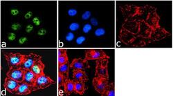
- Experimental details
- Immunofluorescence was performed on fixed and permeabilized A375 cells for detection of BRG1 using Anti-BRG1 Rabbit Polyclonal Antibody (Product # 720129, 1 µg/mL) and labeled with Goat anti-Rabbit IgG (Heavy Chain) Superclonal Secondary Antibody, Alexa Fluor® 488 conjugate (Product # A27034, 1:2000). Panel a) shows representative cells that were stained for detection and localization of BRG1 protein (green), Panel b) is stained for nuclei (blue) using SlowFade® Gold Antifade Mountant with DAPI (Product # S36938). Panel c) represents cytoskeletal F-actin staining using Alexa Fluor® 555 Rhodamine Phalloidin (Product # R415, 1:300). Panel d) is a composite image of Panels a, b and c clearly demonstrating nuclear localization of BRG1. Panel e) represents control cells with no primary Antibody to assess background.

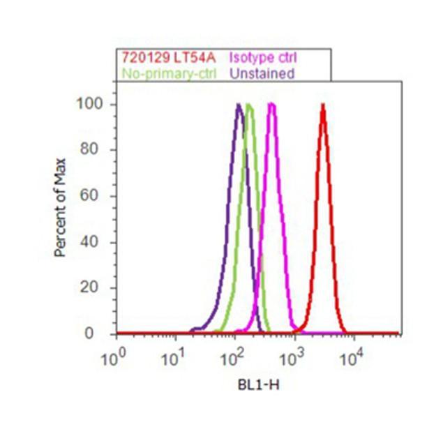
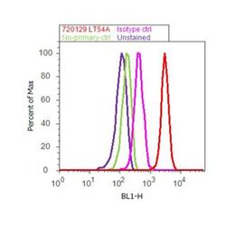
Supportive validation
- Submitted by
- Invitrogen Antibodies (provider)
- Main image
- Experimental details
- Flow Cytometry analysis of BRG1 was performed on HeLa cells labeled with BRG1 Rabbit Polyclonal Antibody (Product# 720129, 2-4 ug/ 1M cells) or with Rabbit isotype control and detected with Goat Anti-Rabbit IgG (H+L) Superclonalª Secondary Antibody, (Alexa Fluor¨ 488 conjugate, Product # A27034, 0.4 ug/ml, 1:2500) as represented by the red and pink histograms respectively. The purple histogram represents unstained control cells and the green histogram represents no-primary-Antibody control. A representative of 10,000 cells were acquired and analyzed for each sample using an Attune¨ Acoustic Focusing Cytometer (4468770).