 Explore
 Validate
 Learn
LearnMAB5738-100
antibody from R&D Systems
Targeting: SMARCA4
BAF190, BRG1, FLJ39786, hSNF2b, SNF2, SNF2-BETA, SNF2L4, SNF2LB, SWI2
 Western blot
Antibody data
- Antibody Data
- Antigen structure
- References [0]
- Comments [0]
- Validations
- Western blot [2]
- Immunohistochemistry [1]
Submit
Validation data
Reference
Comment
Report error
- Product number
- MAB5738-100 - Provider product page

- Provider
- R&D Systems
- Product name
- Human Brg1 Antibody
- Antibody type
- Monoclonal
- Description
- Protein A or G purified from cell culture supernatant. Detects human Brg1 in direct ELISAs and Western blots.
- Reactivity
- Human
- Host
- Rabbit
- Conjugate
- Unconjugated
- Antigen sequence
P51532- Isotype
- IgG
- Antibody clone number
- 2135A
- Vial size
- 100 ug
- Storage
- Use a manual defrost freezer and avoid repeated freeze-thaw cycles. 12 months from date of receipt, -20 to -70 °C as supplied. 1 month, 2 to 8 °C under sterile conditions after reconstitution. 6 months, -20 to -70 °C under sterile conditions after reconstitution.
No comments: Submit comment
Supportive validation
- Submitted by
- R&D Systems (provider)
- Main image
- Experimental details
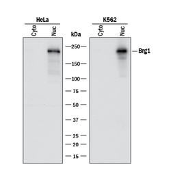
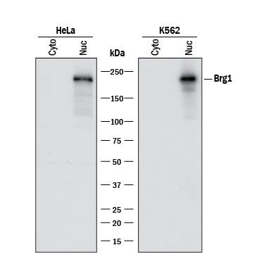
- Detection of Human Brg1 by Western Blot. Western blot shows lysates of HeLa human cervical epithelial carcinoma cell line and K562 human chronic myelogenous leukemia cell line. Gels were loaded with 30 μg of cytoplasmic (Cyto) and 15 μg of nuclear extracts (Nuc). PVDF membrane was probed with 0.5 µg/mL of Rabbit Anti-Human Brg1 Monoclonal Antibody (Catalog # MAB5738) followed by HRP-conjugated Anti-Rabbit IgG Secondary Antibody (Catalog # HAF008). A specific band was detected for Brg1 at approximately 220 kDa (as indicated). This experiment was conducted under reducing conditions and using Immunoblot Buffer Group 1.
- Submitted by
- R&D Systems (provider)
- Main image
- Experimental details
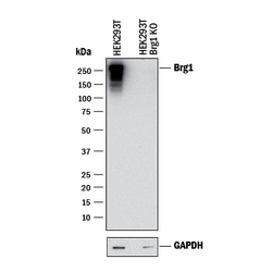
- Western Blot Shows Human Brg1 Specificity by Using Knockout Cell Line. Western blot shows lysates of HEK293T human embryonic kidney parental cell line and Brg1 knockout HEK293T cell line (KO). PVDF membrane was probed with 0.5 µg/mL of Rabbit Anti-Human Brg1 Monoclonal Antibody (Catalog # MAB5738) followed by HRP-conjugated Anti-Rabbit IgG Secondary Antibody (Catalog # HAF008). A specific band was detected for Brg1 at approximately 250 kDa (as indicated) in the parental HEK293T cell line, but is not detectable in knockout HEK293T cell line. GAPDH (Catalog # MAB5718) is shown as a loading control. This experiment was conducted under reducing conditions and using Immunoblot Buffer Group 1.

Supportive validation
- Submitted by
- R&D Systems (provider)
- Main image
- Experimental details
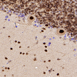
- Brg1 in Human Brain. Brg1 was detected in immersion fixed paraffin-embedded sections of human brain (cerebellum) using Rabbit Anti-Human Brg1 Monoclonal Antibody (Catalog # MAB5738) at 1 µg/mL for 1 hour at room temperature followed by incubation with the Anti-Rabbit IgG VisUCyte™ HRP Polymer Antibody (Catalog # VC003). Tissue was stained using DAB (brown) and counterstained with hematoxylin (blue). Specific staining was localized to neuronal nuclei. View our protocol for IHC Staining with VisUCyte HRP Polymer Detection Reagents.
