 Explore
 Validate
 Learn
Learn Western blot
 Chromatin Immunoprecipitation
Antibody data
- Antibody Data
- Antigen structure
- References [3]
- Comments [0]
- Validations
- Western blot [3]
- Immunocytochemistry [1]
- Immunohistochemistry [1]
Submit
Validation data
Reference
Comment
Report error
- Product number
- MA1-20800 - Provider product page

- Provider
- Invitrogen Antibodies
- Product name
- Nucleolin Monoclonal Antibody (4E2)
- Antibody type
- Monoclonal
- Antigen
- Other
- Description
- Recommended positive controls: Raji cell extract. This antibody does not react with mouse protein.
- Reactivity
- Human, Bovine
- Host
- Mouse
- Isotype
- IgG
- Antibody clone number
- 4E2
- Vial size
- 50 µg
- Concentration
- 1 mg/mL
- Storage
- Store at 4°C short term. For long term storage, store at -20°C, avoiding freeze/thaw cycles.
Submitted references Nucleolin interacts with the rabbit hemorrhagic disease virus replicase RdRp, nonstructural proteins p16 and p23, playing a role in virus replication.
PGRMC1 localization and putative function in the nucleolus of bovine granulosa cells and oocytes.
DGCR8 Acts as an Adaptor for the Exosome Complex to Degrade Double-Stranded Structured RNAs.
Zhu J, Miao Q, Guo H, Tang A, Dong D, Tang J, Wang F, Tong G, Liu G
Virologica Sinica 2022 Feb;37(1):48-59
Virologica Sinica 2022 Feb;37(1):48-59
PGRMC1 localization and putative function in the nucleolus of bovine granulosa cells and oocytes.
Terzaghi L, Luciano AM, Dall'Acqua PC, Modina SC, Peluso JJ, Lodde V
Reproduction (Cambridge, England) 2018 Mar;155(3):273-282
Reproduction (Cambridge, England) 2018 Mar;155(3):273-282
DGCR8 Acts as an Adaptor for the Exosome Complex to Degrade Double-Stranded Structured RNAs.
Macias S, Cordiner RA, Gautier P, Plass M, Cáceres JF
Molecular cell 2015 Dec 17;60(6):873-85
Molecular cell 2015 Dec 17;60(6):873-85
No comments: Submit comment
Supportive validation
- Submitted by
- Invitrogen Antibodies (provider)
- Main image

- Experimental details
- Western Blot analysis using Nucleolin [4E2] antibody Nucleolin Monoclonal Antibody (4E2) (Product # MA1-20800) on HL60 (1) and ZR-75 (2) cell extracts.

- Submitted by
- Invitrogen Antibodies (provider)
- Main image
- Experimental details
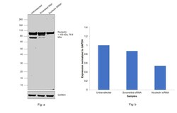
- Knockdown of Nucleolin (NCL) was achieved by transfecting HeLa with Nucleolin specific siRNAs (Silencer® select Product # s9314). Western blot analysis (Fig. a) was performed using Whole Cell Lysate extracts from knockdown cells (Lane 3), non-specific scrambled siRNA transfected cells (Lane 2) and untransfected cells (Lane 1). The blot was probed with Nucleolin Monoclonal Antibody (4E2) (Product # MA1-20800, 1:1000 dilution) and Goat anti-Mouse IgG (H+L), Superclonal™ Recombinant Secondary Antibody, HRP (Product # A28177, 1:4000 dilution). Densitometric analysis of both the observed bands has been done together, this western blot is shown in histogram (Fig. b). Decrease in signal of all the bands upon siRNA mediated knock down confirms that antibody is specific to Nucleolin. Nucleolin is a ubiquitinylated protein, for which the molecular weight is reported to be 76.6 kDa to 100 kDa, which can be observed clearly in the Fig. a.

- Submitted by
- Invitrogen Antibodies (provider)
- Main image
- Experimental details
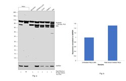
- Western blot was performed using Anti-Nucleolin Monoclonal Antibody (4E2) (Product # MA1-20800). Nucleolin is a ubiquitinylated protein for which the molecular weight is reported to be 76.6 kDa to 100 kDa, which can be observed in the cell lines tested. Whole cell extracts (30 µg lysate) of HeLa (Lane 1), HeLa treated with Heat Shock at 44 degree Celsius for 3 hrs (Lane 2), SH-SY5Y (Lane 3), NTERA-2 cl.D1 (Lane 4), A-431 (Lane 5), PANC-1 (Lane 6) and Raji (Lane 7) were electrophoresed using Novex® NuPAGE™ 4-12% Bis-Tris Protein Gel (Product # NP0322BOX). Resolved proteins were then transferred onto a nitrocellulose membrane (Product # IB23001) by iBlot® 2 Dry Blotting System (Product # IB21001). The blot was probed with the primary antibody (1:1000 dilution) and detected by chemiluminescence with Goat anti-Mouse IgG (H+L), Superclonal™ Recombinant Secondary Antibody, HRP (Product # A28177, 1:4000 dilution) using the iBright FL 1000 (Product # A32752). Chemiluminescent detection was performed using Novex® ECL Chemiluminescent Substrate Reagent Kit (Product # WP20005).

Supportive validation
- Submitted by
- Invitrogen Antibodies (provider)
- Main image
- Experimental details
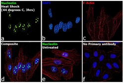
- Immunofluorescence analysis of Nucleolin was performed using HeLa cells. The cells were fixed with 4% paraformaldehyde for 10 minutes, permeabilized with 0.1% Triton™ X-100 for 15 minutes, and blocked with 2% BSA for 1 hour at room temperature. The cells were labeled with Nucleolin Monoclonal Antibody (4E2) (Product # MA1-20800) at 5 microgram/mL concentration in 0.1% BSA, incubated at 4 degree celsius overnight and then labeled with Goat anti-Mouse IgG (H+L) Superclonal™ Recombinant Secondary Antibody, Alexa Fluor® 488 conjugate (Product # A28175) at a dilution of 1:2000 dilution for 45 minutes at room temperature (Panel a: green). Nuclei (Panel b: blue) were stained with ProLong™ Diamond Antifade Mountant with DAPI (Product # P36962). F-actin (Panel c: red) was stained with Rhodamine Phalloidin (Product # R415, 1:300). Panel d represents the merged image showing increase in nuclear staining upon heat shock. Panel e represents untreated cells, showing lower expression of Nucleolin. Panel f represents control cells with no primary antibody to assess background. The images were captured at 60X magnification.

Supportive validation
- Submitted by
- Invitrogen Antibodies (provider)
- Main image
- Experimental details
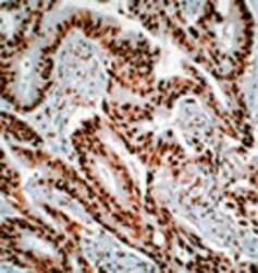
- Immunohistochemistry analysis of nuclear staining of colon cancer sections using Nucleolin Monoclonal Antibody (4E2) (Product # MA1-20800).
